 |
| About Bioline | All Journals | Testimonials | Membership | News |
|
||||||
|
||||||
An anthracyclic glycoside from a member of the Actinoplanaceae R. SAHAI, N.N.GERBER+, C.DeBROSSE*, P. OFFEN* and M.P.LECHEVALIER
Waksman Institute of Microbiology, P.O. Box 759, Rutgers, The
State University of New Jersey, Piscataway, NJ 08855-0759
and
Code Number: AC96001
Sizes of Files:
    Text: 15.8K
    Graphics: line drawings (gif) - 5.0K
Abstract. An actinomycete, tentatively classed within the "spore-dome" (Actinoplanaceae) group, has been demonstrated to produce a red anthracyclic C-glycoside esterified to a dienoic fatty acid (Structure 1). The pigment appears to be similar or identical to a recently-reported streptomycete pigment. During a screening program designed to isolate members of the Actinoplanaceae, a single colony of an actinomycete was isolated from streambank debris collected in Piscataway, NJ, USA. It produced a brilliant soluble red pigment on isolation plates and in certain liquid media. This paper reports on the production and structure of the pigment and the characteristics of the producing organism. Materials and methods Isolation and Characterization of the Actinomycete. The actinomycete (LL-3010) was isolated from leaf debris collected from the banks of a stream. Surface-inoculated dilution plates of crude water agar (2%) amended with actidione and nystatin were incubated for four weeks at 28 C. The isolated strain was maintained on Bennett's agar slants. Morphological observations were carried out directly on water agar and half-strength Bennett's agar plates, using a light microscope with a long working distance condenser. Chemotaxonomic analyses were carried out according to Lechevalier and Lechevalier (1980).
Production of the Pigment. The organism was shaken for six days at 215 RPM at 28 C in Yeast extract (0.4%) Czapek's broth (YCz). The cells were harvested by centrifugation.
Extraction and Chemical Analysis of the Pigment. The methods used will be described in the Results section. Results Taxonomy of the Strain.
Morphological studies of the unknown organism showed that it had rather distinctive properties. On certain agar media, such as Bennett's, it formed colonies of hyphae arranged in radial rosettes and imbedded in a viscous slime. The hyphae bore single rows of spores. When wetted, the terminal spores of each chain became motile, more or less synchronously, and after a few minutes, the next in line also became motile. If the preparation were allowed to dry, then rewetted, the sequential spore release began again until all spores had become motile. On the basis of this morphology, the organism was tentatively assigned to the "spore-dome" group of Willoughby (1969) which has recently been reviewed by Cross and Makkar (1986).
Chemically, the organism was shown to have a cell wall of type III, containing both the meso and L isomers of diaminopimelic acid in approximately equal amounts. The strain has a whole cell sugar pattern of type B (madurose; 3-O-methyl-D-galactose) and a phospholipid pattern of type PI (no nitrogenous phospholipids) (Lechevalier and Lechevalier, 1980).
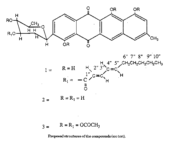
Proposed structures of the compounds (see text).
The pigment was isolated in crude form by extraction of the cells with 80% acetone. Addition of an equal volume of chloroform to the acetone extract and mixing caused the pigment to partition into the chloroform layer. The final step of purification was achieved by chromatography on silica gel in chloroform with gradually increasing concentrations of ethyl acetate. The pure compound was crystallized from chloroform as orange needles (M.p. 208 C; UV (ethanol) max 266,497 nm; IR (KBr) 3400, 2935, 2860, 1715, 1610, 1435, 1395, 1280, 1150, 1090, 850 cm-l). Mass spectroscopic studies indicated the molecular formula C35H3609. The compound gave a positive ferric chloride test suggesting the presence of phenolic hydroxyl groups and was decolorized by sodium borohydride and reoxidised in air to the original chromophore suggesting a rapidly reversible reduced system such as a quinoid system (Gerber and Lechevalier, 1984). The zinc dust distilled product showed UV absorptions similar to that of naphthacene (Scott, 1964). A strong band at 1610 cm^-1 in the IR spectrum supported the conclusion that carbonyls of the quinoid system were hydrogen-bonded to the peri hydroxyl groups (Bloom et al., 1959). The ^1H NMR spectrum had several peaks in addition to what would be expected of an anthracyclic skeleton. The compound could not be hydrolysed even under strong acidic hydrolytic conditions; however, mild alkaline hydrolysis with potassium carbonate at room temperature resulted in a saponified product which was purified by silica gel chromatography using chloroform and increasing concentrations of methanol as eluant. Its UV spectrum was identical to that of original compound and it was crystallized with acetone and methanol as orange needles (m.p. 293 C; IR (KBr) 3360, 2920, 2845, 1610, 1430, 1380, 1260, 1080 cm^-1. The loss of the 1715 cm^-1 peak in the IR spectrum indicated a conjugated ester in the original compound. The mass spectrum of the saponified pigment (Structure 2) showed M+ 450, corresponding to a molecular formula C25H22O8, and its ^1H NMR lacked several peaks present in the ^1H NMR of the original compound, both in the high and low field region. The simplified ^1H NMR of the saponified product showed two ortho-coupled protons at delta 8.15 and 8.08 (J=8Hz). There were three singlets in the aromatic region each integrating for one proton at delta 9.28, 8.00 and 7.20. The lowest field singlet at delta 9.28 was assigned as a peri proton to a carbonyl group. The proton appearing as a singlet at relatively high field at delta 7.2 was assigned to the H-9 proton having a hydroxyl group at the ortho position. The coupling constants and the coupling established by COSY experiments were in conformity with the presence of a deoxy-fucose sugar moiety in the molecule (Table 1).
--------------------------------------------------------------
PROTON  CHEM SHIFT   MULTIPLICITY  INTEGR. COUPLED TO COSY
          (ppm)
--------------------------------------------------------------
1,2    8.15, 8.08    d of d (J=8hz)    2    each other, 1'
6         9.28            s            1    7,8-CH3, 9
7         8.00            s            1    6,8-CH3, 9
8-CH3     2.38            s            3    6,7,9
9         7.2             s            1    6,7,8-CH^3
1'        5.2         d (J=11Hz)       1    1,2,2'eq.*, 2'ax.
2'axial   1.95            q            1    1',2'eq., 3'
2'equit   2.97          d of d         1    1',2'ax**, 3'
3'        4.3             m            1    2'ax.,2'eq., 4'
4'        3.7             t            1    2'ax.,2'eq., 4'
5'        3.88            m            1    4',6'
6'        1.7             d            3    5'

*   = "equit"
** = "axial"
--------------------------------------------------------------
Table 1. ^1H NMR of the saponified pigment in
pyridine-d5
--------------------------------------------------------------
The mass spectrum of the pigment showed a fragment at m/z 115 corresponding to the sugar moiety. The anomeric proton appeared as a doublet (J =11 Hz) at delta 5.2. The presence of a singlet at delta 2.38 integrating for three protons was assigned to an aromatic methyl and a doublet at delta 1.7 integrating for three aliphatic protons was assigned to a fucosyl methyl.
On acetylation with acetic anhydride and pyridine, the saponified product gave a pentaacetate product (Structure 3). The spectrum showed methyls of two aliphatic acetates as sharp singlets at delta 2.02 and 2.08 and that of three aromatic acetates at delta 2.55, 2.56 and 2.58. The acetylation resulted in a downfield shift of two aliphatic carbinolic protons H-3' and H-4' to a^o 4.86 and 4.76 respectively. The aromatic proton ortho to a hydroxyl group showed a downfield shift to delta 7.79. This confirmed the presence of three phenolic hydroxyl groups and two aliphatic hydroxyl groups. Structure 2 best explained the chemical shift, coupling constants and pattern for the saponified pigment.
--------------------------------------------------------------
PROTON  CHEM  MULTIPLICITY  INTEGR.  COUPLED TO   COUPLED TO
        SHIFT                        SHORT RANGE  LONG RANGE
        (ppm)                        COSY         COSY
--------------------------------------------------------------
1, 2     7.8  d of d(J =8Hz)   2        -            1'
6        8.5       s           1        -            -
7        7.6       s           1        -          8-CH3,9
8-CH3    2.4       s           3        -            7,9
9        7.0       s           1        -          7,8-CH3
1'       4.9       m           1       2'ax.   2'ax.,2'eq.,1,2

2'ax.    1.4       m           1    1',2'eq.,3'     1',3'
2' eq.   2.4       d           1       2'ax.        1',3'
3'       4.9       m           1     2'ax.,4'     2'ax.,2'eq.
4'       3.2       m           1        3',5'         -
5'       3.5       t           1        4',6'         6'

6'       1.3       d           3         5'           5'
2"       5.9   d(j=l5Hz)       1         3"           3"
3"       7.2  d of d(J=15,     1        2",4"       2",4"
                 9.7Hz)
4",5"    6.2       m           2        3",6"       3",6"
6"       2.1       t           2          7"          5"
7",8",9" 1.2       m           6        6",10"       10"
10"      0.8       t           3          9"          9"
--------------------------------------------------------------
Table 2. ^1H NMR of the pigment in DMSO-d6
--------------------------------------------------------------
The observation that the deoxy-fucose sugar moiety of the pigment could not be hydrolysed under strong acidic and basic conditions used was suggestive of a C-linked glycoside. Further proof of a C-linked sugar and the position at which it was attached came from a long-range COSY experiment where the H-1' of the sugar was found to have long-range coupling with ortho aromatic protons H-1 and H-2, thereby indicating a C-glycosidic linkage at the C-3 position.
In the IR spectrum of the pigment, the absorption at 1715 cm^- 1 which disappears in the saponified product, was suggestive of a conjugated ester. The mass spectrum of the pigment had fragments m/z 169 and 150 arising from the fatty acid. The additional resonances appearing in its ^1H NMR and absent in the saponified product, could be easily established to be that of a fatty acid with conjugated double bonds (Table 2). The site of acylation was assigned to C-3' from the downfield shift of H-3' to delta 4.9 in the pigment from that in the saponified product at delta 4.3. The low field region showed three additional signals at delta 7.25 (^1H), 6.23(2H) and 5.87(1H) stemming from the conjugated double bonds of the fatty acid. The sharp doublet at delta 5.87(J=15 Hz) was assigned to the terminus of the conjugated system adjacent to the carboxyl carbon trans to the second olefinic hydrogen H-3" at delta 7.25 (d of d, J=15, 9.7 Hz). This proton was coupled to one of the two protons appearing at delta 6.23 (J=9.7 Hz). These protons are coupled to each other in an ABXY2 system, which is in turn coupled to an allylic methylene signal at 2.1 ppm. Decoupling of the methylene protons resulted in a simplified ABX subspectrum centered at delta 6.25. The 15 Hz coupling revealed by analysis of this region suggested a trans stereo- chemistry of the second double bond Silverstein et al., 1981). A triplet at 5 0.8 integrating for three protons was assigned to a terminal methyl and a multiplet at 51.2 was assigned to six protons of internal methylenes of which one is coupled to the allylic methylene(H-5"). This suggested the remaining portion of the acyl chain to be an unbranched hydrocarbon and the fatty acyl component to be trans, transdeca-2,3,4,5-dienoic acid.
Biological Activity. The red pigment studied here belongs to the class of anthracyclic glycosides. Several members of this class have anticancer activity and activity against grampositive bacteria. Biological evaluation of our compound showed weak activity against gram-positive bacteria. It is not known whether it also has antitumor activity. Conclusions The pigment whose chemistry is reported in this paper appears to be almost identical to the pigment recently reported from a streptomycete (Arnone et al., 1988). Both structures are compatible with the data presented; however, considerable degradative chemistry would be required to prove which of the two structures is correct. Acknowledgement. The authors are grateful to the Charles and Jobanna Busch Foundation and the Actinomycete Fund for financial support. References Arnone, A., R.Cardillo, G.Nasini, O.Vajna de Pava & S.Quaroni (1988). A new strain of Streptomyces: an anthracycline containing a C-glucoside moiety and a chiral decanol. Phytochemistry, 1988:3611-3617 Bloom, H, L.H.Briggs & B.Cleverley (1959). Physical properties of anthraquinone and its derivatives. Part I. Infrared spectra. J. Chemical Soc., 1959:178 Cross, T. & N.J.Makkar (1986). Spore dome actinomycetes. In: G.Szabo, S.Biro & M.Goodfellow (eds.) Biological, Biochemical and Biomedical Aspects of Actinzomycetes. Akademiai Kiado, Budapest, Part B, pp. 579-581 Gerber, N.N. & M.P.Lechevalier (1984). Novel benzo (a) naphthacene quinones from an actinomycete, Frankia sp. G2 (ORS 020604). Can.J. Chem., 62: 2818-2821 Lechevalier, M.P. & H.A. Lechevalier (1980). The chemotaxonomy of actinomycetes. In: A.Dietz & D.W.Thayer (eds.) Actinomycete Taxonomy. Society of Industrial Microbiology Special Publication #6, Arlington, Va., USA, pp. 227-291 Scott, A.I. (1964). Intrepretation of Ultraviolet Spectra of Natural Products. Pergamon, Elmsford, NY, USA, p. 127 Silverstein, R.M., G.C.Bassler & T.C.Morrill (1981). Spectrophotometric Identification of Organic Compounds. John Wiley & Sons, New York, Appendix B, p. 223 Willoughby, L.G. (1969). A study on aquatic actinomycetes; the allochthonous leaf component. Nova Hedwigia, 18: 45-113. Copyright 1991 C.E.T.A., The International Centre for Theoretical and Applied Ecology, Gorizia. The following images related to this document are available:Line drawing images[ac91001a.gif] |
| |||||||||