 |
| About Bioline | All Journals | Testimonials | Membership | News |
|
||||||
|
||||||
STREPTOMYCES SP. 1T1, A NEW STENDOMYCIN PRODUCER ISOLATED FROM SOIL B.LOSILLA, M.T.POMMIER and G.MICHEL Laboratoire de Biochimie Microbienne, Universite Claude Bernard-Lyon I, 43 Bd du 11 Novembre 1918, F-69622 Villeurbanne Cedex, France.
Code Number: AC92001
Sizes of Files:
    Text: 18K
    Graphics: Photos (jpg) 74K / Halftones (gif) 46K
ABSTRACT. An actinomycete was found to produce stendomycin, an antifungal peptidolipid, in synthetic organic media. The isolate forms aerial mycelium and there is no fragmentation of vegetative mycelium. The fatty acid composition and the phospholipid pattern are very similar to those, reported for Streptomyces. Cell walls are of type I, they contain LL-diaminopimelic acid and glycine. The results of biochemical, cultural and physiological tests suggest that the strain is related to Streptomyces endus and S. hygroscopicus which are known to produce stendomycin or a related peptidolipid. However the new isolate consistently differs from these Streptomyces by the utilization of carbon sources, tolerance to NaCl and temperature of growth. We have designated the organism Streptomyces sp. 1T1, until it can be allocated to a named taxon. Stendomycin is an antifungal peptidolipid isolated by Thompson and Hughes (1963) from a strain of Streptomyces. The producing strain exhibited properties similar to the Streptomyces species S. endus (NRRL 2339), S. antimycoticus (A 158), S. narbonensis (NRRL B-168), S. arabicus (NRRL B-1733) and S. naganishi (NRRL B-1816), but taxonomic studies showed that it could be considered as a member of the species S. endus (Thompson and Hughes, 1963). Another antifungal peptidolipid, pantomycin, very close to stendomycin, was isolated from cultures of S. hygroscopicus (NRRL 2751) (Gurusiddaiah and Graham, 1980). Both Streptomyces have been described as strains producing antibiotics in complex fermentation media. In the course of our studies on the biosynthesis of stendomycin we needed strains producing stendomycin in simple media in order to determine the influence of organic nutrients on the synthesis of the antibiotic. Our screening for antifungal peptidolipids produced by microorganisms led us to isolate a novel strain of Streptomyces which produces stendomycin in very simple media containing glycerol (1%) and KNO3 (0,2%) or a- amino acid (0,07%) (Losilla et al., 1991). We describe the characteristics of this strain which was studied in comparison with other species of Streptomyces which produce stendomycin or which are taxonomically related to stendomycin-producing species.
MATERIALS and METHODS Strains. The new isolate, strain 1T1, was recovered from a soil sample obtained from Diemoz-village (France). Streptomyces endus (NRRL 2339), S. hygroscopicus (NRRL 2751), S.narbonensis (NRRL B-1680), S.naganishi (NRRL B-1816) and S.arabicus (NRRL B-1738) were obtained from Eli Lilly Laboratories (USA). Cultural and physiological characterization. Cultural and physiological characterizations were carried out according to the methodology suggested by the International Streptomyces Project (ISP) (Shitling and Gottlieb, 1966), using the Colour Index Hue Indication Chart (Society of Dyers and Colourists, Bradford, UK). Carbohydrate utilization was determined according to Pridham and Gottlieb (1948). All the cultures were incubated for 14 dd at 28 C. Morphological characterization. Media used in this study were those recommended for characterization of Streptomyces species: yeast extract-malt extract agar (ISP2), oatmeal agar (ISP3), starch-inorganic salts agar (ISP4), glucose-asparagine agar, Hickey -Tresner agar, nutrient agar, sucrose-nitrate agar (Pridham et al., 1956; Waksman, 1961; Shirling and Gottlieb, 1966). Aerial mycelia were examined by light and by transmission (Jeol 1200 EX) and scanning (Jeol 35 CF) electron microscopy. Chemotaxonomic studies. Cell walls were prepared from washed cells according to the procedure described by Bordet et al. (1972). Purified cell wall preparations were hydrolyzed with 6M HCl, 16 h at 110 C for amino acid analysis, with 3M HCl, 4 h at 100 C or 2M HCl, 2 h at 100 C for amino sugars and sugars respectively. Lipids were extracted with chloroform-methanol (2:1 by vol.). Extracts were washed with the Folch solvent (Folch et al., 1957) and phospholipids were separated by acetone precipitation. The polar lipids analysis was carried out by thin layer chromatography (TLC) on Silica-gel 60 plates (Merck, Germany) in the solvent system chloroform- methanol-water (65:25:4 by vol.) and chloroform-methanol-propan-1-ol-25% KCl-ethyl acetate (25:13:25:9:25 by vol.). Fatty acids were obtained by 2M HCl hydrolysis of lipids. They were extracted with chloroform, esterified with dia- zomethane and analysed by gas chromatography on a SP-2100 capillary columns (25 m x 0.32 mm). Cell wall amino acids were analysed by gas chromatography of N-heptafluoro butyryl butyl esters on Chirasil Val III capillary column (20 m x 0.32 ml (O'Donnell et al., 1982). The chirality diaminopimelic acid was determined TLC on cellulose in the following solvents: methanol-water-pyridine (77:20:10 by vol.) or methanol-water-10M HCl pyridine (80:17.5:2.5:1 by vol.), (Rhuland et al., 1955). RESULTS Chemotaxonomic characteristic The fatty acid composition of total lipid is given in Table 1. In all strains tested, i-C14:0, ai-C15:0,i-C16:0 fatty acids were the major components. Palmitic acid was also found in all strains except S.naganishi. The fatty acid profile of strain 1T1 was very similar to that previously reported for the genus Streptomyces (Kroppenstedt, 1985). All strains possessed phosphatidylinositol, phosphatidylinositomannoside, phosphatidylglycerol, diphosphatidylglycerol and phosphatidylethanolamine (type II phospholipid pattern according to Lechevalier et al., 1977).
-------------------------------------------------------------
            Strain  S. arabicus  S. endus   S. hygroscopicus
Fatty acid   1T1    NRRLB-1733   NRRL2339    NRRL2751
-------------------------------------------------------------
i-C14:0(a)    15        21         9           22
i-C15:0       12        8          9           12
ai-C15:0      14        13         25          13
C15:0         -         10         14          -
ai-C16:0      30        32         19          35
C16:0         21        16         16          18
i-C17:0       -         -          -           -
ai-C17:0      8         -          8           -

-------------------------------------------------------------
             Strain     S. naganishi    S. narbonensis
Fatty acid    1T1       NRRLB-1816      NRRLB-1680
-------------------------------------------------------------
i-C14:0(a)    15           9                13
i-C15:0       12           6                10
ai-C15:0      14           23               25
C15:0         -            -                -
ai-C16:0      30           62               32
C16:0         21           -                30
i-C17:0       -            -                -
ai-C17:0      8            -                -
-------------------------------------------------------------
Table 1. Fatty acid composition of the isolate and selected
species: values are given as percentages total peak area from
GLC of methylesters. (a)Shorthand designations for fatty
acids: i-C14:0 is 12 methyltridecanoic acid
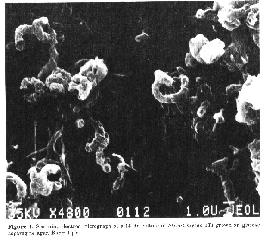
Cell walls of all strains contained the following components of peptidoglycan: D-glucosamine, muramic acid, D-glutamic acid, L-alanine, glycine and LL-diaminopimelic acid, corresponding to chemotype I as defined by Lechevalier characteristic sugars were identified. All these observations support the assignment of the strain 1T1 to the genus Streptomyces (Streptomyces sp. 1T1). Chemotaxonomic studies showed more similarities with two strains: S.endus (NRRL 2339) and S.hygroscopicus (NRRL 2751). Several differences were observed in the polar lipid profiles and fatty acid composition in comparison with the other strains investigated. Morphological characteristics. The vegetative mycelium of strain 1T1 does not fragment. Spore chains (10 or more spores per chain) are spiral (Fig. 1). Spores are cylindrical in shape and 0.4 x 0.3 um in size with smooth spore surface (Dietz and Mathews, 1971). S.endus and S.hygroscopicus also produce spiral spore chains with smooth surfaces 1 x 0.4 u.m and 0.5 x 0.3 u.m in size respec- tively.
-------------------------------------------------------------
Medium       Growth    Colour of Vegetative  Colour of Aerial
                          mycelium               mycelium
-------------------------------------------------------------
Sucrose-nitrate
agar                 moderate    brownish       gray
Inorganic
salts-starch agar    moderate    white      brownish, copious
Oatmeal agar         good        white          abundant
Nutrient agar        moderate    yellow         absent
Glucose-asparagine
agar                 moderate    white          gray
Hickey-Tresner agar  good        yellow
Yeast extract-malt
extract agar         good                       absent
-------------------------------------------------------------
Table 2. Cultural characteristics of strain 1T1
----------------------------------------------------------
                 Strain    S. endus    S. hygroscopicus
Carbon source    1T1       NRRL 2339   NRRL 2751
----------------------------------------------------------
D-glucose         +          ++            +
D-mannose         +          ++            +
sucrose           +          +             +
D-fructose        ++         ++            +
D-xylose          +          ++            ++
L-arabinose       ++         +             +
L-rhamnose        +          ++            ++
meso-inositol     +          ++            ++
salicin           +          +             +
cellulose         -          -             -
----------------------------------------------------------
Table 3. Utilization of carbon sources on basal medium
(Pridham and Gottlieb, 1948) by strain 1T1, S.endus and
S.hygroscopicus. -: No growth; +: growth equivalent to
that with 1% glucose (positive control); ++: growth greater
than that on positive control.Cultural and physiological characteristics. The cultural characteristics of strain 1T1 are shown in Table 2. The organism grows well on most complex and synthetic organic media. Growth is moderate on media containing only inorganic nitrogen sources. Physiological properties were studied in comparison with S.endus and S.hygroscopicus, the results are shown in Tables 3 and 4. DISCUSSION So far stendomycin production has been reported only in a strain of S. endus (NRRL 2339) and a strain of S. hygroscopicus (NRRL 2751) has been found to produce pantomycin, a peptidolipid very close to stendomycin (Bodanski et al., 1969). Comparison of strain 1T1 with S.endus, S.hygroscopicus and with the related species S. narbonensis, S.arabicus and S. naganishi, indicates that, on the basis of fatty acid composition and cell wall type, it is a member of the genus Streptomyces.
-------------------------------------------------------------
Property             Strain   S. endus     S. hygroscopicus
                     1T1      NRRL 2339    NRRL 2752
-------------------------------------------------------------
Melanin formation        -        -          +
Nitrate reduction        -        -          -
Starch hydrolysis        +        +          +
Casein hydrolysis        +        +          v(a)
Gelatin liquefaction     +        +          +
Milk peptonization       v        +          v
Milk coagulation         -        +          -
Growth in the presence
of NaCl (maximum         5%       7%         9%
concentration
allowing growth)
Growth at:
          10 C           -        -           -
          22 C           +        -           -
          28 C           +        +           +
          37 C           +        -           +
          45 C           -        -           +
-------------------------------------------------------
Table 4. Physiological properties of strain 1T1, S.endus
and S. hygroscopicus. (a) v: variable.Streptomyces. a-Hydroxy fatty acids in phosphatidylethanolamine were found in the new strain and in S.endus and S.hygroscopicus, but not in S.arabicus, S. naganishi and S.narbonensis. Like S.endus and S.hygroscopicus, the strain 1T1 has spiral chains of smooth surfaced spores, their size (0.4 x 0.3 um) is smaller than that of S.endus (1 x 0.4 um). The new isolate 1T1 differs from S. endus and S.hygroscopicus in the following properties: coagulation of milk, utilization of sugars as carbon source, melanin formation, tolerance to NaCl and temperature of growth (Table 4). We propose to designated the new isolate Streptomyces sp. 1T1 until it can be allocated to a named taxon. REFERENCES Bodanski, M., l.Izedebski & I.Muramatsu (1969). The structure of the peptide antibiotic stendomycin. J.Amer. Chem. Soc., 91:2351-2358 Bordet, C., M.Karajoli, O.Gateau & G.Michel (1972). Cell walls of Nocardia and related actinomycetes: Identification of the genus Nocardia by cell-wall analysis. Int. J. Syst. Bacteriol. 251-259 Dietz, A. & J.Mathews (1971). Classification of Streptomyces spore surfaces into five groups. Appl. Microbiol., 21:527-533 Folch, J., H.Lees & G.H.Sloane-Stanley (1957). A simple method for the isolation and the purification of total lipids from animal tissues. J. Biol. Chem., 226:497-509 Gurusiddaiah, S. & S.O.Graham (1980). Some chemical and physical characteristics of pantomycin an antibiotic isolated from Streptomyces hygroscopicus. Antimicrob.Agents and Chemother, 17:980-987 Kroppenstedt, R.M. (1985).Fatty acid and menaquinone analysis of actinomycetes and related organisms. In: M. Goodfellow & D.E. Minnikin (eds.) Chemical Methods in Bacterial Systematics. Academic Press, London, pp. 173-199. Lechevalier, M.P., C.De Brievre & H.A. Lechevalier (1977). Chemotaxonomy of aerobic actinomycetcs: phospholipid composition. Biochem. Syst. Ecol., 5:249-260 Lechevalier, M.P. & H.A. Lechevalier (1970). Composition of whole-cell hydrolysates as a criterion in the classification of aerobic actinomycetes. In: H.Prauser (ed.) The Actinomycetales. G.Fischer Verlag, Jena, pp. 211-216 Losilla, B., M.T.Pommier, N.Bonnaveiro, A. Cremieux & G.Michel (1991). Structure activity relationships of stendomycin, a lipopeptide antibiotic from Streptomyces. Microbios (in press) O'Donnell. A.G. D.E.Minnikin. M. Goodfellow and J. H. Parlett (1982). The analysis of actinomycete wall amino acids by gas chromatography. FEMS Microbiol. Lett., 15:75-78 (1982). Pridham,T.G., P.Anderson, C.Foley, L.A. Lindenfelser, C.W.Hesseltine & R.G. Benedict (1956). A selection of media for maintenance and taxonomic study of Streptomyces. Antibiot. Ann., 57:947-953 Pridham, T.C. & D.Gottlieb (1948). The utilization of carbon compounds by some Actinomycetales as an aid for species determination. J. Bacteriol., 56:107-114 Rhuland, L.E., E.Work, R.F.Denman & D.S. Hoare (1955). The behavior of the isomers of a,e-diaminopimelic acid on paper chromatograms. J. Amer. Chem. Soc., 77:4844-4846 Schleifer, K.H. & O.Kandler (1972). Peptidoglycan types of bacterial cell walls and their taxonomic implications. Bacteriol. Rev., 36: 407-477 Shitling, E.B. & D.Gottlieb (1966). Methods for characterization of Streptomyces species. Int. J. Syst. Bacteriol., 16:313-340 Thompson, R.Q. & M.S.Hughes (1963). Stendomycin a new antifungal antibiotic. J. Antibiotics, 16:187-194 Waksman, S.A. (1961). Classification, identification and description of genera and species. In: The Actinomycetes. The Williams and Wilkins Co., Baltimore, vol. 2.
Copyright 1992 CETA
The following images related to this document are available:Halftone images[ac92001a.gif]Photo images[ac92001a.jpg] |
| |||||||||