TAXONOMIC CHARACTERISATION OF ACTINOMYCETE STRAINS WITH
LL AND MESO-DIAMINOPIMELIC ACID IN THEIR CELL WALL.
Z. H. LIU, J. GU^1, J. M. ZHANG, Y. L. SHI and J.-S. RUN
Institute of Microbiology. Academia Sinica and ^1 China-Japan
Friendship Hospital, Beijing, China
Code Number: AC94006
File Sizes:
Text: 16K
Graphics files Line Drawing (Gif) 63K Photo (Jpg) 186K
ABSTRACT. Seven actinomycete strains isolated in the Yunnan
province (South-West China) were investigated and compared with
representatives of the genus Kitasatosporia. On the basis of
a numerical taxonomy study of twenty-nine characters, one strain
was attributed to the genus and one to Microtetraspora. The
remaining isolates are distinct from Kitasatosporia and have
been temporarily placed in a kitasatosporioides group of
the genus Streptomyces.
Following the introduction of the genus Kitasatosporia (Omura et
al., 1982), a number of species has been attributed to
the taxon (Takahashi et al. , 1984; Liu et al., 1986;
Mordarska et al., 1987; Kusakabe and Isono, 1988; Nakamura
et al. , 1989).
According to the original description the organisms show a
streptomycete-like morphology, their cell wall contains LL- and
meso-DAP and are characterised by type II phospholipids and by a
DNA GC mol % of 73.
Taxonomists however were aware that the composition of the
genus was heterogeneous, particularly with reference to
morphological and biochemical features (Kawamoto et al.,
1981; Shearer et al., 1983; Tukahashi et al.,
1985; Liu et al., 1986; Nelson et al., 1986).
Wellington et al. (1992), on the basis of phenotypic and 16S
rRNA analysis, considered Kitasatosporia as a synonym of
Streptomyces and Nakagaito et al. (1992a, 1992b)
proposed the transfer of some Kitasatosporia species to the
'setae' group of Streptomyces.
Seven strains, isolated from soil in the Yunnan province
and characterised by the presence of LL- and meso-DAP', have
been analysed for their morphology, physiology and chemotaxonomy,
in comparison with type species of Kitasatosporia, in order
to clarify the taxonomic status of the genus.
MATERIALS and METHODS
Organisms. Seven strains (Nos. YN 300, 761, 958,1290,1451,
2539 and 3613) were isolated from soil in the Yunnan province
(South-West China). Type strains of K. setae (JCM 3304),
K. papulosa (JCM 7250) and K. grisea (JCM 7249),
obtained from the Japanese Collection of Microorganisms, were used
for comparison, together with strain 33.35-1 of "K. clausa",
isolated in this laboratory. All strains were grown on Sauton s
medium (Lechevalier et al., 1980) at 28 C for two weeks.
Morphological characteristics. Coverslips embedded in
Sauton s agar were examined by light microscopy. Spore morphology
and ornamentation were determined by scanning and transmission
electron microscopy.
Cultural and Physiological Characteristics. These were
determined on modified Sauton s and inorganic salts starch agar,
after 2 weeks growth at 28øC, as suggested by Nakamura et al.
(1989). Colour tables (Anonymous, 1957) were employed.
-------------------------------------------------------------------
SOIL ISOLATES REFERENCE STRAINS
CHARACTERS
YN 300 1451 956 2539 761 2613 1290
Aerial Mycelium + + + + - + -
Mycelium
fragmentation - + - - + +
Spore Chains + - + + - - -
NaCl Tolerance (%) 5 5 5 4 5 5 5
Starch Hydrolysis + + + + + + +
Gelatine
liquefaction - - + - + - -
Milk
peptonisation - + + + - + -
Milk Coagulation - - + - - - -
Nitrate Reduction + - - - - + +
Utilisation of:
L-Arabinose + + + + - + +
D-Xylose + - - - - - +
D-Glucose + + + + + + +
D-Fructose + + - + + - +
Sucrose + - - + + - +
i-Inositol - + + - - + +
L-Rhamnose - + - - - + -
D-Mannitol + - + + - + +
Cellulose - - - - - - +
Cell Wall
sugar type C C C C C C C
Phospholipid
type II II II I II I II
Menaquinone
type MK10 MK9 MK9 MK10 MK9 MK9 MK10
MK9 MK10 MK9 MK9
Mycolate - - - - (+) - -
Mol % G+C 74 73 74 73 73 72 71
Table 1 continued
CHARACTERS
K.grisea K. papulosa K. setae
K.clausa
Aerial Mycelium + + + +
Mycelium Fragmentation - - - -
Spore Chains + + + +
NaCl Tolerance (%) 5 5 2 NT
Starch Hydrolysis + + + -
Gelatine Liquefaction - - - NT
Milk Peptonisation + + + NT
Milk Coagulation - - - >
Nitrate Reduction - - - -
Utilisation of:
L-Arabinose + + + +
D-Xylose + + + -
D-Glucose + + + -
D-Fructose + + - NT
Sucrose - - - NT
i-Inositol - - - -
L-Rhamnose - - - +
D-Mannitol + (+) - +
Cellulose (+) - - -
Cell Wall Sugar Type C C C D
Phospholipid Type II II II I
Menaquinone Type MK9 MK9 MK9 MK9
MK10
Mycolate - - - -
Mol % G+C 72 72 73 72
-------------------------------------------------------------------
Table 1. Characteristics of soil isolates and of reference cultures
used in the numerical study. None of the organisms grows at 0-5 C,
all grow at pH 5.0 to 9.0. None produces melanin or utilises
raffinose. NT: not tested.
-------------------------------------------------------------------
---
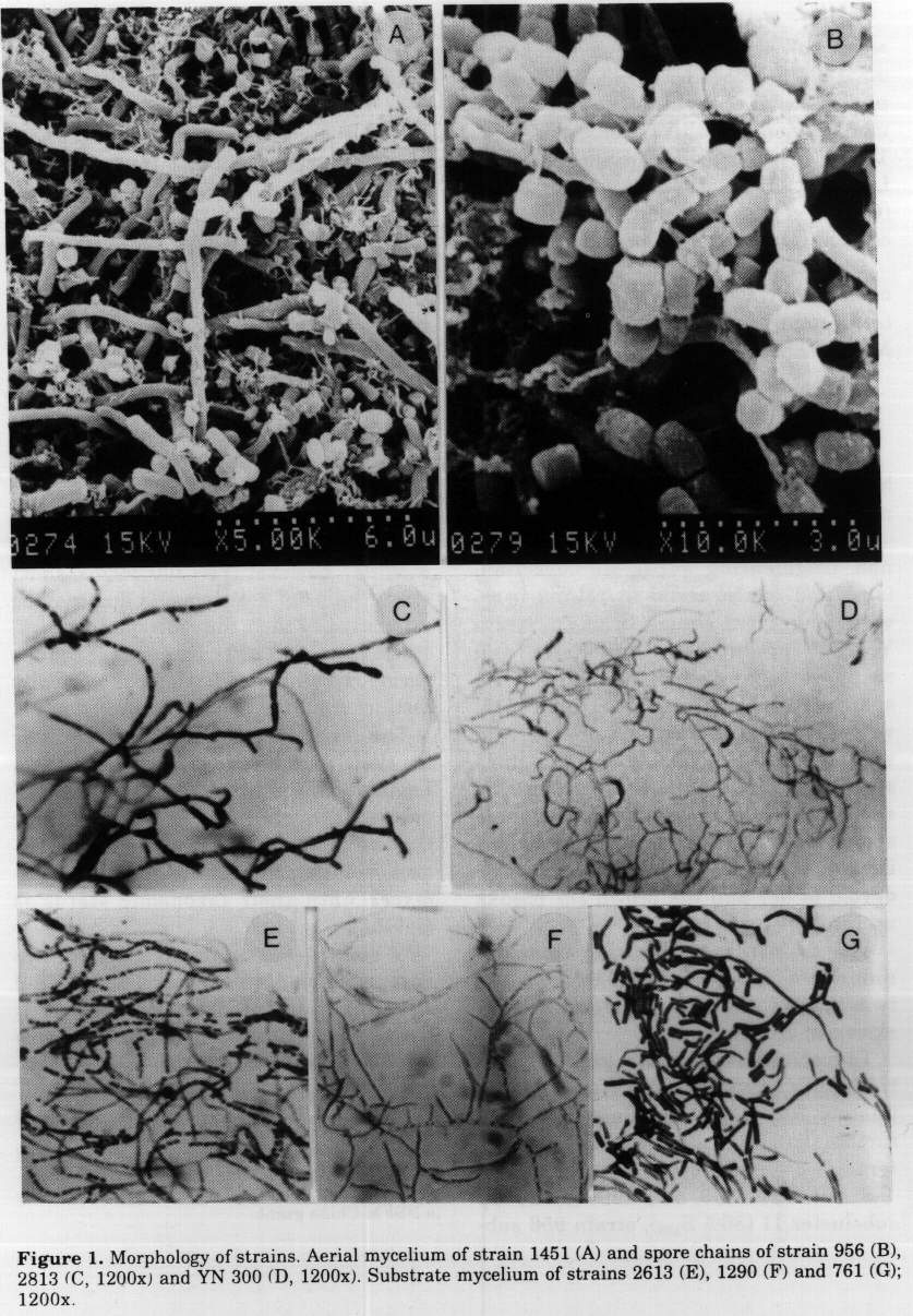
Figure 1. Morphology of strains. Aerial mycelium of strain
1451 (A) and spore chains of strain 956 (B), 2813 (C, 1200x)
and YN 300 (D, 1200x). Substrate mycelium of strains 2613 (E),
1290 (F) and 761 (G);1200x.
Other characteristics investigated were: cell wall composition
(Becker et al. , 1965; Lechevalier et a1. , 1980),
phospholipids (Lechevalier et al., 1980), menaquinones
(Collins, 1985; Wu et a1. , 1989), mycolic acids (Minnikin,
1980; Liu et al., 1990), DNA base composition (Chater et
al., 1982) and DNA GC mol% content (Marmur and Doty, 1962).
Cluster analysis of the results was carried out using the
MINTS system (Academia Sinica Beijing) according to Ma and Zhou
(1989).
RESULTS
All strains are mesophilic and aerobic and their characteristics
are summarised in Table 1.
Strains 956 and 2539 show yellowish, non fragmenting substrate
mycelium (sm ) and white aerial mycelium (am ) with
long spore chains (Fig. 1). Strain YN 300 produces a similar sm
and scant white-grey am with chains of 3-4 spores (Fig.
1). Strains 1451 and 2613, with yellowish fragmented sm and
white grey am, resemble Nocardiopsis species; strain
2613 is characterised by short spore chains, sometimes by single
spores (Fig. 1). Strain 1290 forms yellowish sm, but no
am and strain 761 is also devoid of am and the brown
sm breaks up into irregular rods.
The dendrogram resulting from the numerical analysis (UPGMA)
of the twenty-nine characters investigated is shown in Figure 2.
The eleven strains are grouped into two major clusters (I and
II) at approximately 70% S[sm] level. Cluster I can be further
subdivided into three subclusters.
Strains YN 300 and 1290 make up subcluster I1 (86% S[sm]),
strain 956 subcluster I2 and the three type strains of K.
grisea, K. papulosa and K. setae are recovered in
subcluster I3 (84% [SSm]). The remaining strains, including
the invalidly published representative of "K. clausa , form
Cluster II.
DISCUSSION
Results show the similarity between the named species of the genus
Kitasatosporia and support their placement, together with
strain 956, in the "setae group of the genus
Streptomyces, as suggested by Wellington et al.
(1992) and Nakagaito et al. (1992a, 1992b). Strain 956
may represent a new species of the group.
Strains YN 300 and 1290 show chemotaxonomic characters similar
to those of Ksetae, however YN 300 forms short spore chains
of 3-4 elements and may be attributed to the genus
Microtetraspora. while strain 1290 does not produce aerial
mycelium and is morphologically distinct from the "setae
group.
The other strains investigated differ for their morphological
and chemotaxonomical characters from the "setae group. It
is therefore suggested that they be temporarily placed in a
"kitasatosporioides group of the genus Streptomyces
until their taxonomic status is clarified .
"K. clausa was originally attributed to
Kitasatosporia on the basis of LL- and meso-DAP content.
Recently however it has been reidentified as Microtetraspora
heluata (Nakagaito, 1992b).
ACKNOWLEDGEMENTS. The assistance of J.Ma in numerical
classification. of J. Xie. G. J. Don and X. Zhou in electron
microscopy and of Dr. T. Kudo in providing type cultures. is
gratefully acknowledged. The work was supported by a NSFM China
grant.
Figure 2. Dendrogram from the results (see Table 1) of cluster
analysis based on the S[SM] coefficient and average
linkage.
REFERENCES
Anonymous (1957) Colour Tables. Chinese Committee for
Publications and Translations, Beijing
Becker, B., M. P. Lechevalier & H. A. Lechevalier (1965)
Chemical composition of cell-wall preparations from strains of
various-form genera of aerobic actinomycetes Appl.
Microbiol., 13: 236-243
Chater, K. F., D. A.Hopwood, T. Kieser &C. J. Thompson
(1982). Gene cloning in streptomycetes. Curr. Top.
Microbiol. lmmunol., pp. 93-96
Collins, M.D. (1985). In: M.Goodfellow & D E Minnikin (eds.)
Chemical Methods in Bacterial Systematics. Academic Press,
London, pp. 267287
Kawamoto, I., T. Oka & T. Nora (1981). Cell wall
composition of Micromonospora olivoasterospora and related
organisms. J. Bacteriol., 146: 5 2 7-534
Kusakabe, H. & K. Isono (1988) Kitasatosporia cystarginea
sp.nov., which produces a new antifungal antibiotic cystargin.
J. Antib., 41: 17581762
Lechevalier, M. P. & H. A. Lechevalier (1980) In A.Dietz &
D.W.Thayer (eds.). A University Laboratory Approach. SIM
Spec.Publ., No.6, Arlington, pp 277-284.
Liu, Z., Z. Liu, J. Ruan & X. Yan (1986). A new species of the
genus Kitasatosporia. Acta Microbiol. Sin., 26: 87-89
Liu, Z., J. Su & J. Ruan (1990). Determination of mycolates in some
strains of nocardioform actinomycetes by gas chromatography.
Microbiology, China, 17(5): 307-310
Ma, J. & Y. Zhou (1989). The Progress of Microbial Systematic
Techniques (In Chinese) Guangmin Press, Harbin, pp 340-355
Marmur, J. & P. Doty (1962). Determination of base
composition of deoxyribonucleic acid from its denaturation
temperature. J. Mol. Biology, 5: 109-118
Minnikin, D. E., I. G. Hutchinson, A. B. Caldicott & M.
Goodfellow (1980). Thin-layer chromatography of methanolysates
of mycolic acidcontaining bacteria. J. Chrom., 188:
221-233
Mordarska, H., M. Mordarski & M. Goodfellow (1972)
Chemotaxonomic characters and classification of some nocardioform
bacteria J. gen. Microbiol., 71: 77-86
Mordarska, H., I. Wieczorak, J. Zakrewska-Czerwinska, A. Ziefka &
M. Mordarski (1987) Kitasatosporia setae, an interesting
actinomycete strain isolated from Spitsbergen soil Arch. lmm.
Ther. Experim., 35: 237-250
Nakagaito, Y., A. Yokota & T. Hasegawa (1992a) Three new
species of the genus Streptomyces: Streptomyces cochleatus
sp nov, Streptomyces paracochleatus sp nov, and
Streptomyces azaticus sp nov J. Gen. Appl. Microbiol.,
38: 105-120
Nakagaito, Y., A. Shimazu, A. Yokota & T. Hasegawa (1992b)
Proposal of Streptomyces atroaurantiacus sp.nov and
Streptomyces kifuensis sp nov and transferring
Kitasatosporia cystarginea Kusakabe and Isono to the genus
Streptomyces as Streptomyces cystarginea. J. Gen. Appl.
Microbiol., 38: 627-633
Nakamura, Y., E. Ono, T. Kohda & H. Shibai (1989). Two new
carbapenem antibiotic-producing actinomycetes: Kitasatosporia
papulosa sp. nov and Kitasatosporia grisea sp nov J.
Antib., 42: 18-29
Nelson, R. A., J. A.Pope, G. M.Luedemann, L. E. McDaniel
& C. P. Schaffner (1988) Crisamicin A, a new antibiotic from
Micromonospora. J. Antib., 39: 335-344
Omura, S., Y. Takahashi, Y. Iwai & H. Tanaka (1982).
Kitasatosporia, a new genus of the order Actinomycetales.
J. Antib., 35: 10 13-1019
Shearer, M.C., P. M. Colman & C. H. Nash (1983).
Nocardiopsis mutabilis, a new species of nocardioform
bacteria isolated from soil lnt. J. Syst. Bacteriol., 33:
369-374
Takahashi, Y., Y. Iwai & S. Omura (1984) Two new species of
the genus Kitasatosporia, Kitasatosporia phosalacinea sp.nov
and Kitasatosporia griseola sp nov. J. Gen. Appl.
Microbiol., 30: 377-387
Tukahashi, A., Y.Okami & K.Hotta (1985) Considerations on
the taxonomic position of Nocardiopsis mutabilis.
Actinomycetologica, 47: 8-14
Wellington, E. M. H., E. Stackebrandt, D. Sanders, J.
Wolstrup & N. O. G. Jorgensen (1992) Taxonomic status of
Kitasatosporia, and proposed unification with
Streptomyces on the basis of phenotypic and 16S rRNA
analysis and emendation of Streptomyces Waksman and Henrici.
1943, 339AL Int. J. Syst. Bacteriol., 42: 156-160
Wu, C., X. Liu, M. Gin & J. Ruan (1989) The analysis of menaquinone
compounds in microbial cell walls by HPLC. Microbiology, China.
18(3) 176-178
Copyright 1994 C. E. T. A.
The following images related to this document are available:
Halftone images
[ac94006a.gif]
Photo images
[ac94006a.jpg]
