 |
| About Bioline | All Journals | Testimonials | Membership | News |
|
||||||
|
||||||
A new species of the genus Planotetraspora: Planotetraspora viridis sp. nov. H. Runmao, L. Junying, Y. Guozhu and F. Yi* Sichuan Industrial Institute of Antibiotics, Chengdu, Sichuan and *Pharmaceutical Factory of Bethune Medical University, Changchun, Jilin, P.R. China
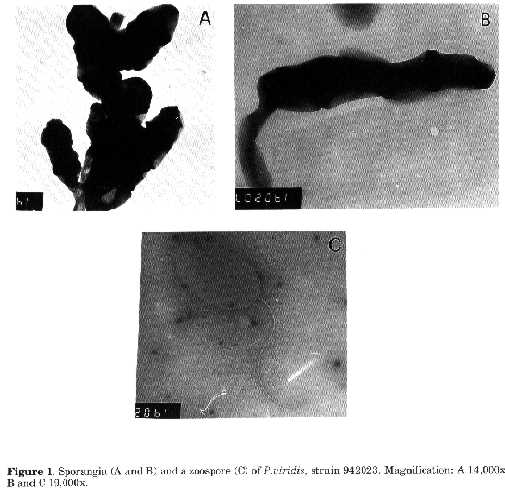
Abstract. A new species of the genus Planotetraspora is described. The organism is characterised by a white aerial mycelium, bearing cylindrical sporangia containing four motile spores in a single row, and by a green vegetative mycelium. The name proposed for the new specie is Planotetraspora viridis sp.nov. Strain 942023 (= ATCC 51498) is the type strain of the species. The genus Planotetraspora was erected in 1993 by Runmao et al. (1993) to describe isolates from soil that produce a white aerial mycelium with cylindrical sporangia at the end of short sporangiophores, each sporangium containing four zoospores in a single row. While investigating actinomycete populations in soils of the eastern part of China, two strains of the genus were isolated. On the basis of chemical and morphological characteristics, they appear to represent a new species of the genus Planotetraspora. Material and methods Microorganisms. Strains 935104 and 942023 were isolated from soil samples collected in the provinces of Guang-Dong in 1993 and Jiang-Xi in 1994 respectively. The type strain Planotetraspora mira, SIIA 9201, was used for comparison purposes. Morphological characteristics. These were examined directly on the surfaces of agar plates by using a 40x long- working-distance light microscopy objective. Sporangial features were determined with a model JEM-100 CXII transmission electron microscope using whole-mount preparations. In order to observe flagella, an agar block containing numerous sporangia was placed in a tube filled with 0.5ml of sterile tap water and kept for 30 to 50 min at room temperature. One drop of the suspension was placed on Formvar coated 300-mesh copper grids, stained with 0.125% phosphotungstic acid and air dried. Cultural and physiological characterisation. Strains were grown on International Streptomyces Project (ISP) media 2 to 9, incubated at 28 C and cultures were observed after 15dd using the methods recommended by Shirling and Gottlieb (1966). Cells were prepared and the chemical composition of cell walls was determined according to Becker et al. (1965). Whole cells were analysed according to Lechevalier (1968). Results and Discussion Description of Planotetraspora viridis sp.nov. Planotetraspora viridis (vi'ri.dis L.adj. viridis, green). Gram positive, non acid fast, aerobic. Long, irregularly branched, vegetative hyphae (0.2 to 0.6æm in diameter) penetrating the agar and forming on the surface of agar media compact, raised, tough, colonies. Figure 1. Sporangia (A and B) and a zoospore (C) of P.viridis, strain 942023. Magnification: A 14,000x, B and C 19,000x.
--------------------------------------------------------------
MEDIUM   SPECIES    GROWTH   REVERSE    AERIAL    SPORANGIUM
(ISP)                                   MYCELIUM  PRODUCTION

2         P.mira    Good     Ivory      Whitish
                                        grey      Good
          P.viridis Poor     Hyaline    Absent    Absent
3         P.mira    Good     Hyaline    White     Good

          P.viridis Good     Dark green White     Good
4         P.mira    Moderate Hyaline    Whitish   Good
          P.viridis Poor     Hyaline    Absent    Absent
5         P.mira    Good     Hyaline    White     Good
          P.viridis Moderate Hyaline    White     Good
6         P.mira    None        -         -        -
          P.viridis Good     Dark green Absent    Good
7         P.mira    None        -         -        -
          P.viridis Moderate Hyaline    White     Good
Table 1. Comparison of cultural characteristics of
P.mira and P.viridis.
-------------------------------------------------------------------------Fragmentation of substrate hyphae usually not occurring. Aerial hyphae (0.2 to 0.8æm in diameter) sparsely branched, rarely septate. Cylindrical sporangia (0.5-0.8 by 3.0-3.4um) formed on short sporangiophores of the aerial mycelium, each containing a single, straight row of four spores (Fig. 1, A and B). Sporangium formation most abundant on oatmeal agar (ISP medium 3) and glycerol-asparagine agar (ISP medium 5). Sporangia formed singly or in groups on the aerial mycelium. By electron microscopy the sporangial envelope appears wrinkled (Fig. 1, A and B). Flooding with water determines the release of ellipsoidal (about 0.6 to 0.8 by 1.0 to 1.4mm) zoospores with a single polar flagellum (Fig.1, C). Cultural and physiological characteristics of the type strain 942023 are shown in Table 1. Additional physiological characteristics of strain 942023 are as follows: milk is slowly peptonised and coagulated, starch is hydrolysed. The strain utilises D-xylose, D- mannitol, L-arabinose, maltose, D-glucose, sucrose, galactose, inulin, galactitol, D-ribose, salicin, L-sorbose and lactose. No growth is observed with D-glucitol, L-rhamnose, melezitose, L-arabitol, melibiose and trehalose as sole carbon sources. Cell wall hydrolysates of strain 942023 contain meso-diaminopimelic acid. Galactose, arabinose, glucose, xylose, mannose and ribose are the sugars detected in whole cell hydrolysates. Strain 942023 (= ATCC 51498) is the type strain of the proposed species. The formation on the aerial mycelium of cylindrical shaped sporangia, containing single, straight rows of four zoospores, together with the cell wall and whole cell hydrolysate characteristics, place strain 942023 in the genus Planotetraspora (Runmao et al., 1993). The only species, Planotetraspora mira, so far described was compared in order to assess differences in morphological, cultural and physiological properties. Differences between P.mira and strain 942023 are summarised in Table 1. In P. mira the colour of the reverse side of colonies is ivory or hyaline, while strain 942023 forms a dark green colour on ISP media 3 and 6. P.mira does not grow on ISP medium 6 and very poorly on ISP medium 7, whereas strain 942023 shows excellent growth with dark green colour of the substrate mycelium. Growth of P.mira is good on ISP media 2 and 4, while strain 942023 shows poor or no aerial mycelium. On the basis of the observations strain 942023 is considered to represent a new species of the genus Planotetraspora, for which propose the name Planotetraspora viridis sp.nov. is proposed. References Becker, B., M.P.Lechevalier & H.A.Lechevalier (1965). Chemical composition of cell-wall preparations from strains of various form-genera of aerobic actinomycetes. Appl.Microbiol., 13: 236-243 Runmao, H., W.Guizhen & L.Junying (1993). A new genus of actinomycetes, Planotetraspora gen. nov. Int.J.Syst.Bacteriol., 43: 468-470 Lechevalier, M.P. (1968). Identification of aerobic actinomycetes of clinical importance. J.Lab. Clin.Med., 71: 934-944 Shirling, E.B. & D.Gottlieb (1966). Methods for characterization of Streptomyces species. Int.J. Syst.Bacteriol., 16: 313-340. Copyright 1995 CECT
The following images related to this document are available:Halftone images[ac95005a.gif]Photo images[ac95005a.jpg] |
| |||||||||