 |
| About Bioline | All Journals | Testimonials | Membership | News |
|
||||||
|
||||||
Actinomycetes, Vol.6, Part 2, 1995 ACTINOPLANES STRAINS ISOLATED FROM ROOTS: MORPHOLOGICAL INVESTIGATIONS
B. PETROLINI, S. QUARONI, M. SARACCHI, P. SARDI and ^*L. FEDELI Institute of Plant Pathology, University of Milan, via Celoria 2, Milan and ^*Pharmacia Farmitalia-C.Erba s.r.l., Nerviano, Italy
ABSTRACT. The isolation of Actinoplanes strains from cortical tissues of roots is reported for the first time. The organisms are rarely encountered in this habitat. Sporangiate genera of actinomycetes are morphologically well definite and their identification can be carried out on the grounds on morphological criteria alone, using as phenetic markers the absence or presence of aerial mycelium bearing reproductive structures and the shape of sporangia and spores. The paper deals with eleven isolates showing different morphological features which allow their separation into five phenetic groups. The isolation of rare microorganisms from unusual ecological niches depends largely on adequate strategies. For example using an air sampling method, novel actinomycetes have been recovered from leaf litter (Petrolini et al., 1992; 1993). Alternatively direct isolation from surface-sterilised roots was carried out to investigate the endophytic actinomycete population. Most isolates obtained from this source belonged to the genus Streptomyces (Sardi et al., 1992) and several to the genus Micromonospora (Williams et al., 1993). However representatives of rare genera may also be found and, among these, some strains of sporangiate actinomycetes. The present report deals with morphological and cultural characteristics of eleven sporangiate isolates with motile spores. They show considerable differences in the sporangial shapes, but all belong to the genus Actinoplanes (Palleroni, 1989), now placed in the emended family Micromonosporaceae Krassilnikov (Goodfellow et al., 1990; Vobis, 1991), with all other genera encompassed into the suprageneric group of actinoplanetes (Vobis, 1989). MATERIALS AND METHODS Isolations were carried out by plating surface-sterilised roots collected from 118 different plant species (131 samples) using a previously described procedure (Sardi et al., 1992). Healthy host plants were from different ecological areas. Eleven motile sporangiate actinomycetes were isolated from the roots of nine plants. They were obtained on 2.5% water agar with 50ppm of cycloheximide and of nystatin to reduce fungal growth (WAA25) and on the same medium, supplemented with tunicamycin or novobiocin (30ppm). Details about hosts and location are given in Table 1. Cultural and morphological characteristics were determined after 7, 14 and 21dd incubation at 26 C on 2.5% water agar (WA25) and on ISP medium 3 (Shirling and Gottlieb, 1966). Micromorphological features and spore motility were detected by light and scanning electron microscopy (Stereoscan 250, Cambridge Sci.Instr., Ltd., UK). Specimens for electron microscopy were air (AD) or freeze (FD) dried (Petrolini et al., 1986, 1993).
------------------------------------------------------------
STRAIN
IPV-                                ISOLATION SOURCE
-------------------------------------------------------------
2878     Cistus monospleliensis    Monte Argentario,Tuscany
2879     Arisarum vulgare           Monte Argentario, Tuscany
2881     unidentified plant (A)    Curitiba (Brazil)
2882     unidentified plant (A)     Curitiba (Brazil)
2883     unidentified plant (B)     Curitiba (Brazil)
2884     Ailanthus glandulosa       Milan, Lombardy
2885     Origanum vulgare           Gallipoli, Apulia

2895     Ailanthus glandulosa       Milan, Lombardy
2896     Pelargonium  sp.           Milan, Lombardy
2897     Sempervivum tectorum       Macugnaga, Piedmont

2908     Arbutus unedo              Monte Argentario,
Tuscany
--------------------------------------------------------------
STRAIN
IPV-       DATE              ISOLATION MEDIUM WAA25 +
--------------------------------------------------------------
2878       Jan. 1993          novobiocin
2879       Jan. 1993          novobiocin
2881       May 1993
2882       May 1993           tunicamycin
2883       May 1993           novobiocin
2884       June 1990
2885       July 1993          tunicamycin
2895       June 1990          tunicamycin
2896       Oct. 1991          tunicamycin
2897       Oct. 1993          tunicamycin
2908       Jan. 1993          novobiocin
--------------------------------------------------------------

Table 1. Origin of investigated strains.

--------------------------------------------------------------
STRAIN    COLONY               COLOUR OF
IPV-      COLOUR               STERILE AERIAL HYPHAE
        WA25         ISP 3             WA25          ISP 3
--------------------------------------------------------------
2878    colourless   pinkish cream     -               -
2879    colourless   pinkish cream     -             pink
2881    colourless   pinkish cream    grey           yellow
2882    colourless   cream             -                -
2883    colourless   pink to
                     brick orange     white          pale pink
2884    colourless   orange to rusty    -               -
2885    colourless   pinkish cream      -               -
2895    colourless   rusty cream        -               -
2896    colourless
        to pale red  purple             -            grey
2897    colourless   pinkish cream      -            grey
2908    colourless
        to cream     purple             -               -
--------------------------------------------------------------
STRAIN     PIGMENT ON                     SPORANGIA
--------------------------------------------------------------
2878           -              globose
2879           -              campanulate to lobate
2881           -              cylindrical
2882           -              globose
2883           -              campanulate to cylindrical
2884       pale brown         small, irregular
2885       pale brown         irregularly globose
2895           -              oval
2896       violet-red         small, irregular
2897           -              campanulate to lobate
2908          red             irregularly globose
-------------------------------------------------------------
Table 2. Cultural characteristics and most frequent
sporangial patterns.Degradation of keratin was carried out according to Vobis (1984). Generic identification of the isolates was carried out according to a simplified determinative key based on morphological criteria (Vobis, 1991). RESULTS Members of the genus Actinoplanes are normal soil and leaf litter inhabitants, preferably colonising plant and animal debris (Cross, 1981), but so far they have never been isolated from roots of living plants. Their frequency in this habitat however is very low; in fact only eleven strains were isolated from nine host plants. It should be pointed out that from each sample at least twenty subsamples (about 1cm long) were examined; in other words the eleven strains were isolated from a total of about 2700 root fragments. All strains produce sporangia on both media employed for morpho-cultural studies; growth is more abundant on ISP medium 3 than on WA25. Cultural characteristics of the isolates are reported in Table 2. Usually aerial mycelium is not formed, but mature colonies of IPV-2879, 2896 and 2897 on ISP medium 3 and of IPV-2881 and 2883 on both media show small areas with a powdery appearance caused by the presence of sterile aerial hyphae growing between the sporangia. Colonies are usually colourless on WA25, on ISP medium 3 the most common colour is pinkish cream, however some strains show rusty-orange, brick-red or purple coloured substrate mycelia. Four isolates produce pale brown, red or violet-red diffusible pigments. None of the isolates decomposes keratin materials; they grow on hair surfaces, however the structure of the latter is never altered. The absence of true aerial mycelium, differentiated into spores, excludes the attribution of the isolates to maduromycetes (Goodfellow, 1989a) and favours their placement within the sporangiate actinomycetes encompassed in the latest edition of the Bergey's Manual of Systematic Bacteriology in the suprageneric group of the actinoplanetes (Vobis, 1989, 1991). Sporangial size and shape of the isolates are quite variable, the latter ranging from globose, irregularly globose, oval, pyriform, cylindrical or campanulate to lobate. Most strains produce sporangia of different shapes on the same colony. Motile sporangiospores, arranged inside the sporangial envelope in more or less regular coils, linear rows or irregular patterns, are globose to pyriform, only in IPV-2895 they are short and rod-shaped. According to the phenetic criteria proposed for actinoplanetes genera identification (Vobis, 1991), all strains belong to the genus Actinoplanes (Palleroni, 1989) on the grounds of spore shape, notwithstanding the wide range of sporangial shapes, some of them similar to those of other actinoplanetes genera. In fact the genus Ampullariella (Vobis and Kothe, 1989) is characterised by distinctly rod-shaped sporangiospores, Pilimelia (Vobis, 1989) includes keratinophilic organisms growing on complex media only and possessing rod-shaped to reniform spores, while Actinoplanes species show globose to pyriform or short rod spores. On the basis of sporangial shape, isolates can be arranged into five groups.
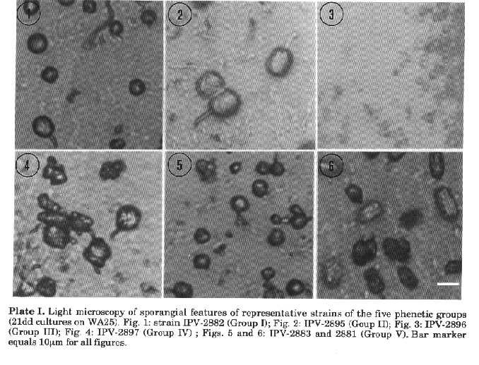
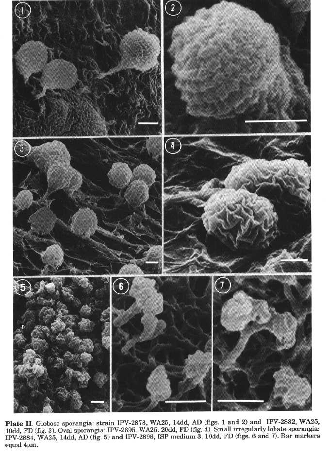
Group I. Strains IPV-2878 and 2882. Only globose sporangia, borne on short sporangiophores, are produced (Pl. I, fig. 1; Pl. II, figs. 1 to 3). The globose spores are arranged in coils, which are regular in strain IPV-2878 (Pl. II, figs. 1 and 2) and less so and usually hardly visible in IPV-2882. The latter strain has a thicker sporangial envelope (Pl. II, fig. 3) and sometimes sessile sporangia, occasionally embedded in a mucilaginous mat. The size of sporangia ranges from 6 to 15um (IPV-2882) and from 9 to 15um (IPV-2878).
Group II. Strain IPV-2895. Oval sporangia (9-15 x 11-21um), rather flattened at the tips and characteristically showing a very thick envelope (Pl. I, fig. 2; Pl. II, fig. 4). Due to the wall thickness, under the light microscope sporangia appear as glistening beads and the spore release is slow and difficult. Short rod-shaped spores are arranged in hardly visible, but regular coils and sporangiophores can be very long and appear septate. These characteristics are similar to those of the genus Pilimelia, but the strain does not require complex media for growth and it is not able to decompose keratin. Sporangia are roughly globose in shape, often sessile and apparently leaning on the mycelial mat at the centre of old colonies.
Group III. Strains IPV-2884 and 2896. Unusually small, irregular to lobate sporangia (Pl. I, fig. 3; Pl. II, figs. 5 to 7). Rarely subglobose with rough surface, they are typically highly irregular, often multilobed (2.5-6.5 x 3-7.5um in IPV-2884; 2-6.5 x 2-7um in IPV-2896). Sometimes they are divided into several parts and the distinct lobes may give the impression of two or three sporangia fused together. Sporogenic hyphae coil irregularly around themselves in several directions to form kinked masses of globose spores. Particularly in IPV-2896 sporangia are often very deeply split along the longitudinal axis (Pl. II, figs. 6 and 7). Sporangiophores are long, however sessile sporangia do occur. Branched sporangiophores in IPV-2896 (Pl. II, fig. 7) and many hyphae coalescing to form a short peduncle in IPV-2884 can be observed. Sporangiospores of IPV-2896 swim slowly.
Plate II. Globose sporangia: strain IPV-2878, WA25, 14dd, AD (figs. 1 and 2) and IPV-2882, WA25, 10dd, FD (fig. 3). Oval sporangia: IPV-2895, WA25, 20dd, FD (fig. 4). Small irregularly lobate sporangia: IPV-2884, WA25, 14dd, AD (fig. 5) and IPV-2896, ISP medium 3, 10dd, FD (figs. 6 and 7). Bar markers equal 4um.
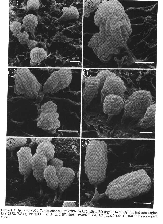
Plate III. Sporangia of different shapes: IPV-2897, WA25, 10dd, FD (figs. 1 to 3). Cylindrical sporangia: IPV-2883, WA25, 12dd, FD (fig. 4) and IPV-2881, WA25, 16dd, AD (figs. 5 and 6). Bar markers equal 4um.
Irregularly globose shapes occur more frequently in strains IPV-2885 and 2908, campanulate to lobate ones in IPV-2879 and 2897. In IPV-2908 lobate sporangia, though rarely occurring, may be multilobed. Except for IPV-2885, sporangiophores are frequently branched to support two, more rarely three sporangia (Pl. III, fig. 3). In IPV-2879 and 2908 they can be very long. All four isolates produce globose spores. Motility in IPV-2879 occurs immediately after release in water.
Group V. Strains IPV-2881 and 2883. More or less elongated cylindrical sporangia, with flattened or slightly rounded distal end and often mound-shaped base, occur in strains IPV-2881 (Pl. I, fig. 6; Pl. III, figs. 5 and 6) and 2883 (Pl. I, fig. 5; Pl. III, fig. 4). In IPV-2881 (4-8 x 10-17um) they are usually more elongated than in IPV-2883 (3-8 x 6-12.5um). Sporangia are mostly single, sometimes the same vegetative hyphae can develop two, rarely three or more, sporangiophores. Globose to pyriform spores are arranged in straight, longitudinal rows (Pl. I, fig. 6; Pl. III, figs. 4 to 6). In spite of the Ampullariella-like linearity, sporangiospores show shapes typical of Actinoplanes species. In addition the two isolates, particularly IPV-2883 (Pl. I, fig. 5; Pl. III, fig. 4), form differently shaped sporangia. IPV-2881 produces mostly cylindrical and elongated sporangia particularly on ISP medium 3. Branched sporangiophores can occur (Pl. III, fig. 6). A septum is often visible about the middle of unbranched sporangiophores (Pl. III, fig. 5). Globose and campanulate sporangia occur; at maturity some undergo a distortion of their shape and become irregularly globose or form one or more lateral bulges, due to the swelling of the sporangial envelope. IPV-2883 rarely develops cylindrical sporangia on WA25, where most fruiting bodies appear subglobose, campanulate or pyriform (Pl. I, fig. 5; Pl. III, fig. 4), as opposed to ISP medium 3 where cylindrical, campanulate and pyriform sporangia are formed, while subglobose ones are scarce. Spore discharge is mostly sudden and violent; often the entire spore mass is pushed out through the top of the sporangium. Spores swimming immediately, though slowly. CONCLUSIONS Due to the peculiarities of the reproductive structures, multispored sporangiate actinomycetes are easily recognisable by light microscopy examination of colonies. However on isolation plates they are rare and often inhibited or obscured by the heavy growth of other microorganisms. Therefore enrichment procedures have to be developed for their isolation (Palleroni, 1980; Vobis, 1991). Plating and incubation on selective agar media of surface-sterilised plant tissues, as employed during this study, succeeded in the detection of root endophytic Actinoplanes strains, whose typical fruiting bodies, emerging as glistening beads from root fragments or from the agar surface, are clearly recognisable by light microscopy. Sporangia-forming actinomycetes are included among actinoplanetes (Vobis, 1989) and maduromycetes (Goodfellow, 1989a), suprageneric groups that can be distinguished using chemical, molecular genetic and phenetic procedures (Goodfellow, 1989b; Goodfellow et al., 1990). The differentiation between the two phylogenetic groups is possible by determining the wall types (Lechevalier et al., 1971). On the other hand, as wall type II is typically associated with the absence of true aerial mycelium in the actinoplanetes and wall type III with the sporulation on the aerial mycelium in the maduromycetes (Cross and Goodfellow, 1973; Goodfellow, 1989a; Vobis, 1989), it is possible to allocate an unknown isolate on morphological grounds alone. The lack of sporogenous aerial mycelium allows the placement of the isolates within the actinoplanetes group and in particular to the genus Actinoplanes on the basis of the features of the sporangia and of the spore shape (Vobis, 1991). Sporangiate actinoplanetes are phenetically diverse and their separation into different genera is based essentially on morphological criteria. On the other hand the close chemotaxonomic and nucleic acid affinities between Actinoplanes, Amorphosporangium and Ampullariella suggest their accommodation in a single genus: Actinoplanes (Cross and Goodfellow, 1973; Goodfellow and Cross, 1984, Stackebrandt and Kroppenstedt, 1987; Szaniszlo and Gooder, 1967). Sporangial shape is determined by the internal disposition of the spores consequent to the extension and branching of sporogenous hyphae within the structureless sac, which can split, undergo distortions or partial digestion giving rise to the great variety of shapes observed. In the last edition of the Bergey's Manual of Systematic Bacteriology (Palleroni, 1989) Actinoplanes and Amorphosporangium are considered synonyms. In addition by using chemical and numerical criteria, Goodfellow et al. (1990) recognised also Ampullariella as a synonym of Actinoplanes. The species concept in Actinoplanes is ill defined and the physiological analysis provides information of limited value. Species differentiation of the genus is at present based on a combination of a few morphological, cultural and physiological properties (Palleroni, 1989), that are in most cases referred to single isolates, with consequent poor information on interspecific differences and intraspecific variations. On the basis of cultural and morphological properties, it is difficult at the moment to identify the isolated Actinoplanes strains with previously described species of the genus. Notwithstanding the low number, the isolates form a heterogeneous population, showing a great variety of sporangial shapes, often overlapping with that of the genus Ampullariella. Few strains differentiate only one type of sporangia, the majority two or more, however morphological features of each isolate are constant. Characteristics of the reproductive structures appear to possess a good discriminative value for differentiating the isolates and for this reason their separation into five phenetic groups on the basis of sporangial shape is proposed. Some strains appear novel and particularly interesting, for example IPV-2895 and 2881 because of the shape of their sporangia and spores. Isolate IPV-2896 forms smaller sporangia than any other of the described species and produces a violet reddish soluble pigment. Further investigations on physiological and chemical properties of the strains are however necessary to clarify the taxonomic relationships of the isolates. ACKNOWLEDGEMENTS. The authors wish to thank Mrs. Jacqueline Rogers for the revision of the English text. REFERENCES Cross, T. (1981). Aquatic actinomycetes: a critical survey of the occurrence, growth and role of actinomycetes in aquatic habitats. J.Appl.Bacteriol., 50: 397-423
Cross, T. & M.Goodfellow (1973). Taxonomy and classification of the actinomycetes. In: G. Sykes & F.A.Skinner (eds.) Actinomycetes: Characteristics and Practical Importance. Academic Press, London, pp. 11-112
Goodfellow, M. (1989a). Maduromycetes. In: S.T.Williams, M.E.Sharpe & J.G.Holt (eds.) Bergey's Manual of Systematic Bacteriology. The Williams & Wilkins Co., Baltimore, Vol. 4, pp. 2509-2551
Goodfellow, M. (1989b). Suprageneric classification of actinomycetes. In: S.T.Williams, M.E. Sharpe & J.G.Holt (eds.) Bergey's Manual of Systematic Bacteriology. The Williams & Wilkins Co., Baltimore, Vol. 4, pp. 2333-2339
Goodfellow, M. & T.Cross (1984). Classification. In: M.Goodfellow, M.Mordarski & S.T. Williams (eds.) The Biology of the Actinomycetes. Academic Press, London, pp. 7-164
Goodfellow, M., L.J.Stanton, K.E.Simpson & D.E.Minnikin (1990). Numerical and chemical classification of Actinoplanes and some related actinomycetes. J.gen.Microbiol., 136: 19-36
Lechevalier, H.A., M.P.Lechevalier & N.N. Gerber (1971). Chemical composition as a criterion in the classification of actinomycetes. Adv. Appl.Microbiol., 14: 47-72
Palleroni, N.J. (1980). A chemotactic method for the isolation of Actinoplanaceae. Arch.Microbiol., 128: 53-55 Palleroni, N.J. (1989). Genus Actinoplanes Couch 1950, 89 (Amorphosporangium Couch 1963, 65), emend.mut.char. 1955, 153^AL. In: S.T.Williams, M.E.Sharpe & J.G.Holt (eds.) Bergey's Manual of Systematic Bacteriology. The Williams & Wilkins Co., Baltimore, Vol. 4, pp. 2419-2428.
Petrolini, B., S.Quaroni & M.Saracchi (1986). Scanning electron microscopy investigations on the relationships between bacteria and plant tissues. I. Comparative techniques for specimen preparation. Riv.Pat.Veg., S. IV, 22: 7-15
Petrolini, B., S.Quaroni, M.Saracchi & P. Sardi (1993). A new genus of the maduromycetes: Planopolyspora gen. nov. Actinomycetes, 4: 8-16
Petrolini, B., S.Quaroni, P.Sardi, M.Saracchi & N. Andriollo (1992). A sporangiate actinomycete with unusual morphological features: Streptosporangium claviforme sp. nov. Actinomycetes, 3: 45-50
Sardi, P., M.Saracchi, S.Quaroni, B.Petrolini, G.Borgonovi & S.Merli (1992). Isolation of endophytic Streptomyces strains from surface-sterilized roots. Appl.Envirom.Microbiol., 58: 2691-2693
Shirling, E.B. & D.Gottlieb (1966). Methods for characterization of Streptomyces species. Int.J.Syst.Bacteriol., 16: 313-340
Stackebrandt, E. & R.M.Kroppenstedt (1987). Union of the genera Actinoplanes Couch, Ampullariella Couch and Amorphosporangium Couch in a redefined genus Actinoplanes. Syst. Appl.Microbiol., 9: 110-114
Szaniszlo, P.J. & H.Gooder (1967). Cell wall composition in relation to the taxonomy of some Actinoplanaceae. J.Bacteriol., 94: 2037-2047
Vobis, G. (1984). Sporogenesis in the Pilimelia species. In: L.Ortiz-Ortiz, L.F.Bojalil & V.Yakoleff (eds.) Biological, Biochemical and Biomedical Aspects of Actinomycetes. Academic Press, Inc., Orlando, pp. 423-439 Vobis, G. (1989). The Actinoplanetes. In: S.T. Williams, M.E.Sharpe & J.G.Holt (eds.) Bergey's Manual of Systematic Bacteriology. The Williams & Wilkins Co., Baltimore, Vol. 4, pp. 2418-2428
Vobis, G. (1991). The genus Actinoplanes and related genera. In: A.Balows, H.G.Truper, M. Dworkin, W.Harder & K.H.Schleifer (eds.) The Prokaryotes. A Handbook on the Biology of Bacteria: Ecophysiology, Isolation, Identification, Applications. Springer-Verlag, Berlin, Vol. II, pp. 1029-1060
Vobis, G. & H.W.Kothe (1989). Genus Ampullariella Couch 1964, 29^AL. In: S.T.Williams, M.E.Sharpe & J.G.Holt (eds.) Bergey's Manual of Systematic Bacteriology. The Williams & Wilkins Co., Baltimore, Vol. 4, pp. 2429-2433 Williams, S.T., R.Locci, A.Beswick, D.I. Kurtb”ke, V.D.Kusnetsov, F.J.Le Monnier, P.F.Long, K.A.Maycroft, R.A.Palma, B.Petrolini, S.Quaroni, J.I.Todd & M.West (1993). Detection and identification of novel actinomycetes. Res.Microbiol., 144: 653-656
Copyright 1995 CETA
The following images related to this document are available:Halftone images[ac95007a.gif] [ac95007c.gif] [ac95007b.gif]Photo images[ac95007c.jpg] [ac95007a.jpg] [ac95007b.jpg] |
| |||||||||