 |
| About Bioline | All Journals | Testimonials | Membership | News |
|
||||||
|
||||||
Taxonomic characterisation of some Promicromonospora-like isolates and description of a new species: Promicromonospora yunnanensis SP. NOV.
L. Zhiheng, Q. Weihong, S. Yanlin and Z. Yamei Institute of Microbiology, Academia Sinica, Beijing, 100080, P.R.China
Code Number: AC97004
Sizes of Files:
    Text: 19.5K
    Graphics: Photographs (jpg) - 159.8K
ABSTRACT. Four actinomycete strains isolated in the Yunnan Province were investigated and compared with the type species of the genus Promicromonospora. Morphological, cultural, physiological and chemotaxonomic characters of the strains are described and a new species, Promicromonospora yunnanensis sp. nov., is proposed.
The genus Promicromonospora, established by Krassilnikov et al. (1961), at present includes three species: P.citrea, which was proposed as the type species of the genus, P.enterophila (Jager et al., 1983) and P.sukumoe (Takabashi et al., 1988).
In the course of a selective isolation of rare actinomycetes, four strains resembling Promicromonospora spp. were isolated. In the present note their characteristics are reported.
MATERIALS and METHODS
Organisms. Strains 126, 321, 1388 and 6828 were isolated from soil samples collected in Yunnan Province, China. The type culture of P.citrea, VKM AC 665, was used as reference strain. Cultures were maintained on Sauton's agar slopes at 4 C and subcultured at 4 week intervals.
Morphology. Observations were carried out on oatmeal and inorganic salt starch agar (Shirling & Gottlieb, 1966) grown cultures (7dd at 28 C) by light (Zeiss) and scanning electron microscopy (Hitachi, S570).
Cultural Observations. Strains were grown at 28 C on media suggested by Shirling & Gottlieb (1966) and by Gordon et al. (1974). A colour standard (Anonymous, 1957) was used for colour determinations. Physiological Characters. Data were recorded after 14dd at 28 C on recommended media (Shirling & Gottlieb, 1966; Kampfert & Kroppenstedt, 1991).
Chemotaxonomy. Cell mass was harvested, after 4-6dd incubation at 28 C, from Sauton's liquid medium (Lechevalier et al., 1980). Cell wall and whole cell components and phospholipids were determined according to Lechevalier et al. (1980) and mycolic acids according to Minnikin et al. (1980) and Liu et al. (1990) by thin layer chromatography (TLC). Menaquinones were extracted and purified (Collins, 1985) and analysed by HPLC (Wu et al., 1989). G+Cmol% was determined by thermal denaturation (Marmur & Doty, 1962) using E. coli as reference.
Molecular Determinations. Strains were grown as for chemotaxonomy. DNA was prepared as suggested by Chater et al. (1982), Hopwood et al. (1985) and Liu et al. (1992). Probe P64 was made of constructed pUC18 (Zakrzewska-Czerwinska et al., 1988) and contains the 5S, 16S and 23S rRNA sequences of Streptomyces lividans. Two micrograms of genomic DNA were digested at 37 C overnight with 1ug BamHI (567604, Boehringer Mannheim GmbH, Germany) in a total volume of 20ug mixture according to the manufacturer's instructions. The restricted DNA fragments were electrophoresed on 1% agarose gel in TE buffer at 90V in a horizontal chamber for 2 hours. A molecular size marker (lDNA/HindI) was used and DNA fragments were transferred to a nylon membrane (Hybond-Nt, Remove Rating 0.45um) by Southern blotting (Sambrook et al., 1982). Labelling, ribotyping and detecting were carried out as suggested by the manufacturer (Boehringer Mannheim, 1992). Results were clustered and the dendrogram was obtained by a trial and error approach.
RESULTS
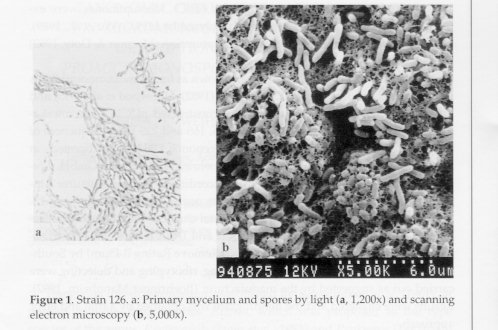
Morphological characteristics of the strains are as follows. In strain 126 the substrate mycelium (0.8-1.0um in diameter) breaks up into fragments. Usually no aerial mycelium is formed except for traces on Bennett and Czapek agar. Non motile, single spores (1.0-1.3um) are borne on a sporophore (Fig. 1).
Figure 1. Strain 126. a: Primary mycelium and spores by light (a, 1,200x) and scanning electron microscopy (b, 5,000x).
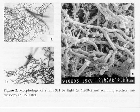
Colonies of strain 321 show a filamentous edge. The vegetative mycelium breaks up into filaments, no aerial mycelium (Fig. 2).
Figure 2. Morphology of strain 321 by light (a, 1,200x) and scanning electron microscopy (b, 15,000x).
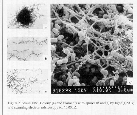
Strain 1388. Colony edge filamentous. The mycelium does not break up into fragments. Spores, single or in very short chains, are smooth surfaced and non motile. No aerial mycelium is formed (Fig. 3).
Figure 3. Strain 1388. Colony (a) and filaments with spores (b and c) by light (1,200x) and scanning electron microscopy (d, 10,000x).
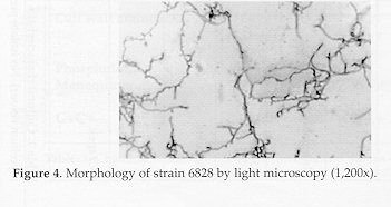
In strain 6828 the edge of the colony is filamentous. The vegetative mycelium breaks up into irregular, non motile rods. No aerial mycelium formation (Fig. 4).
Figure 4. Morphology of strain 6828 by light microscopy (1,200x).
Cultural characteristics are summarised in Table 1. All strains are Gram-positive and aerobic. No soluble pigments are formed.
Table 1. Cultural characteristics of the investigated strains and of the type strain (VKM AC 665) of Promicromonospora citrea (GR: growth; SM: mycelium colour). No isolate produces aerial mycelium (see text); P.citrea shows white aerial mycelium on oatmeal agar.
---------------------------------------------------------------------------
Medium (agar)                        Strain
               ------------------------------------------------------------
                     126                321                 1388
---------------------------------------------------------------------------
Glucose       GR  moderate          moderate light        moderate
asparagine    SM  chrysanthemum     grass green yellow    light yellow
                  bud white

Sauton's      GR  good light        good light            good light
              SM  green yellow      green yellow          green yellow

Inorganic     GR  poor light        poor light            poor light
salt starch   SM  light yellow      light yolk yellow     light yellow

Oatmeal       GR   moderate         moderate              moderate
              SM   hay yellow       light yellow          light yellow

Bennett       GR   good             good                  good
              SM   artemisia yellow light yellow          light brown

Calcium       GR   poor             poor                  poor
malate        SM   colourless       colourless            colourless

Czapek        GR   moderate         moderate              moderate
              SM   light yellow     light yellow          light yellow
---------------------------------------------------------------------------
Table 1. (continued)
---------------------------------------------------------------------------
Medium (agar)                              Strain
               ------------------------------------------------------------
                            6828                       665
---------------------------------------------------------------------------
Glucose asparagine  GR    moderate light           good
                    SM    almond yellow            artemisia yellow

Sauton's            GR    good light               good gold
                    SM    green yellow             thread yellow

Inorganic           GR    poor                     good
salt starch         SM    almond yellow            rape flower yellow

Oatmeal             GR    moderate                 moderate
                    SM    light yellow             wheat straw yellow

Bennett             GR    good                     moderate
                    SM    almond yellow            lemon yellow

Calcium             GR    poor                     poor
malate              SM    colourless               sunflower yellow

Czapek              GR    moderate                 moderate
                    SM    light yellow             light yellow
---------------------------------------------------------------------------
Physiological characteristics are listed in Table 2.
Table 2. Physiological characteristics of the investigated strains and of the type strain (VKM AC 665) of Promicromonospora citrea. No strain produces H2S or tyrosinase. All strains utilise D-glucose and L-arabinose, but not inositol or cellulose; none grows at pH 5.0, or at 10 or 50 C. All strains, except 126, grow in the presence of NaCl (1-5%).
---------------------------------------------------------------------------
                                       Strain
                    -------------------------------------------------------
Character                 126    321    1388    6828    665
---------------------------------------------------------------------------
Milk coagulation           -      +       +       +      +
Milk peptonisation         -      +       -       +      +
Gelatine liquefaction      -      -       -       -      +
Starch hydrolysis          -      -       -       -      +
Nitrate reduction          +      +       -       +      -
Utilisation of:
D-fructose                 +      -       -       -      +
D-xylose                   +      +       -       +      +
D-raffinose                +      +       +       +      -
D-mannitol                 -      +       +       +      -
D-galactose                +      -       +       +
L-rhamnose                 -      +       +       +      +
Growth at pH:
5.5                        +      -       +       +
8.5                        -      +       +       +      +
9.0                        -      +       +       +      +
---------------------------------------------------------------------------
Chemical characteristic are listed in Table 3. All the strains lack mycolic acids and show whole cell type C composition.
Table 3. Chemical characteristics of the investigated strains and of the type strain (VKM AC 665) of Promicromonospora citrea.
---------------------------------------------------------------------------
                                         Strain
                        ---------------------------------------------------
Character                 126         321      1388      6828       665
---------------------------------------------------------------------------
Cell wall amino acids     Gly         Gly       Gly       Gly       Lys

                          Lys                   Lys
                          Asp         Asp       Asp
Phospholipid type          V           I         I         II        V
Menaquinone             MK9(H4)   MK10(H2,4)  MK10(H8)  MK9(H6,8)  MK9(H4)
                                    MK9(H6)
G+C%                     74.0        72.8      74.9       75.0      70-75
---------------------------------------------------------------------------
Hybridisation results are shown in Figure 5 and the homology data, obtained by using rDNA ribotyping, are listed in Table 4.
Table 4. Strain homology by rDNA ribotyping.
----------------------------------------------
 Strain                  Ssm (%)
----------------------------------------------
665                                     100
126                              100     20
321                       100     22     42
1388               100      0     25    100
6828       100      28     11     42     22

Strain    6828    1388    321    126    665
----------------------------------------------
DISCUSSION
All strains investigated appear to be related to some extent to the genus Promicromonospora.
Because of the close similarity, isolate 1388 could be considered as a strain of the type species P.citrea.
On the other hand isolates 321 and 6828 are not closely related to the type species and the determination of their taxonomic status requires further study.
Strain 126 shows morphological, cultural and biochemical characteristics typical of the genus Promicromonospora. Nonetheless it is still quite different from strain VKM AC 665 as also shown by the low (28%) homology level. It appears therefore reasonable to consider it as belonging to a new species of the genus. On the other hand strain 126 also differs from the other two species of the genus, P.enterophila and P.sukumoe, for, among other characters, pigmentation, gelatine liquefaction, starch hydrolysis, sugar utilisation, pH, temperature and NaCl tolerance, menaquinone and phospholipid type, etc. For the above reasons it seems justified that strain 126 should be considered a new species of the genus for which the name Promicromonospora yunnanensis is proposed.
Description of Promicromonospora yunnanensis sp. nov.
Promicromonospora yunnanensis (yun.nan.en sis, M.L.adj. yunnanensis, belonging to the Yunnan Province, one of the South-West provinces of China, where the soil sample was collected).
Gram positive, mesophilic, not NaCl tolerant.
Substrate mycelium breaking up into fragments, single, non motile spores produced.
Aerial mycelium not produced, except for traces on Bennett and Czapek agar, but devoid of spores.
Moderate growth on most synthetic and organic media under aerobic conditions. No growth anaerobically. No soluble pigments.
Cell wall containing lysine, glycine and asparagine (type VI); glucose, galactose, mannitol and ribose as whole cell sugars (type C). MK9(H4) menaquinone; type V phospholipids and no mycolic acids produced.
G+C content 74%.
Isolated from soil.
Strain 126, the type strain of the species, is deposited at the China Committee Culture Collection of Microorganisms (CCCCM) with the accession number AS 4.1333.
ACKNOWLEDGEMENTS. The authors wish to thank Prof. Kalakoutskii, Russia, for providing the strain of P. citrea, Dr. Jola, Poland, for the probe used in this study and the technicians of the Microbiology Institute, Academia Sinica, for the electron micrographs.
REFERENCES
Anonymous (1957). The Colour Standard. Science Publishing House, Beijing Boehringer-Mannheim (1992). DIG System Users Guide for Filter Hybridization. Biochemica, Germany Chater, K.F., D.A.Hopwood, T.Kieser & C.J.Thompson (1982). Gene Cloning in Streptomyces. Curr. Topics Microb. Immunol., 96: 69-95
Collins, M.D. (1985). Isoprenoid quinone analyses in bacterial classification and identification. In: M.Goodfellow & D.E.Minnikin (eds.) Chemical Methods in Bacterial Systematics. Academic Press, London, pp. 267-287
Gordon, R.E., D.A.Barnett, J.B.Handerhan & C.Hor-nay Pang (1974). Nocardia coeliaca, Nocardia autotrophica, and the nocardin strain. Int. J. Syst. Bacteriol., 24: 54-63 Hopwood, D.A., M.J.Bibb, K.F.Chater, T.Kieser, C.J.Bruton, H.M.Kieser, D.J.Lydiate, C.P.Smith, J.M.Ward & H.Schrempf (1985). Genetic manipulation of Streptomyces. A Laboratory Manual. The John Innes Foundation, Norwich Jager K., K.Marialigeti, M.Hanck & G.Barabas (1983). Promicromonospora enterophila sp. nov., a new species of monospore actinomycetes. Int. J. Syst. Bacteriol., 33: 525-531
Kampfert, P. & R.M.Kroppenstedt (1991). Probabilistic identification of streptomycetes using miniaturized physiological tests. J. gen. Microbiol., 137: 1893-1902.
Krassilnikov, N.A., L.V.Kalakoutskii & N.F.Kirillova (1961). A new genus of ray fungi - Promicromonospora gen. nov. Izv.Akad.Nauk SSSR (Ser. Biol.), 1: 107-112 (in Russian) Lechevalier, M.P., A.Dietz & D.W.Thayer (1980). Society for Industrial Microbiology, Special Publication N. 6. Arlington, VA. Liu, Z., J.Ruan, J.Zakrzewska-Czerwinska & M.Mordarski (1992). Analyses of DNA homology and rDNA restriction patterns of some species in the genus Nocardiopsis. Actinomycetes, 3: 51-54. Liu, Z.H., J.Su & J.S.Ruan (1990). Determination of mycolates by gas chromatography. Microbiology, China, 17: 307-310. Marmur, J. & P.Doh (1962). Determination of basic composition of deoxyribonucleic acid from its denaturation temperature, J. Mol. Biol., 5: 109-118 Minnikin, D.E., I.G.Hutchinson, A.B.Caldicott & M.Goodfellow (1980). Thin-layer chromatography of methanolysates of mycolic acid-containing bacteria. J. Chromatog., 188: 221-223
Sambrook, J., E.F.Fritsch & T.Maniatis (1989). Molecular Cloning: A Laboratory Manual. Cold Spring Harbor Laboratory Press, New York, 2nd ed., pp. 304-324; 363-371
Shirling, E.B. & D.Gottlieb (1966). Methods for characterization of Streptomyces species. Int. J. Syst. Bacteriol., 16: 313-340 Takahashi, Y., Y.Tanaka, Y.Iwai & S.Omura (1987). Promicromonospora sukumoe sp. nov., a new species of the Actinomycetales. J. Gen. Appl. Microbiol., 33: 507-519 Wu, C., X.Lu, M.Gin & J.S.Ruan (1989). The analysis of menaquinone compositions in microbial cell wall by HPLC. Microbiology, China, 18: 176-178 Zakrzewska-Czerwinska, J., M.Mordarski & M.Goodfellow (1988). DNA base composition and homology values in the classification of some Rhodococcus species. J. gen. Microbiol., 134: 2807-2813 Copyright 1997 C.E.T.A., The International Centre for Theoretical and Applied Ecology, Gorizia The following images related to this document are available:Photo images[ac97004d.jpg] [ac97004c.jpg] [ac97004b.jpg] [ac97004a.jpg] |
| |||||||||