 |
| About Bioline | All Journals | Testimonials | Membership | News |
|
||||||
|
||||||
ANALYSIS OF RIBOSOMAL PROTEINS BY 2-D GEL ELECTROPHORESIS OF SOME ISOLATES OF PROMICROMONOSPORA-LIKE ACTINOMYCETES L. WICK* and Z.-H. LIU
Institute of Microbiology, Academia Sinica, Beijing, 100080, P.R.C.
Code Number: AC97008
Sizes of Files:
    Text: 10.4K
    Graphics: Photograph (jpg) - 34K
ABSTRACT. Electrophoretic mobility of ribosomal protein AT-L30 has been investigated by two-dimensional polyacrylamide gel electrophoresis in four Promicromonospora-like actinomycete strains isolated from soils of Yunnan Province, China. Although the protein of three isolates exhibit the same electrophoretic mobility as that of the type strain of Promicromonospora citrea VKM AC 655, it does not appear possible to assign, on these grounds, all strains to the genus Promicromonospora. Additional investigations are needed to clarify the taxonomic status of the strains. Ribosomal proteins have lower evolutionary rates than other proteins and for this reason they have been repeatedly used for studying relationships among bacteria, yeasts and fungi. However it is difficult to draw clear-cut conclusions from available data (Adoutte-Panvier et al., 1980; Boeck, 1985; Geisser et al., 1973a, 1973b; Goff & Begueret, 1984).
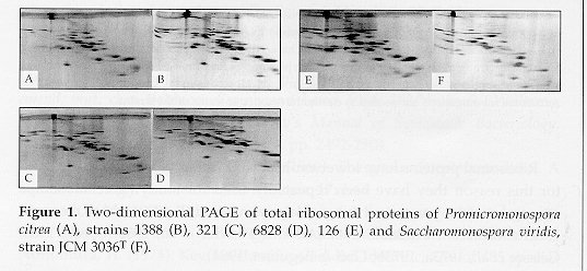
On the other hand it has been shown that two-dimensional PAGE analysis of ribosomal proteins can be helpful in clarifying some aspects of actinomycete taxonomy. In particular the ribosomal protein AT-L30 was used successfully to establish the taxonomic status of several actinomycete genera. The protein exhibits strikingly diverse electrophoretic mobilities in representatives of different genera when run under basic conditions. Amino acid sequences of the protein have also been used in taxonomic studies (Ochi, 1989, 1992, 1995a, 1995b; Ochi & Miyadoh, 1992; Ochi et al., 1991, 1993). In the present investigation the methodology has been applied to determine the taxonomic status of some Promicromonospora-like actinomycetes. MATERIALS AND METHODS Bacterial strains. The strains used in the investigation (Nos. 126, 321, 1388, 6828) were isolated from soil samples collected in the Yunnan Province, China. Promicromonospora citrea VKM AC 665^T (T= type strain) was used as a reference. Strains were grown to the mid-exponential phase in modified Sauton's medium at 28 C. Two-dimensional PAGE. Ribosomal proteins were prepared from 70S ribosomes by extraction with acetic acid (Hardy et al., 1969). The method of Kaltschmidt & Wittmann (1970) was used for the two-dimensional gel electrophoresis. The experiments were carried out according to Ochi (1989). RESULTS Results of two-dimensional PAGE of ribosomal proteins of four PLA and of the type strain of Promicromonospora citrea VKM AC 665^T are shown in Fig. 1. The AT-L30 protein was identified by comparisons with previously published gels (Ochi, 1992; Ochi & Miyadoh, 1992; Ochi et al., 1991, 1993).
Table 1. Electrophoretic two-dimensional mobilities of ribosomal AT-L30 proteins from Promicromonospora citrea, Saccharomonospora viridis and Promicromonospora-like isolates (with reference to S. viridis mobility = 100).
-----------------------------------------------------------------
         ORGANISM                     REM of AT-L30 proteins (mm)
-----------------------------------------------------------------
Promicromonospora citrea VKM AC 665^T    23           29
    Strain 126                         0/32         0/41
    Strain 321                           22           28
    Strain 1388                          22           28
    Strain 6828                          22           28
Saccharomonospora viridis JCM 3036^T     78          100
-----------------------------------------------------------------
Two proteins are present in strain 126, both however show REMs that are different from those of the other strains. With reference to the overall protein pattern, strains 321 and 6828 are most closely related to each other, while 655 and 1388 seem to be the least related. Strain 126 does not show much similarity with any of the others. DISCUSSION From the analysis of ribosomal protein AT-L30 it appears that strain 126 does not belong to the species P. citrea or to the genus Promicromonospora itself, as suggested by chemotaxonomic studies.
The fact that protein REMs are practically identical for strains 655, 321, 1388 and 6828 strongly suggests that they belong to the same genus. However it should be pointed out that a number of actinomycetes have AT-L30 protein REMs failing in the 20 to 30 range (Ochi, 1992). On the other hand chemotaxonomic studies exclude the attribution of strains 321 and 6828 to the genus Promicromonospora (Zhiheng et al., 1997). In this case an amino acid sequence analysis of the AT-L30 protein would be useful (Ochi, 1992).
Ribotyping of rDNA has shown that the type strain of P. citrea is more closely related to 1388 than to the other strains (Zhiheng et al., 1997), the exact opposite of what could be inferred from the present results.
AT-L30 protein analysis has been used successfully in taxonomic studies of some actinomycetes, such as Actinomadura, Microtetraspora, Nocardia, Rhodococcus, Tsukamurella and Gordona. However in the case of Streptosporangium and allied taxa, as well as in the present study, conclusions drawn by the application of the technique are in clear contrast with those derived by rRNA analysis and chemotaxonomic characteristics (Ochi, 1989, 1992, 1995a, 1995b; Ochi & Miyadoh, 1992; Ochi et al., 1991, 1993).
These differences point out that, because of protein pattern complexity, the technique should be used with care and together with other taxonomic methods, as a constituent of a polypbasic taxonomic approach. ACKNOWLEDGEMENTS. We thank Prof. K. Ochi for providing us with his laboratory journal and Prof. L.V. Kalakoutskii, IPBM, Russia, for strain VKM AC 665^T of Promicromonospora citrea REFERENCES Adoutte-Panvier, A., J.E. Davies, L.R. Gritz & B.S. Littlewood (1980). Studies of ribosomal proteins of yeast species and their hybrids: gel electrophoresis and immunochemical cross-reactions. Mol. Gen. Genet., 179:273-282 Boeck, A. (1985). Analysis of ribosomal proteins by two-dimensional gel electrophoresis. Methods Microbiol., 18:109-122 Geisser, M., G.W. Tischendorf, G. Stoeffier & H.G. Wittmann (1973a). Immunological and electrophoretical comparison of ribosomal proteins from eight species belonging to Enterobacteriaceae. Mol. Gen. Genet., 127:111-128 Geisser, M., G.W. Tischendorf & G. Stoeffier (1973b). Comparative immunological and electrophoretic studies on ribosomal proteins of Bacillaceae. Mol. Gen. Genet., 127:129-145 Golf, V.L. & J. Begueret (1984). Immunological comparison of individual ribosomal proteins from in six species of genus Podospora. Mol. Gen. Genet., 193:143-148 Hardy, S.J.S., C.G. Kurland, P. Voynow & G. Mora (1969). The ribosomal proteins of Escherichia coli. 1. Purification of the 30S ribosomal proteins. Biochemistry, 8:2897-2905 Kaltschmidt, E. & H.G. Wittmann (1970). Ribosomal proteins. VII. Two- dimensional polyacrylamide gel electrophoresis for finger-printing of ribosome proteins. Anal. Biochem., 36:401-412 Ochi, K. (1989). Heterogeneity of ribosomal proteins among Streptomyces species and its application to identification. J. gen. Microbiol., 135:2635-2642 Ochi, K. (1992). Electrophoretic heterogeneity of ribosomal protein AT-L30 among actinomycete genera. Int. J. Syst. Bacteriol., 42:144-150 Ochi, K. (1995a). Amino acid sequence analysis of ribosomal protein AT-L30 from members of the family Pseudonocardiaceae. Int. J. Syst. Bacteriol., 45: 110-115 Ochi, K. (1995b). Comparative ribosomal protein sequence analysis of a phy- logenetically defined genus Pseudomonas. Int. J. Syst. Bacteriol., 45:268-273 Ochi, K. & S. Miyadoh (1992). Polyacrylamide gel electrophoresis analysis of ribosomal protein AT-L30 from an actinomycete genus, Streptosporangium. Int. J. Syst. Bacteriol., 42:151-155 Ochi, K., S. Miyadoh & T. Tamura (1991). Polyacrylamide gel electrophoresis analysis of ribosomal protein AT-L30 as a novel approach to actinomycete taxonomy: application to the genera Actinomadura and Microtetraspora. Int. J. Syst. Bacteriol., 41:234-239 Ochi, K., K. Haraguchi & S. Miyadoh (1993). A taxonomic review of the genus Microbispora by analysis of ribosomal protein AT-L30. Int. J. Syst. Bacteriol., 43:58-62 Zhiheng, L., Q. Weihong, S. Yanlin & Z. Yamei (1997). Taxonomic characte- risation of some Promicromonospora-like isolates and description of a new species Promicromonospora yunnanensis sp. nov. Actinomycetes, 8:20-27 Copyright 1997 C.E.T.A., The International Centre for Theoretical and Applied Ecology, Gorizia The following images related to this document are available:Photo images[ac97008a.jpg] |
| |||||||||