 |
| About Bioline | All Journals | Testimonials | Membership | News |
|
||||||
|
||||||
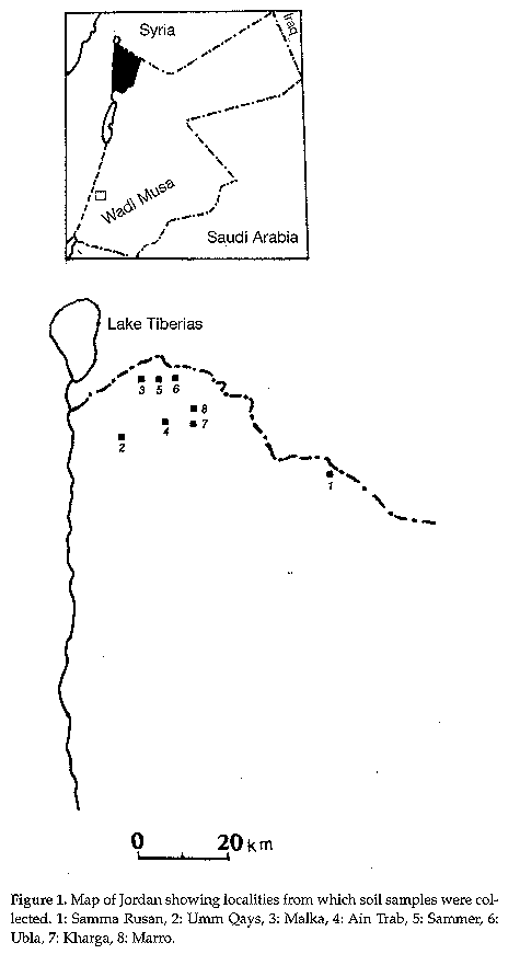
Actinomycetes, Vol. 9, Part 3, 1998, pp.52-60 DIVERSITY OF SOIL STREPTOMYCETES IN NORTHERN JORDAN I. SAADOUN, M. J. MOHAMMAD1, H. I. MALKAWI2, F. AL-MOMANI and M. MEQDAM Dept. Biological Sciences and 1Dept. Natural Resources and Environment, Jordan Univ. of Science and Tech., 2Dept. Biological Sciences, Yarmouk Univ., Irbid-22110, Jordan Code Number:AC98008 ABSTRACT. An investigation was undertaken in order to evaluate the distribution of soil streptomycetes in different locations in northern Jordan. Average microbial counts were 29.2 x 10⁸ CFU/gm dry soil for bacteria and 2.4 x 10⁶ CFU/gm dry soil for streptomycetes. Based on sporophore morphology, 90 isolates were identified as belonging to the genus Streptomyces and 11 to Streptoverticillium. Cultural and morphological characters showed the dominance of grey aerial mycelium species. Antibiotics produced were mostly active against Gram positive bacteria. Since the discovery of actinomycin (Lechevalier, 1982), actinomycetes have produced many commercially important bioactive compounds and antitumor agents in addition to enzymes of industrial interest (Tanaka & Omura, 1990). It has been estimated that approximately two-thirds of the thousands of naturally occurring antibiotics have been isolated from these organisms (Takizawa et al., 1993). Of these, the majority were obtained from the genus Streptomyces (Goodfellow & O'Donnell, 1989). The traditional isolation substrate for the organisms is soil (Labeda & Shearer, 1990). Data concerning the distribution of soil streptomycetes in plant nursery and in non-cultivated soils in Jordan are available (Abussaud & Saadoun, 1988; Saadoun & Al-Momani, 1997a, 1997b). However little is known about the occurrence of these organisms in cultivated soils. In the present note the diversity of streptomycetes in cultivated northern Jordan soils is reported. MATERIALS and METHODS Sampling. Four samples from each of nine locations in northern Jordan (Fig. 1) were collected during the summer of 1996. Samples were taken with an auger (up to 10 cm depth), after removing approximately 3 cm of the soil surface, placed in polyethylene bags and stored in a refrigerator. Figure 1. Map of Jordan showing localities from which soil samples were collected. 1: Samma Rusan, 2: Umm Qays, 3: Malka, 4: Ain Trab, 5: Sammer, 6: Ubla, 7: Kharga, 8: Marro. Soil analysis. Soil samples collected from each site and with a similar plant residue status were combined and thoroughly mixed. The soil pH was measured in 1:1 soil - water suspension and organic matter was determined by the oxidation method (Nelson & Sommers, 1982). Isolation. Samples were mixed thoroughly and sieved (2 mm mesh). Subsamples (1 g) were suspended in 100 ml distilled water in a water-bath shaker (140 rpm, 30 min), serially diluted up to 10-6 and spread (0.1 ml) over the surface of agar plates with sterile L-shaped glass rods. Triplicate plates of nutrient agar and glycerol nitrate casein agar (Küster & Williams, 1964) were used for total bacteria and streptomycete counts, respectively. Plates were incubated at 27°C and the number of colonies was determined after 48 hrs (bacteria) and 10 dd (streptomycetes). Selected colonies were purified by repeated streaking. Characterisation of the isolates. Streptomyces< I> and Streptovertici llium colonies were characterised morphologically and physiologically following the methods given for the International Streptomyces project (ISP) (Shirling & Gottlieb, 1966). General morphology was determined on oatmeal agar plates, incubated in the dark at 27°C for 21 dd, by direct light microscopy examination of the surface of crosshatched cultures. Colours were determined according to Prauser (1964) and isolates were grouped as proposed by Nonomura (1974). Antimicrobial activity. This was tested by the plate diffusion method (Bauer et al., 1966) against Staphylococcus aureus, Bacillus subtilis, Escherichia coli, Candida albicans, and Trichoderma harasmii and Penicillium lilacinum, isolated from olive mill solid products. Isolates were grown on oatmeal agar for 14 days; 3 discs (5 mm in diameter) were cut and placed on nutrient agar seeded with the above test organisms. Plates were incubated at 27°C and inhibition zones determined after 24 hrs for bacteria and after 48 hrs for fungi. RESULTS and DISCUSSION Average microbial counts were 29.2x10⁸ CFU/gm dry soil for bacteria and 2.4x10⁶ CFU/gm dry soil for streptomycetes. The distribution of streptomycetes in each site is summarised in Table 1. Highest values were found in Samma Rusan and the lowest in the Ain Trab site. The quantity of streptomycetes at these sites reflects their edaphic conditions. Table 1. Number of bacteria and streptomycetes and soil characteristics of the sampling sites (CFU: colony forming units, DE: dacayed, MOIST.: moisture, SD: semidecayed; STM:: streptomycetes, UD: undecayed).
Actinomycete numbers in temperate climates range from 106 to 108 per gram of dry soil. Abussaud and Saadoun (1991) reported an average streptomycetes count of 105 to 107/ gm dry soil in Jordan Valley and Msameh (1992) an average of 5.9 x 10⁶/gm dry soil of high lands of Jordan and 5.2 x 10⁴ in Aqaba soils. Streptomycetes account for about 1-20% of the total bacterial count, but in some soils they may dominate (Flaig & Kutzner, 1960; Xu et al., 1996). In a recent study by Valagurova et al (1998), the number of soil streptomycetes at 'Evolution Canyon', Mount Carmel, Israel, was found to vary from 3,000 to 15,000 CFU/gm of dry soil. The number of streptomycetes was related to type of soil and edaphic conditions. In the present investigation 101 streptomycetes were recovered from the samples, 90 isolates were identified as belonging to the genus Streptomyces and 11 to Streptoverticillium. Most streptomycetes were isolated from Ain Trab (19) and Samma Rusan (17) soils (Table 2). Isolates were distributed into series according to the colour of their mature sporulated aerial mycelium (Table 4). Grey, white and yellow colour series were the most representative (32%, 20% and 16% respectively) with the lowest occurrence being that the red series (3%). Table 2. Distribution of the Streptomyces isolates in the different sites (AM: aerial mycelium, GE: green, GY: grey, VAR: variable, pink, orange or violet, Y: yellow, W; white).
Table 3. Table 3. Morphological, cultural and antibiotic characteristics of Streptomyces isolates (FL: flexuous, RA: Retinaculum Apertum, RE: Rectus, SP: spiral).
As shown in previous reports, strains with grey aerial mycelium are usually more abundant (Hamdi et al., 1980; Abussaud & Saadoun, 1988; Msameh, 1992; Saadoun & Al-Momani, 1997a, b; Valagurova et al., 1998). Of the 90 Streptomyces isolates, 27 produce melanin, 61 show distinctive reverse side pigment and 22 produce soluble pigments (Table 3). With reference to the morphology of spore bearing hyphae most isolates show flexuous (48%) sporophores (Table 3). Studies by Hamdi et al. (1980), Coelho & Drozdwicz (1979), Abussaud & Saadoun (1988), Msameh (1992) and Saadoun & Al-Momani (1997a, b) reported a higher frequency of strains with spiral sporophores. Concerning antimicrobial activity, fifty four percent of the isolates were active against one or more of the test organisms, however the percentage of active isolates varies within each colour series, tending to be higher in the red and grey series and lower in the green and white ones. The lowest activity was exhibited against T. harasmii (12%) and P. lilacinum (10%). Streptomyces isolates appear to be mostly active against Gram positive bacteria. Species possessing verticillate sporophores are considered to belong to the genus Streptoverticillium. Morphological, cultural and antibiotic characteristics of the 11 isolates attributed to the genus Streptoverticillium (Locci et al., 1969; Nonomura, 1974) are shown in Table 4. Table 5. Characteristics of the Streptoverticillium strains (AM: aerial mycelium colour, BV: biverticillate, MV: monoverticillate, RC: reverse pigment; SP: soluble pigment)
Streptoverticillium strains are grouped, according to the colour of the aerial mycelium, as grey (8), yellow, pink, and violet (1 each). Nine strains are characterised by biverticillate and two by monoverticillate sporophores (Locci & Schofield, 1989). Seven strains show reverse colour and three produce soluble pigment; however none produces melanin. As regards antibiotic activity, two strains inhibited all tested bacteria and C. albicans. ACKNOWLEDGEMENTS. This research was funded by the Higher Council for Science and Technology, Jordan under the biodiversity of soil microorganisms in Jordan. Appreciation is extended to the Jordan University of Science and Technology for administrative support. REFERENCES Abussaud, M. J. & I. M. Saadoun (1988). Isolation, characterization and taxonomy of Streptomyces sp. isolated from Jordanian soils and antagonistic to Agrobacterium tumefaciens. Egypt. J. Microbiol., 23: 597-609 Abussaud, M. & I. Saadoun (1991). Streptomyces flora of some Jordan Valley soils, characteristics and seasonal distribution. Dirasat, 18B: 66-75 Bauer, A. W., , W. M. Kirby, J. C. Sherris & M. Turk (1966). Antibiotic susceptibility testing by a standardized single disk method. Am. J. Clinic. Path., 45, 493-496 Coelho, R. R. & A. Drozdowicz (1979). The occurrence of actinomycetes in cerrado soil in Brazil. Rev. Ecol. Biol. Sci., 15: 459-474 Flaig, W. & H. J. Kutzner (1960). Beitrag zur Õkologie der Gattung Streptomyces Waksman et Henrici. Arch. Mikrobiol., 35: 105-138 Goodfellow, M. & A. G. O'Donnell (1989). Search and discovery of industrially significant actinomycetes. In: S. Baumberg, I. Hunter and M. Rhods (eds.) Microbial Products: New Approaches. Cambridge University Press, Cambridge, pp. 343-383 Hamdi, Y. A., D. Ahmed & A. M. Al-Tai (1980). Genera and species of actinomycetes isolated from Iraqi soils. Egypt. J. Microbiol., 15: 7-22 Küster, E. & S. T. Williams (1964). Selection of media for isolation of streptomycetes, Nature, 202: 928-929 Labeda, D. P. & M. C. Shearer (1990). Isolation of actinomycetes for biotechnological applications. In: D. P. Labeda (ed.) Isolation of biotechnological organisms from nature. McGraw-Hill Publ. Co., pp. 1-19 Lechevalier, H. (1982). The development of applied microbiology at Rutgers. Rutgers, The State University of New Jersey Locci, R. & G. Schofield (1989). Genus Streptoverticillium Baldacci 1958, 15, emend. mut. char. Baldacci, Farina and Locci 1966, 168AL. In: S. T. Williams, R. E. Sharpe and J. G. Holt (eds.) Bergey's Manual of Systematic Bacteriology. Williams and Wilkins, Baltimore, Vol. 4, pp. 2492-2504 Locci, R., E. Baldacci & B. P. Baldan (1969). The genus Streptoverticillium. A taxonomic study. Giorn. Microbiol., 17: 1-60 Msameh, Y. M. (1992). Streptomyces in Jordan; distribution and antibiotic activity. M.S. Thesis, Department of Biology, Yarmouk University, Irbid, Jordan Nelson, D. W. & L. E. Sommers (1982). Total carbon, organic matter. In: A. L. Pape, R. M. Miller and D. R. Keeney (eds.) Methods of Soil Analysis. Part II. ASA, Madison, Wisconsin Nonomura, H. (1974). Key for classification and identification of 485 species of the streptomycetes included in the ISP. J. Ferm. Tech., 52: 78-92 Prauser, H. (1964). Aptness and application of colour for exact description of colours of Streptomyces. Zeitsch. Allgem. Mikrobiol., 4: 95-98 Saadoun, I & F. Al-Momani (1997a). Streptomycetes from Jordan soils active against Agrobacterium tumefaciens. Actinomycetes, 8: 29-36 Saadoun, I. & F. Al-Momani (1997b). Studies on soil streptomycetes from Jordan. A ctinomycetes, 8: 42-48 Shirling, E. B. & D. Gottlieb (1966). Methods for characterization of Streptomyces species. Int. J. Syst. Bacteriol., 16: 313-340 Takizawa, M., R. R. Colwell & R. T. Hill. (1993). Isolation and diversity of actinomycetes in the Chesapeake Bay. Appl. Environ. Microbiol., 59: 997-1002 Tanaka, Y. & Omura, S. (1990). Metabolism and products of actinomycetes - An introduction. Actinomycetologica, 4, 13-14 Valagurova, H. V., V. E. Koziritskaya, G. A. Iutinskaya, , A. A. Pindrus & K. I. Andreyuk (1998). Diversity of soil streptomycetes at Evolution Canyon, Mount Carmel, Israel. Actinomycetes, 9: 10-18 Xu, L., O. Li, & G. L. Jaing (1996). Diversity of soil actinomycetes in Yunnan China. Appl. Environ. Microbiol., 62: 244-248. Copyright 1998 C.E.T.A., The International Centre for Theoretical and Applied Ecology, Gorizia The following images related to this document are available:Line drawing images[ac98008a.gif] | |||||||||||||||||||||||||||||||||||||||||||||||||||||||||||||||||||||||||||||||||||||||||||||||||||||||||||||||||||||||||||||||||||||||||||||||||||||||||||||||||||||||||||||||||||||||||||||||||||||||||||||||||||||||||||||||||||||||||||||||||||||||||||||||||||||||||||||||||||||||||||||||||||||||||||||||||||||||||||||||||||||||||||||||||||||||||||||||||||||||||||||||||||||||||||||||||||||||||||||||||||||||||||||||||||||||||||||||||||||||||||||||||||||||||||||||||||||||
| |||||||||