 |
| About Bioline | All Journals | Testimonials | Membership | News |
|
||||||
|
||||||
Annals of African Medicine, Vol. 4, No. 2, June, 2005, pp. 77-82 INTRAOPERATIVE VENTRICULAR BIGEMINY: REPORT OF 5 CASES A. S. Ganny and S. A. Eguma Department of Anesthesiology and Intensive Care, Aminu Kano
Teaching Hospital, Kano, Nigeria Code Number: am05019 Abstract
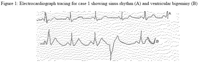
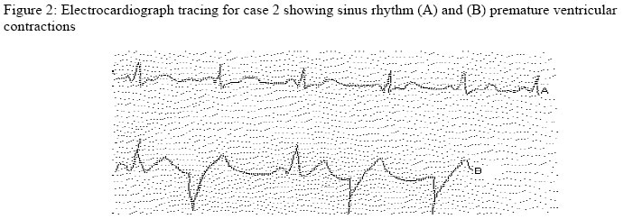
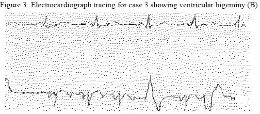
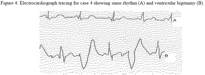
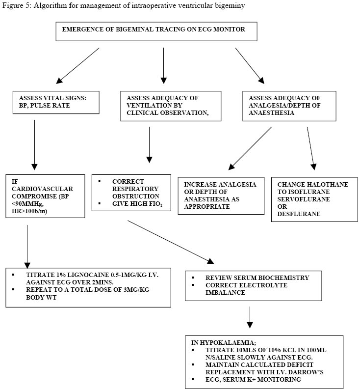
Five patients who had intraoperative ventricular bigeminy while undergoing various orthopaedic procedures are reported. Diagnosis of pulsus bigeminus was established by continuous intraoperative ECG monitoring of lead 11 using a Micromon 7142 (L&T Medical) ECG machine. Causes of these arrhythmias were traced to hypokalaemia, inadequate analgesia, old age, previous rheumatic heart disease and sensitization of the myocardium to catecholamines by halothane. All patients were successfully managed either with intravenous potassium administration, additional analgesia or intravenous lignocaine. The importance of preoperative patient investigations and intraoperative monitoring is highlighted. Key words: Ventricular bigeminy, intraoperative Résumé Cinq patients qui avaient subi la bigéminé intrachirurgicale ventriculaire pendant qu’ils suivaient des procédures orthopédiques diverses sont l’objet de ce raport. On avait déterminé le diagnostic du gigéminus pulsus à travers la surveillance ECG intraopératoires continuelle de plomb 11 avec l’ultilisation de Micromon 7142 (L & T Médical) machine ECG. On a pu établir que les causes de ces arrhythmia étaient hypokalémie, analgésie insuffisance, la vieillesse, maladies cardiaques du rhumatisme précédantes et la sensitisation de myocardium à catécholomine par halothane. Tous les patients ont été traités avec succès soit à travers l’administration du potassium intraveineux, analgesie supplémentaire, soit lignocaine intraveineux. Il s’agit de l’importance d’une étude préopératoire des patients et surveillance intraopératoire. Mots clés : Bigéminé ventriculaire, intrachirurgicale Introduction Ventricular bigeminy is a cardiac arrhythmia characterised by the occurrence of a normal heartbeat followed by an ectopic beat, resulting in the phenomenon known as “coupled beats”. The character of the peripheral pulse felt during such episodes of coupled beats is known as pulsus bigeminus. The incidence of cardiac arrhythmias during general anaesthesia has been put at as high as 60%. 1 Pulsus bigeminus may remain innocuous in so far as it does not induce haemodynamic instability. The main danger however lies in its great potential to degenerate into ventricular fibrillation and cardiac asystole. Correctable factors such as hypokalaemia and light anaesthesia have been implicated in the aetiology2. Conscientious perioperative monitoring of patients would ensure early detection and appropriate management before they progress to cardiac arrests. Five patients who developed pulsus bigeminus while undergoing various orthopaedic surgical procedures under general anaesthesia are reported. The Bain coaxial anesthesia circuit was used in all patients. Each patient was monitored with a Micromon 7142 electrocardiograph (ECG) machine with a freeze facility for the lower tracing. In addition, a BCI 3001 Pulse oximeter and a Dinamap were used to monitor arterial oxygen saturation and non-invasive blood pressure respectively. Case 1A 66year old man weighing 75kg was scheduled for open reduction and internal fixation of a mid-shaft fracture of the right humerus sustained in a road traffic accident. Apart from a long- standing ulcer on his left heel, physical examination revealed no gross abnormality in any of the systems. His chest was clinically clear and heart sounds were normal. His blood pressure was 160/90mmHg, pulse 76 beats/min and regular. Full blood count and urinalysis were found to be within normal limits. Serum biochemistry revealed a potassium level of 3.3mmol/l (mild hypokalaemia). Chest X-ray showed clear lung fields and a cardio-thoracic ratio of 0.5. ECG was normal. He was premedicated with 10mg diazepam orally nocte the night before surgery. Following preoxygenation, anaesthesia was induced with 300mg sodium thiopentone and the trachea intubated after muscle relaxation with 100mg suxamethonium. Anaesthesia was maintained with 70% nitrous oxide in oxygen and halothane 1-2%. Atracurium 25mg was given for muscle relaxation and the lungs manually ventilated by intermittent positive pressure ventilation. Intraoperative analgesia consisted of 10mg incremental doses of pentazocine. About 30 minutes into the surgical procedure, the systolic blood pressure dropped to 110mmHg and ECG tracing showed ventricular bigeminy (Figure 1). The QT intervals were not prolonged and no U waves were seen. 100mg plain lignocaine was slowly titrated intravenously against the ECG tracing with no effect. The dose of lignocaine was repeated and this resulted in restoration of sinus rhythm. Five minutes later there was a recurrence of ventricular bigeminy. 13 mmol of potassium (as KCl) in 20ml normal saline was given slowly intravenously while observing the ECG tracing. This resulted in restoration of normal sinus rhythm. This was followed with slow intravenous infusion of Darrow’s solution about two litres given over twelve hours. Sinus rhythm returned and was maintained till the end of surgery. Arterial oxygen saturation was between 96% and 98% intraoperatively and arterial blood pressure was 140/90mmHg. Serum potassium done 24hours postoperatively was 4.3mmol/l. The patient made an uneventful recovery. Case 2A 60 year old farmer who sustained a fracture of left humerus was scheduled for open reduction and plating under general anaesthesia. General examination revealed a healthy-looking, normotensive elderly man weighing 73kg.Preoperative investigations showed normal serum biochemistry and normal chest X-ray findings. Apart from a slight infero-lateral repolarization abnormality, his preoperative 12-lead ECG was unremarkable. He was premedicated with 10mg diazepam the night before surgery. Pre-induction BP was 160/90mmHg, pulse rate 86/min, regular and SPO2 96%. Anaesthesia was induced with 300mg sodium Thiopentone followed with 100mg suxamethonium and endotracheal intubation. Maintenance of anaesthesia was with 70% nitrous oxide in oxygen and 1-2% Halothane, with the patient breathing spontaneously. About an hour into surgery, a tracing of coupled beats was observed on the ECG Figure 2). 50mg of 1% plain lignocaine was given intravenously. Sinus rhythm returned and was sustained throughout surgery. The patient remained haemodynamically stable post operatively. Case 3This was a 29 year- old woman who sustained multiple simple fractures of the right ilium, left ischium and right neck of femur following a road traffic accident. The patient was unable to sit up or lie down on either side on account of her bilateral hip fractures and therefore had to be given general anaesthesia for plating first of the right ilial fracture. She gave a past medical history of rheumatic heart disease ten years previously for which she was placed on continuous antibiotics. She, however, did not know the exact cardiac pathology and had stopped the antibiotics on her own, having been asymptomatic for a long time. On examination, she was afebrile, not pale and weighed 87kg. Her pulse rate was 80b/min and blood pressure 120/70mmHg. She had normal heart sounds and was haemodynamically stable. Preoperative urinalysis, haemogram and serum biochemistry results were essentially normal. ECG did not reveal any significant abnormality. She received 10mg diazepam nocte orally for premedication. Anaesthesia was induced with 400mg sodium thiopentone and tracheal intubation with a size 8.0mm Portex endotracheal tube facilitated with 100mg Succinylcholine. 70% nitrous oxide in oxygen as well as 1-2%halothane was used to maintain anaesthesia. 10mg increments of intravenous pentazocine were given for intraoperative analgesia. At skin incision the patient manifested episodes of bigeminal tracings on the ECG (Figure 3) lasting about5-10 seconds despite sustained haemodynamic stability. No treatment was given due to the brevity of the arrhythmia. Four days later the patient had to have a repeat anaesthetic for insertion of dynamic hip screw and plating of her right femur. Since neuraxial blockade was not feasible general anaesthesia was repeated. The patient manifested the same ventricular bigeminy as in the previous anaesthetic. Again no treatment was given and she had an uneventful recovery. Case 4A 34year old female police officer sustained extensive abrasions on her forehead, right upper eyelid and conjunctiva as well as bilateral Colles’ fracture and fracture dislocation of her right ankle in a road traffic accident. There was a brief loss of consciousness following the accident. She was scheduled for debridement, examination of the eyes under anaesthesia and reduction of her fractures. On examination, she was conscious and had no associated chest or abdominal injuries. She was pregnant and fundal height was 12-week size. Preoperative skull and chest X-rays were unremarkable and haemogram and serum biochemistry were normal. The risk of spontaneous loss of pregnancy was explained to the patient and informed consent for surgery and anaesthesia obtained. She received full preanaesthetic antacid prophylaxis. Preoperative vital signs were arterial BP 120/80mmHg and pulse rate 80/min Heart sounds were normal. No preoperative ECG was done. Rapid sequence induction of anaesthesia and endotracheal intubation was done and anesthesia maintained with 50:50 nitrous oxide, oxygen and halothane 1-2%. As the orthopaedic surgeon applied traction and thenar grip to reduce the right Colle’s fracture, the patient developed a bigeminal tracing on the ECG (Figure 4) which lasted for about 2minutes.There was tachycardia with a heart rate of 102/minute. 10mg of pentazocine was given intravenously which resulted in restoration of sinus rhythm. Ventricular bigeminy was again observed on ECG during attempts to reduce the left Colles’ fracture. Another 10mg intravenous pentazocine abolished the arrhythmia. Case 5 A 40year old man with non-union of old left femoral fracture required anaesthesia for removal of K-nail and plating of his femoral fracture. Following routine and normal preoperative examination and investigations he was premedicated with 10mg oral diazepam the night before surgery. There was no preoperative ECG. He was given a subarachnoid block using 3mls of 0.5% heavy bupivacaine through the L3-L4 interspace. This provided good sensori-motor blockade up to theT10 level and surgery commenced. After about 90minutes the patient complained of unbearable pain at the operation site and general anaesthesia was therefore induced by inhalation of 70% nitrous oxide in oxygen and halothane. About 30minutes later, the sinus rhythm changed to a ventricular bigeminal tracing on the ECG screen with about six premature ventricular contractions every minute. 100mg of 1% plain lignocaine was given intravenously but the coupled beats persisted. The systolic blood pressure also dropped from 110mmHg to 90mmHg and heart rate was 96beats /minute. At this point, halothane was tailed off and isoflurane was introduced gradually. Normal sinus rhythm returned and persisted till the end of surgery under isoflurane inhalation. Discussion Ventricular bigeminal rhythm is said to occur when a premature ventricular contraction (PVC) follows every normal heartbeat. Premature ventricular contractions are bizarre extra ECG complexes triggered by an ectopic focus. On the ECG, coupled beats manifest as a normal QRS complex followed immediately by an abnormal one and a prolonged pause of electrical activity, then by a normal QRS complex. They can be felt as extra peripheral pulse beats. Sometimes due to inadequate ventricular filling, they may be perceived as missed beats. PVCs are common, increase with age and are generally benign. They can however be an indication of some underlying cardiac pathology. During ventricular bigeminy, there is a PVC for every normal sinus beat, which nearly doubles the patient’s original heart rate on the ECG screen. There is an increased potential for ventricular fibrillation or intraoperative cardiac asystole. The presence of more than 5 PVCs in one minute is said to increase cardiac risk in the perioperative period. 3 Intraoperative cardiac arrhythmias arise from ectopic foci in the ventricle and may serve as markers for myocardial ischaemia. Pre-existing heart disease is a known cause of perioperative cardiac arrhythmia and this may have accounted for the bigeminal rhythm observed in our second and third patients. Another cause of intra-operative cardiac arrhythmia is disturbance in potassium balance.4,5 Hypokalaemia is associated with ST segment depression, T wave depression and prominent U waves on ECG.6 Hyperventilation during anaesthesia may result in a respiratory alkalosis, reducing serum potassium concentration and precipitating arrhythmias in an already hypokalaemic patient. Our first patient had a long-standing leg ulcer from where he may have been losing potassium. Although his preoperative ECG did not reveal any changes consistent with hypokalaemia, his preoperative serum potassium concentration was 3.3mmol/l, which is just below the lower limit of normal (Normal=3.5-5.5mmol/l). Although Wong et al5 in their review of hypokalaemia and its anaesthetic implications did not find hypokalaemia to be an independent risk factor for cardiac arrhythmias, the fact that the ventricular bigeminal rhythm was abolished in our patient by the administration of potassium lends credence to the implication of hypokalaemia as the cause of the ventricular bigeminy noticed in this patient. The relaxant technique was not used because neostigmine was not available for reversal of residual muscle paralysis. Volatile anaesthetic agents have been implicated in the causation of abnormal cardiac rhythms during anaesthesia.7, 8, 9 Halothane has been shown to increase the sensitivity of the myocardium to circulating catecholamines thereby precipitating premature ventricular contractions.7 This it does by prolongation of conduction through the bundle of His and Purkinje system to facilitate the re-entry phenomenon.10 It also stimulates alpha1 adrenergic receptors in the heart.11 This is thought to explain the bigeminal rhythm observed in our fourth and fifth patients. The arrhythmia in our fourth patient was abolished with the administration of intravenous analgesia. Inadequate analgesia would result in the patient perceiving pain and thus eliciting the stress response with out-pouring of adrenaline into the circulation. Premature ventricular contractions are common in the elderly and may have contributed to the bigeminy in cases 1 and 2. Other causes of abnormal cardiac rhythms include hypoxia, hypercapnia, administration of atropine, digitalis toxicity, alcohol, mitral valve prolapse, congestive cardiac failure, prolonged Q-T syndrome and the transfusion of stored blood.12, 13 Hypoxia as a cause of ventricular bigeminy in uor patients could not be ruled out because, although arterial oxygen saturation was monitored and remained normal, end tidal CO2 was not monitored intraoperatively to confirm the adequacy of ventilation. An algorithm for management of intraoperative venricular bigeminy is shown in figure 5. Premature ventricular contractions may be abolished by the administration of 0.5-1mg /kg plain lignocaine intravenously over 2 minutes, then 0.5mg/kg every 2-5minutes, to a maximum of 3mg/kg. Lignocaine can also be given as an infusion. Other drugs that can be used for treatment are intravenous amiodarone150mg given over 10 minutes, procainamide 20mg/70kg/min or intravenous bretylium tosylate 5-10mg/kg over 5-10 minutes. The definitive treatment however is to treat the underlying cause such as correction of hypokalaemia, administration of adequate analgesia and withdrawal of the offending volatile agent and its replacement with a less arrhythmogenic agent as was done for our patients. If ventricular fibrillation occurs, synchronized direct current cardio version may be indicated although its place in the management of ventricular bigeminy remains to be evaluated. 14 If there is life- threatening haemodynamic instability, overdrive cardiac pacing should be considered. For ventricular arrhythmias due to congenital cardiac disease corrective surgery is indicated. 15 For our patients, synchronized direct current cardio version was not necessary as prompt recognition and pharmacological treatment of the cause restored sinus rhythm. Cardiac arrhythmias during anaesthesia may be precipitated by various factors. We found ventricular bigeminy to be the commonest arrhythmia in our orthopaedic patients who had general anaesthesia and these were caused by hypokalaemia, rheumatic heart disease, inadequate intraoperative analgesia and the use of halothane. Detection of cardiac arrhythmias was made possible by intraoperative monitoring of the cardiovascular system. Perioperative patient assessment and monitoring is important for timely detection and management of cardiac arrhythmias occurring during anaesthesia. References
Copyright 2005 - Annals of African Medicine
The following images related to this document are available:Photo images[am05019f4.jpg] [am05019f5.jpg] [am05019f1.jpg] [am05019f3.jpg] [am05019f2.jpg] |
| |||||||||
