 |
| About Bioline | All Journals | Testimonials | Membership | News |
|
||||||
|
||||||
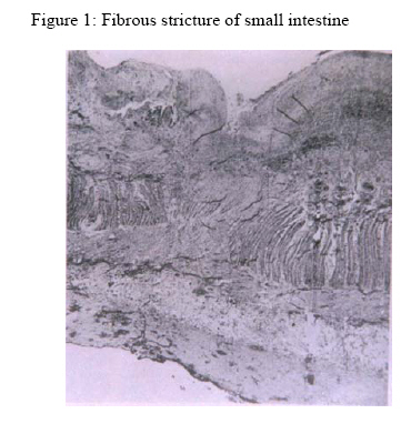
Annals of African Medicine, Vol. 5, No. 1, 2006, pp. 56-58 Fibrous Stricture of the Small Intestine Following Strangulated Inguinal Hernia: Report of Two Cases A. Ahmed Department of Surgery, AhmaduBelloUniversity Teaching Hospital, Zaria, Nigeria Reprint requests to: Dr. A. Ahmed, Department of Surgery, Ahmadu Bello University Teaching Hospital, Zaria, Nigeria. E-mail: mrahmed1010@yahoo.com Code Number: am06013 Abstract Strangulated inguinal hernia is a common surgical emergency with potentially life threatening consequences. Following reduction of an obstructed hernia the reduced intestine usually returns to normal function. We present two patients each of who presented with obstructed right inguinal hernia of more than 8 hours duration. Following release of the obstructions the intestines were judged viable and returned to the peritoneal cavity and herniorrhapies done. They represented to us several months later with features of intestinal obstruction necessitating laparatomy. Fibrous strictures obliterating the lumen of the intestines were found at the site where their obstructed hernias were previously released. The strictures were resected and end-to-end anastomosis done. Key words:Hernia, obstruction, strangulation, ischaemia, stricture Résumé L'hernie inguinale étranglée est une chirurgie de secours commune avec potentiellement des consequences qui peuvent menacer la vie. Suite à la réduction d’une hernie obstruée, l'intestin réduit revient habituellement à la fonction normale. Nous présentons deux malades qui chacun a été présenté avec la bonne hernie inguinale obstruée de plus de 8 heures de durée. Suite au dégagement des obstructions, les intestins ont été jugés viable et retournés à la cavité et les herniorrhapies péritonéaux faites. Plusieurs mois plus tard, Ils ont représenté à nous des dispositifs d’ obstruction intestinale nécessitant une laparatomie. Des rétrécissements fibreux effaçant le lumen des intestins ont été trouvés à l'emplacement où leurs hernies obstruées ont été précédemment libérées. Les rétrécissement ont été réséquées et l’anastamosis de bout à bout ont été faits. Mots clés: Hernie, obstruction, étranglement, ischémie, rétrécissement Introduction External hernia is still the commonest cause of intestinal obstruction in developing countries with strangulation rate of about 5% and 30% for inguinal and femoral hernia respectively.1, 2In strangulated hernia there is both luminal occlusion and interference with blood supply. The severity of bowel injury in a strangulated hernia depends on the degree of ischemia and may range from sudden death to virtually no damage at all.3 The first event in a strangulated hernia is venous obstruction causing congestion of the gut wall and haemorrhage in to the lumen which may be followed by venous thrombosis. In due course the arterial supply is occluded either by direct pressure at the constricted neck or thrombosis. Infarction results and the necrotic bowel is at once colonized by putrefactive saprophytes native to the faeces resulting in gangrene.3 Therefore, it is of the highest importance to distinguish strangulating from non-strangulating intestinal obstruction because if the former is not relieved by urgent operation gangrene follows quickly. Acute intestinal ischemia insufficient to cause massive infarction may never the less lead to infarction of the mucosa and inner muscular layer which may under go necrosis and ulcerate.3,4. Healing by resolution usually occurs with complete return to normal function.3 However, occasionally healing occurs by granulation tissue with scarring and stricture formation which may result in intestinal obstruction. 1, 3,4This type of fibrous stricture is one of the rare intestinal complications seen in patients who habitually reduce their obstructed hernias. This is a report two such cases. Case 1 A 56- year old man was first admitted in 1994 with an obstructed right inguinal hernia (RIH). He was operated upon 8 hours after the onset of his symptoms. The hernia contained a 15cm loop of ileum, 28cm from ileo-caecal junction. The obstructed bowel was judged viable, returned to the abdomen and the hernia repaired. Four months later he presented to a rural hospital with recurrent colicky abdominal pain, nausea and vomiting. A diagnosis of postoperative intestinal obstruction was made which was treated non-operatively. His intermittent colicky abdominal pains continued. He represented to us 2 years later with features of acute intestinal obstruction, which was thought to be due to postoperative adhesions. Plane abdominal radiographs showed multiple air fluid levels. Complete blood count and serum electrolytes and urea were normal. A non-operative management was instituted but when there was no improvement in his clinical state laparotomy was effected. A solitary 2cm ring-like stricture 28cm from ileo-caecal junction was found. This was resected and end-to-end anastomosis done. No mesenteric lymphadenopathy or ulcerations were found. Other abdominal viscera were normal. Histopathology of the resected bowel revealed a fibrous stricture. There was mucosal necrosis with transmural fibrosis and destructions of muscularis propria (Figure 1). The patient did well post operatively. Case 2 A 17-year-old man was admitted in 1999 with obstructed RIH of 20 hours duration. He had surgery 6 hours later after adequate resuscitation. At surgery a 10cm loop of obstructed ileum 30cm from ileo-caecal junction was released. This was judged viable and returned to the abdomen and hernoirrhapy was done. Nine months later he presented to us with a progressively worsening recurrent colicky abdominal pain, nausea and vomiting which stated one month after surgery. There was no cough or contact with any person with chronic cough. The abdomen was distended and tender. There was no ascites. Rectal examination was normal. Complete blood count and serum electrolytes and urea were normal. At laparotomy a 5cm fibrous stricture was found at the site of previous obstruction. Resection of the strictured segment and end-to-end anastomosis was done. Histological examination of the resected segment revealed that the mucosa had necrosed while the muscular and serosal layers were infiltrated by fibrous tissue. Discussion Bowel stricture following release of strangulated hernia was first described in the English literature by Thompson in 18875 and has now been eponymised as Barry stricture.6 The pathogenesis of Barry stricture is multifactorial. When the blood flow to an ischemic intestine is restored there is ischemia- reperfusion injury that involves a sequence of various cellular, biologic and immunologic components. During the period of reduced blood flow there is conversion of xanthine dehydrogenase to xanthine oxidase. 7, 8 With the onset of reperfusion there is a concomitant increase in reactive oxygen species formed by xanthine oxidase. These free radicals cause direct tissue damage and attraction of neutrophils that cause local tissue destruction by release of proteases and free radicals. 7, 8 There is also local generation of thrombin, which converts fibrinogen to fibrin thereby causing microvascular thrombotic obstruction. 8 The intestinal injury occurs as a continuum ranging from mild reversible postischaemic dysfunction to permanent tissue damage characterized by intestinal necrosis. 3, 5 The capacity of the intestine to heal depends largely on the integrity of the submucosal plexus which is the major collateral circulation within the bowel wall9. In these patients, the submucosal plexus of the macroscopically normal bowel is compromised. These changes lead to necrosis of the mucosa and damage to the submucosal layers. 3, 4 However, because the mucosa has immense powers of regeneration, it is usually completely restored. In a few cases this does not occur and the deeper submucosal layers are exposed to continuous infection from the local flora of the intestinal tract. This prevents complete resolution leading to healing by scar tissue and fibrous stricture formation. 1, 3, 4 Barry stricture can follow the reduction of any type of obstructed hernia irrespective of the duration of symptoms, as the presence of the usual criteria for viability of the reduced bowel is an unreliable guide to the state of the mucosa. 4 In our patients as in others6 viability of the relieved intestine was established by presence of normal peritoneal luster, improvements in color, peristalsis and pulsation of mesenteric vessels. It is probable that thrombosis of mesenteric or intramural vessels may proceed after the bowel has been replaced in the abdomen. 7, 8 However, this should not alter the traditional management of obstructed bowel that is judged viable at operation. Bloody diarrhea and colicky abdominal pains may suggest the non-return of normal function following reduction of obstructed bowel as was experienced by our patients. This may be followed by features of intestinal obstruction. Rarely the intestine perforates proximal to the stricture and the patient presents with features of generalized peritonitis. At operation, the strictured segment can be treated by resection and end-to–end anastomosis. In developing countries the more common hyperplastic tuberculosis of the ileum should be excluded. The rare Crohn’s disease should also be considered. Microscopically the differentiation offers little difficulty. In ischemic strictures there is formation of a uniform type of granulation tissue that extends to the muscularis propria and peri-intestinal tissues with associated fibrosis and scarring. Haemosiderin laden macrophages are also frequently seen in the granulation tissue base. 3 With the present improved medical facilities and ease of successful surgical repair of hernia, the incidence of intestinal stenosis following strangulated hernia should be rare. References
Copyright 2006 - Annals of African Medicine The following images related to this document are available:Photo images[am06013f1.jpg] |
| |||||||||
