 |
| About Bioline | All Journals | Testimonials | Membership | News |
|
||||||
|
||||||
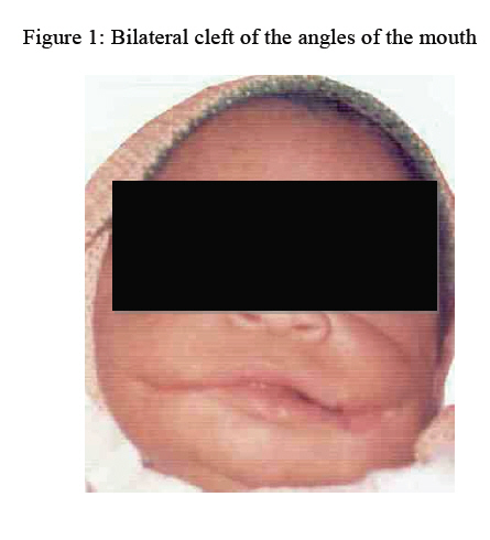
Annals of African Medicine, Vol. 6, No. 1, 2007, pp. 39-40 Bilateral Transverse Facial Cleft as an Isolated Deformity: Case Report 1V. I. Akinmoladun ,2F. J. Owotade, and 1A. O. Afolabi Oral-Dental Health Centre, State Specialist Hospital, Akure, Ondo State and 2Obafemi Awolowo University Teaching Hospital, Ile-Ife, Osun State, Nigeria Reprint requests to: Dr. V. I. Akinmoladun, Department of Oral/Maxillofacial Surgery, University College Hospital, Ibadan, Nigeria. E-mail: viakinmoladun@yahoo.com Code Number: am07010 Abstract Transverse facial clefts are rare deformities, these mostly occur as part of syndromes such as facial dysostosis and branchial arch syndrome. This is a report of a case of isolated, asyndromic bilateral facial cleft seen at a semi-urban specialist hospital. Congenital facial defects remain sources of mental and social stress to the families. Infanticide, perhaps a thing of the past in the developed world may still be practiced in cases of congenital deformities in the developing countries, hence the need for early involvement of social workers and clinical psychologist in management. Key words:Macrostomia, transverse facial cleft, infanticide Résumé Facials fendus transverse sont des déformités rares, le plus souvent, ils arrivent comme une partie des syndromes tels que facial dysostose et syndrome branchial arch. Il s’agit d’un rapport d’un cas isolé, facial fendu asyndromique bilatérial vu dans un hôpital specialiste semi-urban. Défauts facials congénitaux est toujours les sources du stress social et mental pour des familles. L’enfanticide, peut-être une chose du passé dans le monde développé pourrait être en pratique dans les cas des déformités congénitales dans les pays en voie de développement, donc, le besoin pour une participation précoce des ouvriers social et psychologiste clinique dans la prise en charge. Mot-clés : Macrostomie, facial fendu transverse, infanticide Introduction Transverse facial cleft (Tessier type 7) 1 or congenital macrostomia is a rare congenital anomaly1-3 which results from failure of the maxillary and mandibular portions of the first branchial arch to unite.4, 5 It seldom occurs alone but normally associated with deformities of other structures developed from the first and second branchial arches, and it is thought to be part of the manifestation of hemifacial microstomia, the second most common congenital craniofacial anomaly.2 An incidence of about 1 in 60,000 births to 1 in 300,000 live births has been recorded.5 Anderson’s (1965) series of 3988 cases of facial clefts treated in Denmark over a period of 30 years gave a figure of 13 patients with macrostomia, an incidence of 0.3% of the total series.5 Almost all of these patients had other associated anomalies making isolated cases a rarity. At seven weeks of gestation the lips separate from the alveolar areas with the formations of a vestibule and the maxillary and mandibular swellings then merge laterally to form the cheeks. Incomplete union here results in macrostomia, which could be unilateral or bilateral. Other etiopathogenesis have been given including that of Mckenzie and Craig7 who believe the defects of the first branchial arch arise from inadequate arterial blood supply occurring during a period of rapid and critical facial growth and development. It could vary from slight widening of the mouth, to a cleft extending back to the ear, they are usually unilateral and do not extend beyond the anterior border of the masseter. 8 Case report A full term, two-day old female patient was referred to the Oral-Dental Health Centre from a rural health centre, through the paediatric unit of the hospital with a diagnosis of isolated macrostomia. The mother was a teenage primigravida attending a local secondary school in a rural community, the level of antenatal care received could not be ascertained, but she claimed to have had an uneventful pregnancy and labour and delivery were routine. It appeared the baby was an unwanted one by both parents and their family due to the congenital defect and the fact that it was the product of an unplanned pregnancy that was bound to disrupt the mother’s education. Father was a school mate to the mother of the patient. Both parents were of poor background. On examination, there was a wide mouth and bilateral extension of the angles of the mouth to the masseter area (Figure 1). The clefts were lined with skin externally and buccal mucosa internally. A clear line of demarcation was noticeable where the lips ended and the defect began. Further physical examination did not show other abnormalities. The mother and grandmother who brought the patient were counseled on the need to bond with the baby and ensure adequate feeding, more so that baby was otherwise healthy and will require adequate feeding for a relatively easy surgical repair to be carried out. They were asked to report in the clinic the following week, but they failed to attend. After several weeks of failing to report, they were traced and found, the patient was said to have fallen ill before the scheduled appointment and died two days later at a private clinic. The mother was reluctant to give details, and she refused to give the address of the said private clinic where the baby died, insisting that it was a chapter of her life that was closed and should remain permanently so. Figure 1: Bilateral cleft of the angles of the mouth Discussion Macrostomia is a relatively rare congenital craniofacial defect. It is more commonly unilateral than bilateral. It is not surprising that the condition is usually associated with other defects because of the many facial structures developing simultaneously. Almost always present are malformation of the mandible and or the ear, 5 though this was not seen in this case presentation. The factors that could be responsible for the development of macrostomia are genetic and environmental, in individual cases however, as in the case presented here, it is often impossible to identify a specific aetiological factor. In this case there was no history of medication, use of traditional medications, illnesses or nutritional deficiencies in pregnancy and no evidence of attempted abortion was established. Although a family history of facial cleft was also negative, this could not be relied upon. In this part of the world such information is often concealed due to the fear of stigmatization. The circumstances surrounding the death of the patient were suspicious and would suggest infanticide as baby appeared clinically otherwise healthy at presentation. In our environment, congenital defects especially facial (whether amenable to surgery or not) are still sources of mental and psychological stress. Sometimes such defects are associated with evil forces and undocumented observations have confirmed the deliberate elimination of such babies. In the case being presented, the grandmother kept referring to the baby as the strange one. Bilateral transverse facial cleft remains a rare congenital malformation to which the attention of practitioners needed to be drawn. Though a relatively easy surgical intervention was required, the need for adequate counseling of family members cannot be over emphasized. It is hoped that with public campaign and enlightenment activities, societal attitudes to children with congenital deformities will change. It is also hoped that the authorities will enact and enforce laws to ensure the rights of such babies, especially right to care and life. References
Copyright 2007 - Annals of African Medicine The following images related to this document are available:Photo images[am07010f1.jpg] |
| |||||||||
