 |
| About Bioline | All Journals | Testimonials | Membership | News |
|
||||||
|
||||||
Annals of African Medicine, Vol. 7, No. 2, 2008, pp. 77-81 Description of the Normal Variants of the Anatomical Shapes of the Sella Turcica Using Plain Radiographs: Experience from Sokoto, Northwestern Nigeria A. D.Zagga1 , H.Ahmed2 , A. A.Tadros1 , and S. A.Saidu3 1Departments
of Anatomy, College of Health Sciences, Usmanu Danfodiyo University, Sokoto, Nigeria Code Number: am08017 Abstract Background: The anatomy of the sella turcica is variable in size
and shape. It has been classified into three types: round, oval and flat. It
can also be deep or shallow in both children and adults. The floor of the sella
turcica which in most cases is concave may be, flat or even convex. In both
anatomical and radiological practice in Nigeria, normal data in relation to the
description of the normal variants of the anatomical shapes of the sella
turcica are based on Caucasian studies. Key words: Description, normal variants, anatomical shapes, sella turcica Résumé Arrière Plan: L’anatomie de sella turcica varie selon la forme et
la plat. Elle peut aussi être profonol ou plat, aussi chez les enfants que chez
les adults. Le planche de sella turcica, qui est généralement concave peut être
convexe, ou plat de forme. Bien en anatomie et en radiologie practiques au
Nigéria. Les données relatives à la description des variants normales de formes
anatomiques de sella turcica sont basées sur des études caucasiennes. Le plancher de sella de plus commun pour les sujets africains étudiés est
concave et la différence en frequence entre sella a plancher cancave sella à
plancher convexe is statistiquement ties élevée. (PL 0.001).pour les deux cas
de formes plancher de sella turcica et les différents types de plancher de
sella turcica, en ce qui est du sex des sujets étudiés, la différence en
frequence entre les mâles et les femelles est statistiquement très élevée. (PL
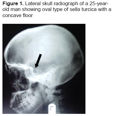
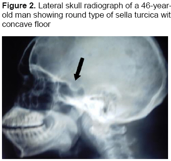
0.001). Mots clés: Description, variantes normales, formes anatomiques, sella turcica Introduction The sella turcica (‘Turkish saddle’)1 is the superior saddle shaped concavity on the intracranial surface of the body of the sphenoid bone.2 It contains the central hypophyseal or pituitary fossa which lodges the hypophyses cerebri or pituitary gland. Anteriorly its bony landmarks include the planum sphenoidale, the limbus sphenoidale, the chiasmatic sulcus, and the tuberculum sellae. Anterolateral landmarks include the optic canal, the anterior clinoid processes, and the optic strut, which forms the floor of the optic canal. The floor of the sella is the roof of the sphenoidal air sinus. Posteriorly, the sella is bounded by the dorsum sellae and the posterior clinoid processes; its lateral margins are the carotid sulci, and its superior boundary is the diaphragma sellae.3 The anatomy of the sella turcica is variable in size and shape. It has been classified into three types: round, oval and flat.4 It can also be deep or shallow in both children and adults. In children, 70% of sella turcica are round. In adults only 24.4% are round, whereas 58% are oval and 17.2% are flat.5 In profile, the sella at times has a somewhat high concave appearance caused by what appears to be an excavation beneath the anterior clinoids. This is frequently described in children and has no pathological significance. The floor of the sella turcica which in most cases is concave may be, flat or even convex.6 The sella turcica is usually demarcated by a dense thin white line in lateral radiographs. It is sometimes more important to recognize this feature than to estimate the size of the fossa.7 In both anatomical and radiological practice in Nigeria, normal data in relation to the description of the normal variants of the anatomical shapes of the sella turcica are based on Caucasian studies. There is therefore the need to have base line databased on studies from indigenous population. This study was, therefore, designed to describe the normal variants of the anatomical shapes of the sella turcica using normal lateral radiographs of Nigerians examined in the department of Radiology, Usmanu Danfodiyo University Teaching Hospital (UDUTH), Sokoto, from 2002 to 2004. Materials and Methods Selection of materials All available lateral radiographs of subjects over a 3-year period from 2002 to 2004 were retrieved from the Radiology Department of the Usmanu Danfodiyo University Teaching Hospital, Sokoto for the study. Of the three hundred and fifty (350) lateral radiographs only 228 satisfied the inclusion criteria. Sample size was determined using the formula proposed by Oyejide.8 All the radiographs were ascertained to have been taken by trained radiographers in a standardized condition/manner (focus to film distance/target to film distance (FFD/TFD) of 40 inches (100cm).9 Radiographs studied were of good quality and clearly showed the anatomical features of the sella turcica. Only radiographs interpreted by experienced radiologists were studied. Thus, radiographs for study were selected on the basis of good lateral positioning, without patient rotation and having no visible evidence of pathology, which might be related to the sella turcica. Inclusion criteria Selection of radiographs for the study was based on the following: (a) Perfect superimposition of the clinoid processes, to rule out tilting of the skull during positioning of the patient. (b) Clear visualization and recognition of the dorsum sellae and tuberculum sellae. (c) Distinct sella turcica floor so that the shape of the fossa and its floor could be classified. Exclusion criteria (a)Thirty two radiographs showed abnormal sella turcica and were excluded from the study. (b)Twenty six radiographs were excluded from the study because of poor quality. (c) Sixty four radiographs were excluded because they were not interpreted by experienced radiologists. Description technique Radiographs were mounted on the viewing boxes and variants of the anatomical shapes of the sella turcica were studied and classified according to the methods adapted by Jones et al4 and Isadore5 (for the shapes of the sella) and Bruneton et al.6 (for the shapes of sella floor). Statistical methods Data was initially sorted out manually and tabulated and then entered into the computer using Microsoft Word, Microsoft Excel and Minitab 13.1 statistical package. χ2 (with Yates correction) was used for comparison of proportions. Results The results obtained are summarized in Tables 1-4. A total of 228 subjects were involved in this study. Of this number, 171(75%) were males, and 57(25%) were females (m: f ratio=3:1). The various anatomical shapes of the sella turcica seen in the study are shown in Table 1.The predominant shape of sella in the Nigerian subjects studied is oval (Figure 1), and the difference in frequency of oval shaped sella and that of round (Figure 2) or flat (Figure 3) types is highly statistically significant. (χ2=257.1579; df=2; P<.001). With regards to the various anatomical shapes of the sella turcica (oval, round and flat) in relation to the sex of the subjects, this study revealed males to be predominant for each of the three types of shapes of the sella turcica as shown in Table 2. The difference in frequency of male and female subjects is highly statistically significant (χ2=57.0000; df=1; P<.001). Table 3 shows the different shapes of the floor of the sella turcica. The commonest type of sellar floor in Nigerian subjects studied is concave (Figures 1 and 2), and the difference in frequency of concave shaped sella floor and that of flat or convex (Figure 3) types is highly statistically significant (χ2=180.0263; df=2; P<.001). Table 4 shows the types of sella turcica floor (concave, flat and convex) in relation to sex of the subjects studied. It shows that male subjects predominated for each type of sella turcica floor. The difference in frequency of male and female subjects is highly statistically significant. (χ2=57.0000; df=1; P<.001). Table 1. The various anatomical shapes of the sella turcica
χ2=257.1579; df=2; P<.001. Table 2. The various anatomical shapes of the sella turcica in relation to sex
χ2=57.0000; df=1; P<.001 Table 3. Types of sella turcica floor
χ2=180.0263; df=2; P<.001. Table 4. Types of sella turcica floor in relation to sex
χ2=57.0000; df=1; P<.001 Discussion This study assessed the normal variants of the anatomical shapes of the sella turcica from normal lateral cephalometric radiographs of Nigerian subjects, a means that had also been used previously by Jones et al,4 Isadore5 and Bruneton et al6 in Caucasians. The three types of shapes of sella turcica (round, oval and flat) reported by Jones et al4 have also been observed in this study. Although, Jones et al.4 did not report the percentage prevalence of each of the anatomical type of sella turcica, we found the oval type to be the commonest in our study, 83%. This was followed by the round variety, 11%. The flat was the least in occurrence, 6%. However, this study compares favorably with that of Isadore5 who reported 58% oval, 24.4% round and 17.2% flat in adult Caucasians. The number of subjects (228) used in this study is similar to the number (200) used by Bruneton et al.6 The incidence of normal variants of anatomical shapes of the sella turcica floor observed in both series are similar in relative frequency; ie, concave (commonest) followed by flat and then convex, but differ in terms of their prevalence rates. In this study, the prevalence of concave type of sella turcica floor is 75% which is higher than the 58% reported by Bruneton et al.6 In both series, flat type of sella turcica floor appeared second, with a prevalence of 32.5% reported by Bruneton et al6 which is higher than in the present study. However, the prevalence rates of the convex type of sella turcica floor (which appeared with the least frequency in both studies) are very similar. Bruneton et al6 reported a prevalence of 9.5% for convex sella floor, similar to our study. From the foregoing, it follows that the prevalence and the relative frequencies of the normal variants of the anatomical shapes of the sella turcica reported in this study on Nigerian subjects is similar to those reported in Caucasians. Further studies on a larger scale are needed to corroborate our findings. References
Copyright 2008 - Annals of African Medicine The following images related to this document are available:Photo images[am08017f3.jpg] [am08017f2.jpg] [am08017f1.jpg] |
| |||||||||
