 |
| About Bioline | All Journals | Testimonials | Membership | News |
|
||||||
|
||||||
Annals of African Medicine, Vol. 7, No. 3, September, 2008, pp. 141-148 Opinion Achalasia: What is the Best Treatment? Adamu Ahmed Division of General
Surgery, Department of Surgery, Ahmadu Bello University Teaching Hospital,
Zaria, Nigeria Code Number: am08031 Abstract Background: Achalasia is an infrequent primary motility disorder
of the esophagus. Because of uncertain etiology, treatment is only palliative
and is directed at decreasing lower esophageal sphincter pressure, improving
esophageal emptying and relieving the symptoms of achalasia. Current treatment
options include pharmacological, endoscopic and surgical. We undertook a
systematic literature review of the management strategies currently available
for achalasia. Key words: Achalasia, Heller’s esophagomyotomy, pneumatic dilatation Résumé Contexte: L’achalasie est une rare anomalie motrice de
l’œsophage. En raison de son étiologie incertaine, le traitement est seulement
palliatif et vise la réduire la pression sphinctérienne du bas œsophage,
l’amélioration de la vidange et le soulagement des symptômes de l’achalasie.
Les options thérapeutiques courantes font appel aux moyens pharmacologiques,
endoscopiques et chirurgicaux. Nous avons effectué une revue systématique de la
littérature relative aux stratégies thérapeutiques actuellement disponibles
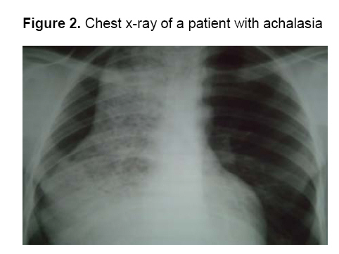
pour l’achalasie. Mots clés: Achalasie, esophagomyotomie de Heller, dilatation pneumatique Introduction The original description of achalasia was first made in 1674 by Sir Thomas Willis when he used whalebone to dilate the esophagus of a patient who was unable to swallow because of failure of the lower esophageal sphincter to relax.1Achalasia is a benign idiopathic disorder caused by progressive neuronal degeneration of the myenteric plexus of Auerbach.2, 3 It is the commonest primary esophageal motility disorder. The pathophysiology of the process represents a selective loss of inhibitory nerves that result in unopposed stimulation of the smooth muscle fibers of the LES. The etiology of primary achalasia remains controversial. Histological examination of the esophagus in achalasia suggests that the reduction in intramural ganglion cells may be a secondary change probably due to inflammation triggered by autoimmune mechanism or a chronic degenerative process of the central or peripheral part of the vagus nerve.4 The primary lesion could also be a severe myopathy of the smooth muscle cells.4,5 Familial occurrence and association with class II HLA antigens also suggest genetic predisposition.6,7 A secondary form of achalasia is Chagas disease, an infectious disease cause by the parasite Trypanasoma Cruzi. It seen mainly in Central and South America where an estimated 11 million people are infected.8 In developed countries the incidence of achalasia is 1 in 100000 individuals.9 In sub-Saharan Africa, 3 to 4 patients are seen per year. 10-12 Among other esophageal diseases, achalasia is observed in 3.1% - 20% of cases and is second to gastrooesophageal reflux (GER) as the commonest functional esophageal disorder requiring surgical therapy.13,14 In developing countries the peak incidence is in the third decade, a decade lower than in developed countries.10,13 Typically, patients present with long-standing progressive dysphagia for both liquid and solid. Regurgitation of undigested food is common and may result in aspiration and recurrent respiratory tract infection. Retrosternal pain is a prominent feature and may result from GER.15 More commonly it results from food stagnation and bacterial growth in the distal esophagus or fermentation of retained food.15,16Weight loss is seen in 68% of patients and may be severe.17In developed countries manometric diagnosis is obtained before therapeutic intervention.18 The most important finding is aperistalsis of the body of the esophagus in the distal smooth muscle segment of the esophagus.14,19Other manometric findings that are characteristic but not necessary for the diagnosis of achalasia are failure or incomplete relaxation of the LES with swallowing, sustained hypertension of the LES and elevated intra esophageal body pressure compared to pressure within the stomach.14,18,19Barium swallow is an important diagnostic tool in patients with symptoms suggestive of achalasia. Fluoroscopy reveals repetitive non-peristaltic contractions of the esophagus.14, 19 As the disease progress there is continuous dilatation of the esophagus until it become tortuous or even sigmoid in appearance (Figure 1). Twenty-four hour pH monitoring is also routinely used.20 Oesophageal manometry, barium swallow and pH- metry are also used for objective evaluation of outcome of treatment. Other diagnostic evaluations include esophagogastroduodenoscopy, trans-abdominal and high frequency intraluminal esophageal ultrasonography.20, 21Chest x-ray may show mediastinal widening, presence of air-fluid level in the mid-esophagus, absence of gastric air bubble and abnormal pulmonary markings due to chronic aspiration (Figure 2). In our setting as in others with limited facilities, diagnosis of achalasia is made to a high degree of accuracy given the symptom complex of long-standing dysphagia, regurgitation and weight loss with barium swallow showing delayed emptying, dilatation of esophageal body and the classic bird beak deformity of the distal esophagus.10-12 Modalities of Treatment Since the early treatment of achalasia in the 17th century, surgeons have made a significant advancement in the treatment of this disease and the resultant outcomes have improved. Because the etiology of achalasia is not known therapy is directed at relieving dysphagia and preventing stasis related complications by reducing the outflow resistance caused by the dysfunctional LES so that force of gravity would be enough to transport food into the stomach. Medical treatment, botulinum toxin (BTox) injection, pneumatic dilatation (PD) and surgical myotomy are the modalities currently available to achieve this goal. Medical treatment Calcium channel blockers and nitrates are the drugs commonly used to treat achalasia.22-24 Calcium channel blockers inhibit cellular calcium uptake and as calcium is necessary for contraction, relaxation occurs. Nifedipine given at a dose of 20 mg reduces the LES pressure by 30 to 40%.24, 25Nitric oxide is the measure neurotransmitter release from the myentric neurons that induces swallow and esophageal distension induced LES relaxation.22Nitrate acts by compensating for the decrease in the inhibitory neurotransmitter.22 Both nifedipine and isosorbide dinitrate are given sublingually 30 to 60 minutes before meals. These drugs have variable results in alleviating the symptoms of achalasia with initial improvement rate of 50 to 90%.26 However, long term use of these drugs is associated with tolerance which severely decreases their effects over time.25 Nifedipine has induced long term remission and even normalization of physiology in a few patients.26 Unfortunately the common side effects of hypotension, headache and peripheral edema occur in about 30% of patients taking calcium channel blockers or nitrates and this limits their clinical use.14,22 Sildenafil has been considered for the treatment of achalasia but because of significant side effects its clinical use has been abandoned.27 It inhibits phosephodiasterase type 5, which prevent the destruction of nitric oxide-stimulated cyclic guanisine monophosphate.22,27 This action leads to decrease in the resting LES pressure in patients with achalasia but to a lesser extent than nifedipine.14 Medical treatment has limited efficacy in patients with achalasia. It is recommended only to patients as a bridge to more definitive treatment or to those in whom other treatments are contraindicated.14, 22 Endoscopic botulinum toxin injection Botulinum toxin is a neuro toxin that causes sustained inhibition of neurotransmitter release at cholinergic terminals. The toxins specifically bind the presynaptic membrane and enter the cystol of the nerve terminal where they cleave different proteins involved in neuroexocytosis.28,29 Botulinum toxin- A which is used for the treatment of achalasia cleaves the SNAP-25 molecule of the presynaptic membrane, thus blocking acetylcholine (ACH) release.29,30 In the early nineties, Pasrich and colleagues evaluated the usefulness of intrasphincteric injection of BTox in achalasia patients.31,32 The rationale was that the selective loss of inhibitory nerves in achalasia upset the excitatory cholinergic influences on the LES.29 Thus by blocking the release of ACH, locally injected BTox might reduce the LES pressure and improve passive esophageal emptying.29,33 The toxin is injected through a standard schelerotherapy needle during an upper endoscopy performed under conscious sedation.34 Usually, 80 to 100 units of BTox- A are injected in each quadrant of the LES in 0.5 to 1ml aliquots.28 This improves the symptoms of achalasia, decrease LES pressure, increase lower esophageal aperture and improve esophageal emptying.31 The clinical effects of a single injection are short lived with relapse in more than 50% within 6 months.28 However, repeated injections may confer clinical benefit in 70% to 90%.32 In one report, 21% of newly diagnosed achalasia patients were treated with BTox as the initial modality of treatment with an average duration of effect of 6 months.33 This waning of efficacy over time is may be related to development of autoimmune response with production of antibodies against BTox.31 Predictors of good response to BTox injection are age less than 50 years, vigorous achalasia, an initial clinical response to BTox injection and decrease in LES pressure in response to BTox injection.22,35 Endoscopic injection of BTox is remarkably safe. About 10% of patients develop chest or epigastric pain shortly after injection but usually does not require specific treatment.14, 28 Treatment with BTox confers beneficial effects on subsequent pneumatic dilatation which is actually enhanced.22, 31 However, repeated injection of the toxin makes subsequent surgical myotomy more difficult due to adhesions of the muscular layers to mucosal plane with an increased danger of mucosal perforation.14, 33Given its safety profile, BTox injection may be given to patients who are not candidates for PD or surgery and those whose life expectancy does not exceed 2 years.30, 35 Pneumatic dilatation Historically, dilatation was the first attempt at treatment of achalasia.1 The principle is to weaken the LES by tearing its muscle fibers by generating radial force from within the esophagus. In the past, a variety of mercury filled buggies, hydrostatic dilators and metallic devices were used with varying degree of success. Modern dilators consist of expanding balloons that forcefully dilate the LES. Pneumatic dilatation is currently the most effective non- surgical treatment of achalasia.14,17 However variations exit as to the type of dilators, fluoroscopic or endoscopic positioning of the balloon and the degree and duration of dilatation.34,36,37 Pneumatic dilatation is performed in the endoscopy suite under conscious sedation. Single dilatation with Rigiflex dilator resulted in 60% reduction in LES pressure and resolution of symptoms in 70% of patients.34 However, graduated dilatation results in a higher and more durable clinical response.37,38 A report from Cleveland clinic showed that 41% of newly diagnosed achalasia patients were initially treated with PD.33 The clinical response was 86% in these patients with improved esophageal emptying of 54%.33 Pneumatic dilatation has the advantage of achieving symptom relief while avoiding the risk associated with invasive surgery. In addition, it can be safely used when BTox injection or surgery have failed.22 The major problem of PD is the relatively high mortality rate of 1 to 2%.14,34 Gastrooesophageal reflux is seen in 4% to 16% of patients.34 Perforation occurs in 1% to 3% of patients many of who would require emergency surgery.38 The risk of perforation is highest during the initial dilatation as oppose to subsequent dilatation especially in patients with vigorous achalasia.38 Predictors of good outcome of PD include age above 50 years and more than 50% reduction in LES pressure.22,38 Overall, PD provides a safe and effective way of achieving long-term control of symptoms in patients with achalasia. Surgical treatment Historically, the treatment of achalasia by cardiomyotomy was first designed by Gottstein in 1901.39 However, in 1913 Heller acclaimed the procedure by performing it abdominally on the anterior and posterior esophageal walls.40 Currently, the universally accepted surgical treatment of achalasia is the Heller myotomy, as modified by Zaaijer.41 Myotomy became the surgery of choice to treat achalasia because of it simple technique on the anterior wall of the esophagus accessed either by the abdominal or thoracic root, by open or minimally invasive surgery. In either case, the abdominal root is preferred because the surgical set-up is easier and anesthesia is simpler because single lung ventilation is not required.10 Postoperative management of the patients is easier and they leave the hospital earlier. The abdominal approach also provides opportunity to diagnose and treat associated intra abdominal diseases.42-44 Laparoscopic esophagomyotomy was first described in 1991.45 This procedure is associated with significant decrease in dysphagia and low complications rate. It provides symptoms relief in about 90% of patients with more than 80% remaining dysphagia free at 5 years.18,46 However, the high cost of this approach, access to reference centers, the need of surgical skills that are not readily available and surgeon learning curve are still an issue. Recently, computer enhanced robotic telesurgery provides further improvement over laparoscopic Heller myotomy.47 Open esophagomyotomy is associated with relapse rate of less than 10% at 9 years and is currently the modality with which other forms of treatment are compared.44 In developing countries where facilities for laparoscopic approach are not readily available Heller myotomy performed by laparotomy represent a reliable and effective surgical treatment for achalasia, with good to excellent palliation of dysphagia in more than 80% of patients.11,12 The methods of assessing completeness of division of the LES fibers during myotomy are intra operative endoscopy and manometry.14,48 Endoscopic view allows precise identification of the squamocolumnar junction and guides the proximal and distal extent of the myotomy and also detects perforations that can be repaired immediately.14 Manometry on the other hand gives a more functional assessment of the myotomy and can be use to locate areas of residual high pressure zone.48 The role of antireflux procedure following esophagomyotomy remains controversial. Opponents of antireflux procedure argue that a good postoperative LES pressure with low incidence of reflux symptoms can be achieved without fundoplication by avoiding excessive posterior dissection and limiting the myotomy to 0.5 to 1cm onto the stomach.10,49A recent meta-analysis on 21 studies involving 601 patients concluded that the rates of GER was not statistically different in patients who had myotomy with partial fundoplication compared to those without fundoplication.50 Lopushinsky and Urbarch also did not find any difference in the pattern of antireflux medication use between patients who had antireflux procedure as part of a surgical myotomy and those who did not.38 Therefore, while Heller myotomy and fundoplication may be safe the need for the antireflux procedure is unknown. It is generally accepted that a complete 360° wrap Nissen fundoplication should be avoided because it creates a high pressure zone in the distal esophagus which negates the intent of the operation.50 The commonest antireflux procedure added to Heller myotomy is the Dor anterior 180° fundoplication.14,46,51 This is associated with low rate of GER as determined by 24-hour pH monitoring.46 Suturing the gastric wall to the edges of the myotomy according to the Dor technique also maintains the edges of the myotomy open, thus preventing the consequences of scar tissue repair and covers any undetected perforation of the esophageal mucosa thus preventing any significant postoperative morbidity.14,51 Many surgeons consider the Dor technique an inadequate procedure and instead use a posterior 270° Toupet fundoplication.19,22 This provides excellent relief of dysphagia with low rate of postoperative reflux.19 Choice of Treatment In deciding to treat a patient with BTox injection, PD or surgical myotomy, there are a number of considerations including the risk of the procedure, its relative effectiveness in treating the symptoms of achalasia and the risk of treatment consequences such as GER. Several randomized trials have compared these 3 accepted modalities of treatment and clarified the best treatment for achalasia. They all concur that a myotomy or PD are superior to BTox injection.2,28,33,52,53 These studies have shown that BTox and PD are equally effective in the short-term but long-term relief of symptoms requires repeated injection of BTox.52,53 Comparison of surgical myotomy and BTox injection also revealed that the probability of being symptom-free at two years was 87.5% after surgery and 34% after BTox.33 Surgical myotomy has better long-term control of symptoms and fewer complications when compared to PD. A recent population based study reported that both Heller myotomy and PD provided good relief of symptom.38 However, the cumulative risk of subsequent intervention for achalasia at 10 years was 63.5% for patients initially treated with PD compared to 37.5% for patients initially treated with surgical myotomy.38 Although many studies indicate that surgical myotomy is superior to PD in providing long term relief of symptoms, a few physicians still favor PD because of the relatively higher risk associated with open surgical myotomy.33,38,44 However, surgical myotomy has regained primacy because of the immense success of laparoscopic Heller myotomy which is associated with low morbidity, shorter hospital stay, faster convalescence and less postoperative pain.52 When cost analyses were performed, PD was found to be the most cost-effective treatment in otherwise healthy populations.54 In patients with other co-morbidities that decrease the life expectancy to less than 2 years, BTox is more cost-effective.22,54 Although surgery is the most effective treatment for achalasia it is the most costly over the initial period of treatment.33,54 Dysphagia following cardiomyotomy can be either persistent or recurrent. Following incomplete myotomy or the addition of a tight fundoplication, dysphagia remains practically unaltered soon after operation or there may be only minor improvement.55,56 Dense adhesions and fibrotic healing at the site of myotomy are the causes of relapse of dysphagia in the relatively late postoperative period.56 Persistent or recurrent dysphagia can be treated by endoscopic pneumatic dilatation or reoperative cardiomyotomy.57 In patients with prolonged symptoms the esophagus may be sigmoid shape which is thought to represent the most advanced stage of disease. Some surgeons recommend myotomy as the first line treatment and reserve esophageal resection for patients with persistent symptoms.19,55,56 Others recommend esophagectomy as the first-choice treatment believing that marked esophageal dilatation and redundancy predict the impossibility of improving emptying by means of myotomy.57,58 Furthermore achalasia is considered to be precancerous with a reported incidence of 0.2%-2.0%.59 The chronic irritation in achalasia leads to squamous hyperplasia which progress to dysplasia and eventually to carcinoma.59 In addition, a p53 protein mutation study of the esophagus in patients with achalasia suggests that the entire esophagus may be in a precancerous state.60 We believe that every effort at preserving the native esophagus should be made before deciding to perform esophagectomy for a benign disease. Conclusion A lot of improvements have been made on the understanding of the etiology, pathophysiology and treatment of achalasia in the last two decades. Although different treatment options are available definitive cure is lacking. The choice of treatment involves the consideration of several clinical and economic factors. On the basis of evidence-based medicine, laparoscopic Heller’s myotomy is currently the best treatment and should be considered as the initial treatment for most patients with achalasia. In developing countries where facilities are not readily available, open surgical myotomy remains an effective therapeutic option. Because of the high probability of GER, it is considered prudent and reasonable to combine the myotomy with an antireflux procedure, preferentially a Dor or Toupet fundoplication. Pneumatic dilatation is an effective form of treatment and appears to be the most cost-effective alternative but its long-term efficacy is less than that of surgical myotomy. Endoscopic BTox injection is safe and effective but the effect diminishes over time and the need for multiple repeat injections must be considered. This may be the optimum treatment in patients with co-morbidities or those whose life expectancy does not exceed 2 years. The usefulness of nifedipine and nitrates is minimal. These drugs should be used only on temporary basis, while waiting for more effective therapeutic options. Continued surveillance of treated patients is necessary because a few of them may require additional treatment irrespective of the initial therapeutic modality. References
Copyright 2008 - Annals of African Medicine The following images related to this document are available:Photo images[am08031f1.jpg] [am08031f2.jpg] |
| |||||||||
