 |
| About Bioline | All Journals | Testimonials | Membership | News |
|
||||||
|
||||||
Annals of African Medicine, Vol. 7, No. 4, December, 2008, pp. 163-167 Electropherotypes and Subgroups of Group A Rotaviruses Circulating among Diarrhoeic Children in Kano, Nigeria 1 A. ADzikwi, 1 J. U.Umoh, 1 J. K. P.Kwaga, 2 A. A.Ahmad, 3 M. de Beer and 3A.D Steele 1Department
of Veterinary Public Health and Preventive Medicine, Ahmadu Bello University, Zaria, Nigeria Code Number: am08034 Abstract Background: It is estimated that about 600 000 children die annually as a result of severe dehydrating diarrhea caused by
rotaviruses. The virus is a double stranded RNA (dsRNA) virus with 11 segments.
Group A rotaviruses show a characteristic 4-2-3-2 pattern following
electrophoresis. The VP6 subgroups, I and II exist. This work was carried out
to study the prevalence of rotavirus infection among children 0-5 years with
diarrhea in Kano, and to determine the circulating subgroups and
electropherotypes and of the rotavirus isolates. Key words: Electropherotypes, Subgroups, Group A, Rotavirus, Nigeria Résumé Fond: On l'estime qu'environ 600.000 enfants meurent annuellement
en raison de la diarrhée de déshydratation grave provoquée par des rotaviruses.
Le virus est un virus bicaténaire d'ARN (dsRNA) avec 11 segments. Groupez
l'exposition de rotaviruses d'A un modèle 4-2-3-2 caractéristique après
l'électrophorèse. Les sous-groupes VP6, l'I et l'II existent. Ces travaux ont
été menés à bien pour étudier la prédominance de l'infection de rotavirus parmi
des enfants 0-5 ans avec la diarrhée dans Kano, et pour déterminer les
sous-groupes et les electropherotypes de circulation et des isolats de
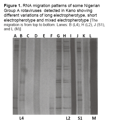
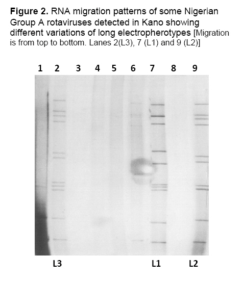
rotavirus. Mots clés: Electropherotypes, sous-groupes, groupent A, Rotavirus, Nigéria Introduction Rotavirus is the single most important cause of severe diarrheal illness in infants and young children in both developing and developed countries.1 The virus also infects the young of other mammals and avians.2 It composes a genus in the family Reoviridae and is a medium sized (70nm) and non enveloped virus. 3 A conservative estimate of 600,000 children die because of infection with this virus, most of this figure being from Asia and Africa. The most cost effective means of intervention is vaccination; therefore the epidemiology of this disease is important to ensure proper intervention with regards to the circulating strains in different regions of the world. There also exist many reports of emerging reassortants.4, 5 In Nigeria, reports on the prevalence and characterization of the virus in young children exist.6-9 VP6 is the most abundant protein.10 It is stable and has conserved epitopes which makes it a major target in diagnostic assays.11 Furthermore, it is highly immunogenic and antigenic therefore it may be important in protective immunity.12 The objective of this study was to provide additional information from an area not previously. Materials and Methods Collection and handling of specimens A total of 218 fresh faecal specimens were collected from 198 diarrhoiec children and 20 non-diarrhoiec controls 0-5 years of age in sterile bottles. Information was obtained on the ages and sexes of the children. Six hospitals within Kano metropolis namely Hasiya Bayero Paediatrics Hospital, Classic Clinics, Sir Sanusi Specialist Hospital, Assumpta Clinics, International Clinics and Abdullahi Wase Specialist Hospital served as collection centres. These hospitals were selected on satisfaction of the following criteria: Consent of the authorities, availability of a paediatrics unit and a reasonable number of patients visiting per day (approximately 50). The specimens were appropriately labeled and stored at -20°C at the collection centres prior to transportation in a cold box to MRC/MEDUNSA where they were analysed. Group A ELISA A 10-20% suspension of each faecal specimen was made using Bartex water and screened for group A rotavirus by double sandwich ELISA using commercially available DAKO kits (IDEIATM, Dako Cambridgeshire, UK) according to manufacturer’s instructions. Results were read spectrophoto-metrically at 450nm. PAGE Only ELISA positive specimens were subjected to PAGE to confirm the presence of Group A rotaviruses and to determine the circulating electropherotypes. Viral RNAs were extracted as described.13 Briefly, 50µl prewarmed sodium acetate (NaAc) containing 10% sodium dodecyl sulphate (SDS) was added to approximately 400µl stool suspension, incubated at 37°C in a water bath after which 500ul phenol/chloroform (1:1) was added to release the RNA. These were mixed in a vortex machine for 1 minute and incubated at 56°C for 15 minutes in a water bath. Air pressure was released by quickly opening and closing back the Eppendorf tube. The contents were mixed and centrifuged at 12,000rpm for 3 minutes to sediment. The upper aqueous phase was collected in a clean Eppendorf tube to which about 40µl 3M NaAc and 1ml absolute ethanol were added, gently mixed and incubated overnight at -20°C. The RNAs pellets left in the tube, after being centrifuged and the supernatant poured off were air-dried. PAGE was carried out using the discontinuous buffer system as described elsewhere.14 Slab gels (10%) and 3% spacer gels were used. About 30µl of each sample was loaded onto the gels and electrophoresis carried out at 100v for 18 hours at room temperature. Silver staining of the gels was done as described elsewhere.15 VP6 Subgroup ELISA The VP6 subgroups of rotavirus strains were determined using the VP6 monoclonal antibodies.16, 17 An ELISA protocol developed by MRC/MEDUNSA Diarrheal Pathogens Unit was used as described below. Microtitre plates were coated with rabbit anti-human rotavirus overnight at 4°C and repeatedly washed six times in phosphate buffered saline/tween 20 (PBS/T). About 50µl stool suspension was added, followed by 100 µl PBS/T/EDTA into three consecutive wells and incubated at 4°C overnight. Positive and negative controls consisted of standard rotavirus strain and distilled water respectively. A 1:5000 dilution of rotavirus Group antigen (A3M4),16 Subgroup I (mouse # 255/60/125/14) and Subgroup II (mouse #631/9/104/56)17 monoclonal antibodies were done in PBS/T/Bovine Serum Albumen (BSA). PBS/T/BSA was prepared by dissolving 5 g BSA in 1 litre PBS/T at pH 7.2 stored at -20°C. The monoclonal antibody (10 µl) was diluted in 10 ml PBS/T/BSA and 100 µl dispensed into every third row. Horseradish peroxidase conjugate obtained (Zymed, San Francisco, USA) was used at a dilution of 1:1000 with PBS/T/BSA to detect activity. About 100 µl was dispensed per plate and incubated at 37 °C for 2 hours. The plates were washed in PBS/T. To each well, 100 µl of a mixture of hydrogen peroxide and 3, 3¢5, 5¢-tetramethylbenzidine (TMB) was added and plates incubated for 10 minutes in the dark. The reaction was read visually then 5% sulphuric acid was added to stop the reaction. The plates were read spectrophotometrically at 450nm and the optical densities determined. Results Group A ELISA Twenty seven (12.4%) specimens were positive out of 218 specimens screened. Of the positive specimens, one came from a non diarrhoeic child. Children 0-12 months of age had the highest rate of infection and the mode of 18.57% came from the 7-12 months age group. PAGE Of the 27 ELISA positive specimens, 22 were analysed by PAGE. The remaining five were not tested. Seventeen yielded electropherotype patterns while five did not yield enough RNA bands on the gel to permit their electropherotype determination. Nine (40.9%) exhibited long electropherotypes of four variations while three (13.6%) exhibited short electropherotypes. Two samples (9.1%) were of mixed infection with more than 11 segments each. The electropherotypes of the remaining three (13.6%) could not be determined. Figures 1 and 2 show the migration patterns on polyacrylamide gel. VP6 Subgroups Five (22.72%) of the ELISA-positive samples belonged to subgroup I, nine (40.90%) belonged to subgroup II. Two (9.09%) belonged to subgroup I/II and coincidentally both of them appeared faint on gel. Three (13.63%) of the samples belonged to neither subgroup I nor II. The subgroup of three others could not be determined (Table 1). L1 and S1 electropherotypes exhibited subgroup I specificity while L2 were of subgroup II specificity. The only L4 belonged to subgroup I/II (Table 1). Table 1: Frequency of electropherotypes and subgroups of Rotavirus circulating among diarrhoeic children in Kano
The long profile was more common among males and among the age-group 7-12 months, while the short profile was more common among females and the 0-6 months age-group . The mixed electropherotypes occurred only among females of not more than 12 months (Table 2). Table 2. Distribution of electropherotypes in relation to sex and age among diarrhoeic children in Kano
Nd: not determined; * Percentages are in parenthesis Discussion This work reports the dominance of the long electropherotypes of rotavirus. This is similar to observations that the long electropherotype of rotavirus dominates in many African Countries18 and in Nigeria.8 Although the typical group A pattern is the 4-2-3-2, atypical ones may arise due to shift, drift or rearrangement.11 All the group A ELISA positive isolates gave the characteristic pattern. However, there was a lone short type (S1) reported in this work. Electropherotypes provide information on genetic diversity of rotavirus, heterogeneity of circulating rotaviruses and are useful in tracing spread through a population.10 Viruses of same serotype can exhibit different electropherotypes and those of same electropherotypes, different serotypes.10,19 In this study, 23% of the ELISA positive specimens exhibited no bands on PAGE while 14% were of the short type. There is a report of a similar finding of 26% and 10% of the PAGE negative and short types respectively (personal communication). The absence of the band could be due to too little RNA or its destruction during extraction by phenol/chloroform.8 The implication of mixed infection is that reassortants are likely to emerge since there is simultaneous infection of an individual with different isolates.20 The short and mixed electropherotypes were observed in children 0-12 months of age. At this point it is not clear why this age group alone in this study exhibited these electropherotypes. It may be necessary for more studies to explain this occurrence. The short electropherotype were of SI as expected. The long electropherotypes even though were expected to be SII exhibited SI, SII and others that could not be determined. VP6 subgroup specificity testing by ELISA yielded a dominance of subgroup II (41%) over subgroup I (23%). This agrees with the findings in South Africa, Tunisia, and Nigeria. 4, 8, 9, 21 Two (10%) of the samples reacted to both subgroup I and subgroup II monoclonal antibodies while three (14%) reacted with the group but neither subgroup I nor II monoclonal antibodies. A possible explanation for this may be that the stool specimens probably contained low level of the virus.21 These two specimens appeared very faint on PAGE, which indicates low RNA level. This work shows evidence of great diversity of rotavirus strains circulating at the same time within a single metropolis. Kano is heavily populated and suggests rich variety of strains within this type of locations. This study provides information on the genomic diversity of RNA electropherotypes even though a clear-cut pattern for subgroups and electropherotypes did not exist. Acknowledgements The authors acknowledge WHO and MRC/MEDUNSA for sponsoring this work. The work was done during the WHO African Rotavirus Workshop held MEDUNSA in 2002. The authorities and staff of the hospitals in Nigeria where the specimens were obtained are also acknowledged. References
Copyright 2008 - Annals of African Medicine The following images related to this document are available:Photo images[am08034f1.jpg] [am08034f2.jpg] | |||||||||||||||||||||||||||||||||||||||||||||||||||||||||||||||||||||
| |||||||||
