|
| About Bioline | All Journals | Testimonials | Membership | News |
|
||||||
|
||||||
Australasian Biotechnology, Vol. 11 No. 1, 2001, pp. 32-34 BIOTECH RESEARCH A MODIFIED AND SIMPLE METHOD FOR THE EXTRACTION OF GENOMIC DNA FROM A WIDE VARIETY OF ANIMAL AND PLANT TISSUES Diana Sara Leal-Klevezas, Juan Pablo Martínez-Soriano Diana Sara Leal-Klevezas, Centro de Investigación Biomédica de Occidente, Instituto Mexicano del Seguro Social, Mexico, and Juan Pablo Martínez-Soriano, Unidad de Biotecnología e Ingenieréa Genética de Plantas. Centro de Investigación y de Estudios Avanzados, Mexico. Code Number: au01013 AbstractEvery DNA extraction procedure must break the cell by disrupting membranes and cell walls, inactivate DNAses and remove other macromolecules. Many techniques have been described to isolate DNA from specific types of cells, but they do not necessarily apply to different types of cells. Here we are reporting a DNA extraction method that yields DNA in good amounts and purity to perform PCR, RAPD, AFLP, restriction analysis and cloning. This protocol has proved to be useful not only from small amounts of samples from a wide variety of organisms and tissues alone, but also in complex cell mixtures and food products, by reducing the presence of residual inhibitors in the reactions. The described method not only is very versatile, saves time and avoids using fridge and bench space to store endless DNA extraction solutions, but is also simple, inexpensive and yields DNA that is stable for years. Key words: DNA extraction, animal, insect, fungi, virus, dairy products. IntroductionThe choice of the DNA extraction method depends upon the source and purpose of the target DNA. The chosen procedure must be able to break the cell by disrupting membranes and cell walls (such as in plant, yeast and bacterial cells), inactive DNAses, remove proteins, polysaccharides and other macromolecules. Although procedures vary greatly from source to source, a rule of thumb is that longer procedures yield cleaner and more stable DNA than shorter ones. Several techniques have been described to isolate DNA from specific types of cells, but they do not necessary apply to other different cells or samples (e.g. from animal to bacteria or plant cells). When someone starts working with a new organism, one must find the right DNA purification method, starting from a bibliographic research, preparing the proper solutions and trying one or more of such methods. Eventually, any lab could end up using precious fridge and bench space to store numerous DNA extraction solutions. We have developed a simple, fast and inexpensive DNA extraction procedure that we have being using for more than 5 years now. Every newcomer to our labs that have worked with a different organism has tried it with success every time. Table 1. Performance of the method using blood and bacteria.
Materials and Methods Unless otherwise stated, all chemicals were Sigma Chemical Co. (Gaithersburg, MD, USA) or Fisher Scientific. Enzymes were from GIBCO-BRL (Springfield, NJ, USA). Glassware and distilled water used in preparing the solutions was autoclaved at 121ºC for 20 min. Thermocycling reactions were performed on a Thermal Cycler (Perkin Elmer) (Foster City, CA, USA), a PTC 150 (MJ Research) (Watertown, MA, USA), or a GTC-2 (Applied Research) (Grayslake, IL, USA). Primers were synthesized by Bio-Synthesis, Inc. (Lewisville, TX, USA). Electrophoresis, gel visualization and photography were performed using Fotodyne equipment (Hartland, WI, USA). Culture collections were from: Central Veterinary Laboratory (CVL) (Waybridge, UK) Virginia Maryland Regional College of Veterinary Medicine (VMRC) (Blacksburg, VA, USA); Instituto Nacional de Referencia Epidemiológico (INDRE) (Mexico City, Mexico); Escuela Nacional de Ciencias Biológicas (ENCB) (Mexico City, Mexico); Stratagene (La Jolla, CA, USA); American Type Culture Collection (ATCC) (Rockville, MD, USA); Centro de Investigación sobre Fijación del Nitrógeno (CFN); (Cuernavaca, Mexico); Instituto Nacional de Investigaciones Forestales y Agropecuarias (INIFAP), (Palo Alto, Mexico); Universidad Autónoma de Chapingo (UACh), (Texcoco, Mexico); Universidad Autónoma de Nuevo León (UANL) (Monterrey, Mexico); Culture Collection, University of Göteborg (CCUG), (Göteborg. Sweden). Biological MaterialsClinical samples.Sputum from tuberculosis positive patients, cervical scrapings from humans suspected of carrying Human Papilloma Virus (HPV), bone marrow, blood containing heparin or EDTA as anticoagulant from healthy and Brucella-infected humans, goats, dogs, sheep, horses, bovines, and pigs. Organs of Brucella-infected and deceased goats and their miscarried fetuses (spleen, brain, heart, liver, lung, kidney, cotyledon, lymphatic node, mammary gland, salivary gland, eye, gull bladder, uterus, urinary bladder), fluids (spinal, sinovial, and ocular liquids, bone marrow, saliva and amniotic fluid), exudes (eye, nose, vaginal, and saliva), animal products (milk, cream and freshly made cheese). Blood from Borrelia burgdorferi borne dogs and their ticks (Acari: Ixodidae) and insects (Homoptera: Cicadellidae) harboring phytoplasmas. Walled eukaryotic cells.Saccharomyces cereviseae, filamentous fungi (Aspergillus flavus), plant material such as Catharantus roseus, Cocos nucifera, Citrus sinensis, and Capsicum annum and microorganisms harbored by those, such as Phytoplasma and viruses. DNA extractionBacterial strains. Bacteria were suspended in TE buffer (10 mM Tris-HCl pH 8.0 and 1 mM Na2EDTA) from isolated colonies or pelleted bacteria collected from 400 µl of liquid culture. Samples were collected by centrifugation and the DNA of decanted pellets was then extracted with the universal DNA extraction procedure described here. Isolated bacteria did not need Proteinase K treatment, although when this reagent was added, it did not interfere with DNA yield or quality. Exudes, body fluids, and fatty top layer of raw goat milk (cream). These samples were processed directly to extract DNA. Cells were not pelleted and decanted as described above and extraction buffer was added directly to 400 µl of each sample. Whole cow’s milk and cheese. It was processed as before, only 200 µg of Proteinase K (20 µl of Proteinase K from a frozen stock containing 10 mg/ml). Blood, cervical scrapes and bone marrow. Four hundred µl of bone marrow, cervix scrape in saline solution or blood collected in Vacutainer™ (Becton-Dickinson) tubes containing sodium heparin (green stopper) or EDTA (lavender stopper), were placed in 1.5 µl Eppendorf tubes and added 1 ml of erythrocyte lysis solution (155 mM NH4Cl; 10 mM NaHCO3; 100 mM Na2EDTA pH 7.4), mixed and centrifuged as before. Treatment with erythrocyte lysis solution was repeated until the white cell pellets lost all reddish coloring. Pellets were treated with the protocol described here. Sputum. Samples were concentrated and decontaminated as described elsewhere (Kinyoun, 1914) before processing. Solid animal tissues. Samples were smashed (SEWARD stomacher 80) in the presence of an equal volume (v/w) of sterile saline solution. Four hundred µl of the sample were then processed by the DNA extraction procedure described here. Walled eukaryotic cells. One g of each samples or 2 ml of pelleted saturated cultures were frozen and reduce to powder with mortar and pistil in the presence of liquid nitrogen, to latter proceed with the DNA extraction. Once the samples were prepared as described above, four hundred µl of lysis solution (2% Triton-X 100, 1% SDS, 100 mm NaCl, 10 mm Tris-HCl pH 8.0) and 10 µl of Proteinase K (10 mg/ml) were added, thoroughly mixed and incubated for 30 min at 50ºC. Four hundred µl of saturated phenol were added, mixed thoroughly and centrifuged for 5 min at 8,000 Xg. The aqueous layer was transferred to a fresh tube and an equal volume of chloroform-isoamyl alcohol was added (24:1); the tubes were mixed thoroughly and centrifuged for 5 min at 8,000 Xg. The upper layer was again transferred to a fresh tube and 200 µl of 7.5 M ammonium acetate were added and mixed thoroughly. Samples were kept on ice for 10 minutes, and then centrifuged for 5 min at 8,000 Xg and aqueous content transferred to a fresh tube. Two volumes of 95% ethanol or 1 volume of isopropanol were added, mixed and stored overnight at -20ºC. DNA was recovered by centrifuging the samples for 5 min at 8,000 Xg; pellets were rinsed with 1 ml of 70% ethanol, air-dried and resuspended in 20 µl of TE buffer. DNA concentrations were determined by further absorbancy at 260 nm and samples were stored at 20ºC until their processing. Ribonuclease treatments of the samples were found to be unnecessary but harmless to the DNA. The amount of DNA was judged by spectrophotometry at 260 nm and gel visualization. The quality of the DNA was assessed by comparing OD readings at 260 nm vs. 280 nm, appearance on 0.8% agarose gel, or performance on PCR and digestion with the endonucleases.
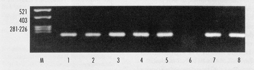
PCR proceduresThe PCR assays were performed for microorganisms or samples as described elsewhere for Brucella spp (Leal-Klevezas et al., 1995), M. tuberculosis (Wilson et al., 1993), phytoplasmas from infected plants (Martínez-Soriano et al., 1999), Borrelia burgdorferi (Lebech & Hansen, 1992), human β-globin and human papilloma virus (Bauer & Manos, 1993), fungi 5.8 S rDNA, including Internal Transcribed Sequences (Nazar et al., 1991). Results and DiscussionOur method has also proved to be simple, inexpensive, able to obtain DNA of good quantity and quality that has remained stable for years. The tested method was qualified as a strong protocol according to four basic concepts: 1) Excellent amount of DNA obtained; 2) High quality of DNA, as judged by the spectro-photometric ratios at DO280; 3) Strong and consistent PCR signals when the extracted DNA was used as template; 4) Endonuclease digestions were fairly complete. Data showing yield and quality of our universal method is shown on Table 1. The average yields of several DNA extractions are expressed in µg/µl (final volume, 20 µl) from Brucella spp. and Escherichia, human, goat, and bovine DNA samples. Performance of extracted DNA on PCR also reflected high quality of DNA, achieving amplification from omp 2 sequences on Brucella strains and infected tissues and samples (Figure 1); Mycobacterium tuberculosis strains and contaminated; Aspergillus spp. strains ITS 1-5.8-ITS 2 sequences; Phytoplasma’s infected coconut palm and Catharantus roseus, as well as potato and Phytoplasma-carrying insects. Borrelia burgdorferi from infected dogs and their ticks; and β-globin sequences from human blood, cervical samples, and bone marrow (data not shown). The high quality of the DNA obtained by our method permitted the detection of minute traces of DNA. As reported earlier for Brucella where we detected the presence of this pathogen from as little as 2.5x10⁻¹¹µg of DNA (Leal-Klevezas et al., 1995). Furthermore, the DNA extraction method described here has allowed us to detect minute amounts of Brucella DNA on patients under treatment and on milk containing 1-10 bacterial cells per ml of sample (data not shown). AcknowledgementsWe wish to thank Drs. M. Corbel, G. Schurig, A. López-Merino, E. Martínez, E. Díaz-Aparicio, B. Osterman, and F. Enevold for kindly providing the strains used. We are grateful to E.M. González-García, I.H. Almeyda-León, E.I. Cortés-Gutiérrez, M.I. Dávila-Rodríguez, C. Lara-Campos, A. Morales-Loredo, P. Becerril-Montes, I.O. Martínez-Vázquez, and M. Bères for trying our procedure. We also thank J.A. Luna-De-la-Rosa for helping with the photographic material. This research was supported by Consejo Nacional de Ciencia y Tecnología-Mexico (grant 264100-5-1382PN), SIHGO-CONACyT (Alim-18/97) and ICAMEX-Mexico. References
Copyright 2001 - AusBiotech |
| |||||||||