Australasian Biotechnology,
Volume 6 Number 5, September/October 1996, pp.281-282
Branched DNA Technology as a New Tool for Nucleic Acid
Quantitation.
Stuart Rodda,
Chiron Mimotopes Pty Ltd, 11 Duerdin Street, Clayton, Vic
3168
Code Number: AU96013
Size of Files:
Text: 4.9K
Graphics: Photographs (jpg) - 72.4K
[FIGURES AT END OF TEXT]
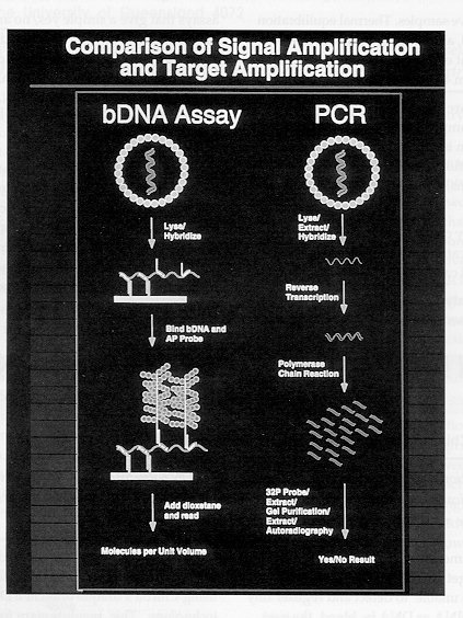
Nucleic acid detection with high sensitivity is achieved in
the bDNA approach by amplification of output signal, rather
than amplification of target nucleic acid prior to output
signal generation (as in conventional PCR). The article
describes the main features of the bDNA approach developed at
Chiron.
Early methods of measuring serum viral levels depended on
target amplification techniques. Since assays were unable to
detect and register tiny amounts of viral RNA or DNA in
blood, the next best thing was to duplicate the "target" (RNA
or DNA) present over and over again until enough was present
to be "seen" by available detection methods.
Rather than reproducing additional target, the ideal way to
track disease progression would be to measure viral RNA/DNA
as it appears in the serum. This approach would be to
amplify the signal generated from whatever amount of target
is present (direct measurement of target) rather than
increasing the amount of target. In short, signal
amplification (direct measurement) allows one to "find the
needle in the haystack", while target amplification "makes a
haystack out of needles" .(Figure 1)
In order to make practical the quantitation of viral nucleic
acid (RNA or DNA) by direct detection, Chiron has developed
an assay system based on capture of the viral target by
hybridization, followed by detection of the captured target
using a system with a high amplification factor, enabled
using Chiron's unique branched DNA (bDNA) technology. This
revolutionary form of signal amplification not only finds the
virus in the blood but reliably and directly quantifies it as
well, allowing clinicians a new 'window' for viewing viral
disease and measuring success of therapy.
How does bDNA work ?
In order to increase the sensitivity of direct detection,
bDNA molecules are attached to the target viral RNA (HIV,
HCV) or viral DNA (HBV). Once attached to target, these
molecules in turn bind to substrate (alkaline phosphatase)
molecules which trigger another substrate (dioxetane) to
produce light. The amount of light produced is directly
proportional to the amount of viral RNA/DNA present in the
sample, and can be recorded using a Luminometer.
The Quantiplex (TM) assay is a unique solution-phase
hybridization assay coupled with signal amplification using
bDNA. It uses an ELISA-like format which is easy to run on
any benchtop without fear of contamination (can be run in any
routine laboratory). In detail, attached to the bottom of
each microwell are capture probes. Initially, serum is added
to the microwell along with reagents that release viral DNA
or RNA directly. This reagent also contains target probes,
which bind to the DNA/RNA, then link (hybridize) to the
capture probes, attaching the target DNA/RNA to the
microwell. The capture probes contain small sequences of
nucleotides which are "mirror images" or "matching base
pairs" to the most highly conserved regions of the target
nucleic acids.
After washing, a different set of target probes is attached
to the target DNA or RNA strand. Next, the branched DNA
probes are added. Assuming that the target virus is present
in the sample, and the first two steps have occurred, the
branched DNA molecule now attaches to the target probes via
hybridization.
The bDNA molecule is essentially a "tree" which binds to the
target complex. Each "tree" contains 15 branches, which are
staggered to allow maximal binding of the chemiluminescent
(light producing) substrate molecules to be added in the last
step of the assay. Next the addition of alkaline phosphatase
to the complex (three alkaline phosphatase molecules) can
bind to each bDNA branch. When dioxetane substrate is then
added, light is released (as a result of enzymic
dephosphorylation of this substrate) which is read on a
luminometer and is directly proportional to the amount of
virus target in the sample (based on a standard curve that
covers the dynamic range of the assay).

