 |
| About Bioline | All Journals | Testimonials | Membership | News |
|
||||||
|
||||||
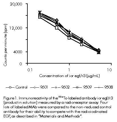
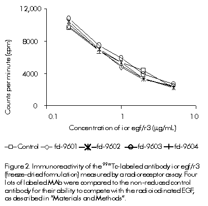
Biotecnologia Aplicada 2000; Vol. 17 No. 1, pp. 39-44 Freeze-dried Formulation for the Direct 99mTc-labeling of ior egf/r3 Monoclonal Antibody: Equivalence Studies Alejo Morales,1 Niuvis Pérez,1 Gilda Núñez,1 Idania Caballero,1 Jorge Ducongé,2 Eduardo Fernández,2 Ana Veloso,1 Aida de la Paz,1 Normando Iznaga1 1Center of Molecular Immunology. PO Box 16040, Havana 11600, Cuba. Fax: (53-7) 33 5049; E-mail: alejo@ict.cim.sld.cu . 2Institute of Pharmacy and Food (IFAL). University of Havana. Calle 222 y ave. 23, Rpto. La Coronela, Ciudad de La Habana, Cuba. Received in December ,1998. Accepted for publication in August, 1999. Code Number: BA00009 Abstract Monoclonal antibodies (MAb) have been useful for immunoscintigraphic applications in clinical diagnosis since they were introduced in the nuclear medicine practice. The direct method for radiolabeling reduced MAb with 99mTc in the presence of methylene diphosphonate (MDP), developed by Schwarz and Steinstrasser and modified by Mather and Ellison, has proved to be a good procedure to obtain high labeling efficiency and stable labeled MAbs. The ior egf/r3 MAb is a murine highly specific IgG2a isotype that recognizes the human epidermal growth factor receptor (hEGF-r). It was obtained and produced in the Center of Molecular Immunology in Havana, Cuba, by standard hybridoma techniques. Based on the direct method referred above, a freeze-dried kit formulation is described for immunoscintigraphic applications of tumors of epithelial origin: 3 mg of reduced MAb, 15 mg of glucose, 200 µg of MDP, 0.13 µg of SnF2, and 80 µg of sodium p-amino-benzoate. The aim of the present report is to show the equivalence between the 99mTc-labeled antibody in solution and the freeze-dried formulation. Effects on immunoreactivity and stability of labeled antibody are reported. Purity, fragmentation, labeling efficiency, radiocolloids, immunoreactivity, biodistribution and pharmacokinetics in rats showed the high quality of the freeze-dried kit formulation and the equivalence with the product used in clinical trials. Keywords: 99mTc, antibody, biodistribution, equivalence, freeze-dried, radiolabeling Resumen Formulación liofilizada para el marcaje directo del anticuerpo monoclonal ior egf/r3 con 99mTc: estudios de equivalencia. Los anticuerpos monoclonales (AcM) han sido ampliamente utilizados en el diagnóstico clínico por inmunogammagrafía en las unidades de medicina nuclear. Estos anticuerpos han sido marcados con isótopos radiactivos como el tecnecio-99m, que es el de mayor preferencia. El método directo de marcaje reportado por Schwarz y Steinstrasser, y modificado posteriormente por Mather y Ellison, ha demostrado que es posible obtener una elevada eficiencia de marcaje de AcM, los cuales, a su vez, muestran una gran estabilidad de enlace. El AcM ior egf/r3 es un anticuerpo murino de la subclase IgG2a, que reconoce de manera específica el receptor del factor de crecimiento epidérmico humano (hEGF-r). Este anticuerpo producido por el Centro de Inmunología Molecular, en La Habana, Cuba, fue obtenido mediante la tecnología estándar de producción de hibridomas. El presente trabajo tiene como objetivo demostrar la equivalencia entre el producto en solución utilizado en ensayos clínicos para el diagnóstico por inmunogammagrafía de tumores de origen epitelial, y la formulación liofilizada del anticuerpo lista para el marcaje. Basado en el método de marcaje mencionado previamente, se describe una formulación liofilizada del AcM ior egf/r3: 3 mg del AcM reducido, 15 mg de glucosa, 200 µg de MDP, 0,13 µg de SnF2 y 80 µg de p-amino-benzoato de sodio. Estudios realizados demostraron la equivalencia entre los productos en solución y la formulación liofilizada establecida. La elevada calidad de la formulación liofilizada descrita está avalada por su pureza, la eficiencia de marcaje, la cuantificación de radiocoloides, la inmunoreactividad del AcM, su biodistribución y su farmacocinética en ratas. Palabras claves: 99mTc, anticuerpos, biodistribución, equivalencia, liofilización, radiomarcaje Introduction The clinical use of monoclonal antibodies (MAb) has gained more significance in the last few years. For that reason, a wide range of MAbs is produced for therapeutic and in vivo diagnostic use. The development of a product with clinical application is a time-consuming process in most cases and usually the effectiveness of the molecule is not demonstrated in the final formulation. After demonstration of its utility, some changes in the manufacturing technology could be necessary for improving performance or stability. Most of these, even minor qualitative and quantitative changes, require a new application for the second form of the antibody according to legal regulations [1]. Usually, a complete characterization is already available for the first form of the antibody. The manufacturer should have the possibility to use the previous characterization on the first form of the antibody by demonstrating the product equivalence for both forms obtained before and after the introduction of changes in the manufacturing process. Parameters like isotype, subclass, affinity, microheterogeneity, molecular weight, primary and secondary structures, structural integrity of the antibody molecule, glycosylation pattern, specificity, cross-reactivity, and biological potency, should be evaluated by appropiate methods as recommended in the guidelines [2-7]. The ior egf/r3 MAb has been successfully used in the diagnosis of tumors of epithelial origin [8, 9]. The antibody recognizes an epitope located in the extracellular domain of the human epidermal growth factor receptor (hEGF-r), expressed in normal and malignant tissues (i.e. glioma, meningioma, malignant fibrous histiocytoma, neurofibrosarcoma and lung adenocarcinoma). The direct method for radiolabeling the reduced MAb with 99mTc in the presence of methylene diphosphonate (MDP), developed by Schwarz and Steinstrasser [10] and modified by Mather and Ellison [11], has proved to be a good procedure for obtaining high labeling efficiency and a stable labeled ior egf/r3 MAb. The method of 99mTc-MDP uses reduction of disulfide bonds in the molecule by adding an excess of the reducing agent 2-mercaptoethanol (2-ME). After purification to eliminate the excess of 2-ME the reduced antibody was labeled with technetium via Sn2+ reduction of pertechnetate, using MDP as a weak competing ligand. In this paper, we describe an evaluation of the product equivalence of two formulations of the ior egf/r3 MAb obtained before and after the introduction of some changes in the manufacturing process. Changes were introduced in order to scale up the whole process and to obtain a more stable product using new additives, during the lyophilization process that obviously modifies the final product formulation. The equivalence study is necessary to reduce the extent of the testing procedure for the second modified form of the antibody, especially the extent of the in vivo testing procedure, including clinical trials in humans. Materials and Methods Monoclonal antibody The ior egf/r3 MAb is a highly specific murine IgG2a isotype that recognizes the hEGF-r. It was obtained and produced in the Center of Molecular Immunology (Havana, Cuba) by standard hybridoma techniques as previously described [12]. Sterile and apyrogenic neutral solution with an antibody concentration of 5 mg/mL was used. Antibody reduction and radiolabeling Product in solution. The MAb (1 mL, 5 mg/mL) in phosphate-buffered saline solution (PBS: 137 mM NaCl, 2.7 mM KCl, 8.1 mM Na2HPO4, 1.5 mM KH2PO4, pH 7.4) was reduced with a molar excess of 2-ME in a relation 2000:1 (2-ME:MAb), at room temperature for 30 min. The reduced antibody was purified using a PD-10 column (Sephadex G-25M, Amersham Pharmacia Biotech, Sweden). PBS purged with nitrogen was used as mobile phase. The ability of the reduced MAb to be labeled with 99mTc was assessed as follows. After reconstitution of the Amerscan Medronate II Bone Agent MDP kit with 5 mL of saline purged with nitrogen, 150 µL of this solution were added to aliquots of 3 mg of the reduced MAb to be labeled with 40 mCi (1480 MBq) of 99mTcO4- eluted from the 99mMo/99mTc generator system (Elumatic II, Amersham, UK). Freeze-dried kit formulation. After the reduction step, the MAb (50 mL, 5 mg/mL) was purified on an XK 50/60 (1 L) G-25 Sephadex column. Then, glucose was added to a final concentration of 5 mg/mL and the concentration of the MAb was adjusted to 2 mg/mL. Three milligrams of antibody (1.5 mL) were dispensed per vial and frozen in liquid nitrogen. A 0.2-mL aliquot of the MDP solution was added to each vial and frozen again in liquid nitrogen. Then, the product was lyophilized for 24 h. Vials were stoppered under vacuum and kept at 4 ºC until use. Radiolabeling of this formulation was performed using 2 mL (40 mCi) of 99mTcO4- eluted from the 99mMo/99mTc generator system. Labeling efficiency The labeled products were subjected to ascending chromatography on Whatman 3MM paper (Whatman, USA). Strips (0.5 x 8.0 cm) of paper were used as stationary phase and saline was used as mobile phase to separate free pertechnetate and Tc-MDP that moved with the solvent front [9]. Separation of pertechnetate alone was achieved using acetone as the mobile phase where it moved with the solvent front [13]. After developing and drying, each strip was cut at one-third (saline) and one-half (acetone) of the distance between the origin and the solvent front. Each part was counted separately in a gamma counter (LKB, Clinigamma Model 1272, Wallac, Finland). Radiocolloid determination To determine the quantity of radiocolloids that may have been formed, both formulations were analyzed using human serum albumin (HSA)-impregnated ITLC-SG (Instant Thin Layer Chromatography Silica Gel, Gelman Sciences, USA) and NH4OH:ethanol:water (1:2:5) as the mobile phase to separate radiocolloids which remained at the origin [14]. Antibody immunoreactivity The immunoreactivity of the antibodies was measured by a radioreceptor assay [15] using placental membrane extract, which has a high expression of hEGF-r. Both formulations were compared for the ability to compete with radioiodinated murine epidermal growth factor (125I-EGF). EGF was iodinated with 125I to a specific activity of 0.25 mCi/µg using the chloramine T method [16]. After 1 h at room temperature, tubes were centrifuged at 4500 rpm at 4 ºC during 30 min and the supernatant was discarded. The radioactivity of the pellets was counted. Different antibody concentrations were used for both formulations and for a non-reduced control antibody. Exponential curves were fitted by a non-linear least square regression. Structural integrity, aggregates and purity Sodium dodecyl sulfate-polyacrylamide gel electrophoresis (SDS-PAGE). Denaturing electrophoresis was performed using the Phast System (Amersham Pharmacia Biotech, Sweden) in 7.5% acrylamide gels. Samples were run as recommended by the manufacturer, either reduced, non-reduced or in native electrophoresis. The sample concentration was 1 mg/mL. Gel Filtration. Samples were chromatographed on a Superose-12 FPLC column (Amersham Pharmacia Biotech, Sweden) using PBS as mobile phase. The column was equilibrated with 50 mL of PBS. Performance conditions were: sample volume of 200 µL, sample concentration of 1 mg/mL, and flow rate of 38.2 cm/h. Retention volume was measured as a parameter related to the molecular weight of the different compounds present in the samples. Isoelectric focussing Charge heterogeneity was assessed by isoelectric focussing (IEF). Samples were focused using the Phast System and pH 3-9 gels (Amersham Pharmacia Biotech, Sweden), as described by the manufacturer. A reference sample was used. Biodistribution and pharmacokinetic studies in rats Biodistribution. Wistar normal male rats (200-310 g) from the Center for the Production of Laboratory Animals (CENPALAB, Havana, Cuba), were used in this study. A total volume of 200 µL of the labeled antibody (1 mg/200 µCi) was administered intravenously via tail vein to 18 rats. After injection, animals were perfused and blood samples were taken at time intervals of 1, 4, 8, 16, 24, and 48 h (n = 3 for each time). The right kidney, pancreas, spleen, stomach, small intestine, lungs, liver, brain, and cerebellum were excised, rinsed out of residual blood and weighed. Samples were kept at 4 ºC until they were counted for radioactivity in an automatic gamma counter (LKB, Clinigamma Model 1272 PAIS) with the window setting set to detect the 140 keV photopeak of 99mTc. A sample of 350 µL of the solution prepared at the same time was used to determine the counts per minute (cpm) of the injected dose. The results of the organ biodistribution were expressed as percentages of the injected dose per gram of tissue (% ID/g). Pharmacokinetics. Forty five rats were selected, including those chosen for biodistribution studies. One and a half milliliter of the blood samples were taken at time intervals of 5, 15, 30, and 45 min, and at 1, 2, 4, 8, 12, 16, 20, 24, 28, 36, 42, and 48 h. Serum was separated from blood and the radioactivity in cpm was determined in a gamma counter (LKB, Clinigamma Model 1272 PAIS). The pharmacokinetics of the post-injection products is best described by the use of a biexponential function according to the expression (dependent model): C = A eat + B ebt where: t: tiempo C: MAb concentration a and b: constants determined by the linear least square regression according to the distribution and elimination phases A and B: coefficients determined by the intercepts of a and b phases of the biexponential function The first (T1/2a) and second (T1/2b) phase half-lives were calculated from standard pharmacokinetic equations using the first-order rate constant: T1/2a = ln(2)/a and T1/2b = ln(2)/b The parameters derived from the biexponential fit were used to calculate model-dependent pharmacokinetic parameters as previously described [17-19]. The other pharmacokinetic parameters, the maximal activity concentration (Co), the apparent volume of the steady state (Vss), the systemic clearance (CL) and the area under the curve (AUC), were calculated according to standard methods [20]. Statistical significance of the difference between pharmacokinetic parameters for both formulations was determined by the software Statistic for Windows version 3.0 (1993) using Student's t test when appropriate, being considered significant the difference for p < 0.05. Results Labeling efficiency ITLC of labeled MAb in acetone showed that approximately 1.5% or less free pertechnetate migrated with the solvent front. When chromatogram was obtained in saline, more than 98% of the activity remained at the point of application and only 2% or less migrated with the solvent front, which was presumably Tc-labeled MDP and free pertechnetate (Table 1). MDP labeled with 99mTc was determined by the difference between the results of saline and acetone. It showed to be the major impurity as expected because there is a competition between the reduced antibody (-SH groups generated) and MDP to bound 99mTc. Results for the freeze-dried kit formulation were similar to those of the product in solution. The presence of glucose in the final product and the scaling-up of the process did not alter the radiolabeling of the antibody. Table 1. Labeling efficiency for ior egf/r3 antibody measured by ascending chromatography on Whatman 3MM paper, and radiocolloids determination by HSA-impregnated ITLC-SG. (Data for both formulations)
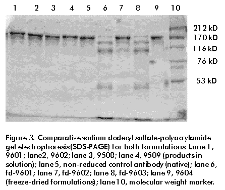
Radiocolloids determination Results of radiocolloids are shown in Table 1. For the lyophilized products, radiocolloids level was a little higher than the products in solution, but the HSA-impregnated ITLC showed only 1.5% or less radioactivity remaining at the origin for all preparations. Radiocolloids determination is especially important for the freeze-dried formulation because the lyophilization process could induce colloid formation by changes in the oxidation state of Sn2+. Antibody immunoreactivity Figure 1 shows the results of antibody immunoreactivity obtained for four lots of the product in solution. In a radioreceptor assay, both reduced antibody and non-reduced control antibody were able to compete in a similar way with radioiodinated EGF to bound hEGF-r. Results were plotted on a semilog paper showing activity on a linear scale and antibody concentration on a log scale. Similar immunoreactivity curves for antibodies clearly demonstrated that the incubation for 30 min with 2-ME at a molar ratio of 2000:1 with respect to the antibody, does not show any measurable loss in immunoreactivity of the antibodies. Similar results were obtained for four lots of the freeze-dried formulation, compared with the non-reduced control antibody (Figure 2). Figure 1. Immunoreactivity of the 99mTc-labeled antibody ior egf/r3 (product in solution) measured by a radioreceptor assay. Four lots of labeled MAb were compared to the non-reduced control antibody for their ability to compete with the radioiodinated EGF, as described in "Materials and Methods". Figure 2. Immunoreactivity of the 99mTc-labeled antibody ior egf/r3 (freeze-dried formulation) measured by a radioreceptor assay. Four lots of labeled MAb were compared to the non-reduced control antibody for their ability to compete with the radioiodinated EGF, as described in "Materials and Methods". SDS-PAGE Antibody purity was assessed by SDS-PAGE of the antibodies. Under reduced conditions (7.5%) light and heavy chains were separated and no difference was found between both formulations. Under non-reduced conditions (7.5%) a typical pattern of antibody bands was obtained for the product in solution, which is similar to the pattern obtained with the non-reduced control antibody. Different results were obtained for the freeze-dried kit formulation. Two lots of this product (fd-9601 and fd-9603) showed a very high fragmentation (at least seven bands) when they were reconstituted with 99mTc and stored at 4 ºC until total 99mTc decayed (four days) (Figure 3). This fragmentation was not obtained in the same lots when they were reconstituted with saline and immediately tested by electrophoresis. This is not a real fragmentation of the lyophilized antibody. It has been reported that the use of SDS-PAGE for this type of product can induce the fragmentation of the antibodies during the sample preparation, which involves SDS and a high temperature (100 ºC) during 5 min. In order to obtain more details about fragmentation, a native electrophoresis (heterogeneous gels 4%-15%) was performed for all samples and no fragments were found in the lot for both formulations. A typical antibody band pattern was obtained similar to the pattern obtained with the non-reduced control antibody (data not shown). Figure 3. Comparative sodium dodecyl sulfate-polyacrylamide gel electrophoresis (SDS-PAGE) for both formulations. Lane 1, 9601; lane2, 9602; lane 3, 9508; lane 4, 9509 (products in solution); lane 5, non-reduced control antibody (native); lane 6, fd-9601; lane 7, fd-9602; lane 8, fd-9603; lane 9, 9604 (freeze-dried formulations); lane 10, molecular weight marker. Superose-12 Using gel filtration chromatography on Superose-12 FPLC, no fragmentation was obtained when both formulations were chromatographed. These results were found not only when the samples were reconstituted with Tc and allowed for Tc to decay during four days at 4 ºC, but also when they were chromatographed immediately after reconstitution with saline. In general, two peaks were obtained with a retention volume of 10.2 and 20.4 mL, corresponding to the antibody and to the MDP solution, respectively. Non-reduced control antibody and MDP solution were used as control and were run in separate chromatographies under the same conditions (Table 2). Antibody purity was higher than 95% for the freeze-dried formulations, except for the lot fd-9603 (90.02%). Table 2. Gel filtration chromatography on a Superose-12 FPLC column as purity criteria for both formulations. Non-reduced control antibody and MDP solution were used as control.
*Percentage of the peak area corresponding to the antibody over
the total area under the curve. Isoelectric focussing The results of IEF showed a typical antibody band pattern for both formulations, similar to the pattern obtained to the non-reduced control antibody. No significant difference was found between both formulations. Biodistribution and pharmacokinetics The biodistribution of 99mTc-ior egf/r3 in normal rats at 1, 4 and 24 h post-injection is presented in Table 3 for the two products. The results show that there was only a low accumulation of the products in liver and kidneys as the main organs in their biotransformation, catabolism and elimination. The kinetic study did not show any specific localization in organs or tissues in healthy mice. Table 3. Biodistribution studies in Wistar normal rats for 99mTc-labeled antibody ior egf/r3 expressed in percent of injected dose per gram of tissue (% ID/g, data for both formulations).
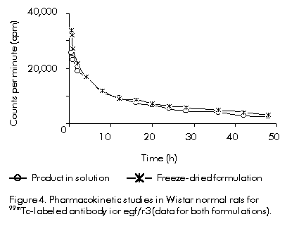
The results of the pharmacokinetic study are shown in Figure 4 and the calculated parameters are presented in Table 4. The radioactivity content of rat serum, expressed as concentration of activity per milliliter of serum, was plotted against the time of blood drawing in a linear plot. Serum time-activity curves were best fitted to a biexponential equation with a correlation coefficient of 0.9412 and 0.9382 for the product in solution and for the freeze-dried formulation, respectively. Despite the little numeric differences shown for both formulations, the main parameters do not vary significatively. These results were confirmed by the statistical analysis performed using the linear least squares regression method, and the Student's t test with confidence intervals of 95% and a probability higher than 0.05. Table 4. Pharmacokinetic parameters for 99mTc-labeled antibody ior egf/r3 (data for both formulations). The statistical analysis (Student's t test) showed no significant difference between the main parameter (significant difference for p < 0.05).
*Significant difference between both formulations was found for a < 0.05. Figure 4. Pharmacokinetic studies in Wistar normal rats for 99mTc-labeled antibody ior egf/r3 (data for both formulations). Discussion To use the results of previous studies with the ior egf/r3 MAb, we evaluated the equivalence of two formulations obtained before and after introduction of changes in the manufacturing process. Changes included increases in scaling-up, modification of the formulation by adding glucose, and lyophilization to obtain a stable product. A battery of physicochemical and biological assays was developed which demonstrated the equivalence of these two formulations. The freeze-dried kit formulation has some advantages over the product in solution. It includes a stable reduced antibody for achieving quantitative radiolabeling of the MAb with 99mTc at the time it is used just by mixing the kit with Tc-pertechnetate. Labeling efficiency for both formulations was higher than 98% and no purification step or addition is needed but the addition of a required amount of pertechnetate. Since the content of the kit is lyophilized, no special storage conditions or transportation are required. The high labeling efficiency and the very low level of radiocolloids (less than 1.5%) guarantee the high quality of the product. An important issue of the in vitro characterization of the MAb is the comparison of the biological activity of the antibody obtained prior and after the introduction of changes during the manufacturing process. For this reason, a radioreceptor assay was performed which demonstrated that changes did not affect the immunoreactivity of the antibodies. It was also demonstrated that the use of 2-ME to reduce the antibody and to generate sulfhydryl groups to bound Tc does not alter the biological activity of the antibody. The use of glucose as an additive to obtain a more stable compound in the lyophilized product does not interfere in the radiolabeling of the antibody. The hydroxyl groups present in the molecule (glucose) are low-affinity, high-capacity sites and cannot compete with the thiol groups generated in the molecule, which are high-affinity, low-capacity sites [21]. The use of glucose in the new formulation was established in previous studies where glucose enhanced the stability of the lyophilized product (data not shown). Changes in culture, purification or formulation procedures could lead to increasing aggregates or fragments. Therefore, it should be demonstrated that the changes undertaken do not influence the structural integrity of the product. To determine the molecular integrity of the antibody produced by the modified manufacturing process, tests which detect aggregates, dimmers and fragments in the products should be performed. We selected column chromatography (gel filtration in Superose-12, FPLC), electrophoresis (SDS-PAGE and native electrophoresis), and isoelectric focussing for microheterogeneity. The results confirmed the presence of only one protein in both formulations, which has the characteristics and the physicochemical and biological behavior of an antibody. No aggregates, dimmers or fragments were found. Data of biodistribution and pharmacokinetics also demonstrated the equivalence of the two formulations obtained in the in vitro studies. No significant difference was found between the calculated parameters for the two formulations. Significant difference was found only for the distribution half-life (T1/2a) for both formulations. Nevertheless, no significant difference was found for the elimination half-life (T1/2b). Equivalence is assumed taking into account that the elimination half-life is more important than the distribution half-life. Firstly, the elimination half-life determines the accumulation behavior for the product, which is a very important issue for diagnosis. Secondly, it represents more than 80% of the area under the curve. In our opinion, these are the most important parameters to be studied for these particular formulations. Parameters like isotype, subclass, primary and secondary structure, glycosylation pattern and specificity are unnecessary because there is no change in the production process of the MAb. This study has demonstrated the equivalence between the product in solution labeled with 99mTc and used in clinical trials, and the freeze-dried kit formulation presented. References 1. Schaffner G, Haase M, Gress S. Criteria for investigation of the product equivalence of monoclonal antibody for therapeutic and in vivo diagnostic use in case of introduction of changes in the manufacturing process. Biologicals 1994;23: 253-9. 2. Note for guidance: Production and quality control of monoclonal antibodies of murine origin. The rules governing medicinal products in the European Community. 1989;3:53-72. 3. Note for guidance: production and quality control of human monoclonal antibodies. The rules governing medicinal products in the European Community. 1990;3:41-56. 4. Notice for applicants for marketing authorization for medicinal products for human use in the Member States of the European Community. The rules governing medicinal products in the European Community. 1993;IIA. 5. Note for guidance: Pre-clinical safety testing of medicinal products derived from biotechnology. The rules governing medicinal products in the European Community 1990;3:73-88. 6. Points to consider in the manufacture and testing of monoclonal antibody products for human use. Department of Health and Human Services, Center for Biologics Evaluation and Research, FDA, 2 August 1994. 7. Proposed requirements for monoclonal antibodies for clinical use in humans. WHO. Geneva. 22-29 October 1991. 8. Morales A, Zayas F, Núñez G, Iznaga N, Pérez N, Izquierdo JC. Technetium-99m direct radiolabeling of monoclonal antibody ior egf/r3. Nucl Med Biol 1998;25: 25-30. 9. Rodríguez N, Torres O, Ramos M, Gómez JA, Marrero I, Catalá M, et al. Efectividad diagnóstica inmunogammagráfica del anticuerpo monoclonal (AcM) ior egf/r3 en pacientes con tumores malignos de origen epitelial. Rev Esp Med Nuc 1995;14:285. 10. Schwarz A, Steinstrasser A. A novel approach to Tc-99m-labeled monoclonal antibodies. J Nucl Med 1987;28:721. 11. Mather SJ, Ellisson D. Reduction-mediated technetium-99m labeling of monoclonal antibody. J Nucl Med 1990;31: 692-7. 12. Fernández A, Pérez R, Macías A, Velandia A, Álvarez I, Ramos M, et al. Generación y caracterización primaria de anticuerpos monoclonales contra el receptor del factor de crecimiento epidérmico. Interferón y Biotecnología 1989;6: 289-94. 13. Kowalsky RJ, Perry J. Radiopharmaceuticals in nuclear medicine practice. p 125. Appleton and Lange, Norwalk, Conn/Los Altos Calif; 1978. 14. Thrall JH, Freitas JE, Swanson D, Roger WL, Clare JM, Brown ML, et al. Clinical comparison of cardiac blood pool visualization with Tc-99m red blood cells labeled in vivo and with Tc-99m human serum albumin. J Nucl Med 1978;19:796-803. 15. Macías A, Pérez R, Lage A. Estudios sobre el factor de crecimiento epidérmico (EGF) II: desarrollo de un radioreceptor análisis para la determinación de cantidades picomolares. Interferón y Biotecnología 1985;2:115-27. 16. Greenwood FG, Hunter WM, Glover JS. The preparation of 131I-labeled human growth hormone of high specific radioactivity. Biochem J 1963;89:114-23. 17. Yamaoka K, Nakagawa T, Uno T. A nonlinear multiple regression program, MULT(2)(Bayes), based on a Baye(s) algorithm for microcomputers. J Pharmacobio Dyn 1978;8:246-56. 18. Yamaoka K, Nakagawa T, Uno T. Application of Akaike´s Information Criterion (AIC) in the evaluation of linear pharmacokinetic equations. J Pharmacokinet Biopharm 1978;6:165-75. 19. Yamaoka K, Nakagawa T, Uno T. Statistical moments in pharmacokinetics. J Pharmacokin Biopharm 1978;6:547-58. 20. Gibaldi M, Perrier D. Pharmacokinetics. In: Gibaldi M, Perrier D, editors. Pharmacokinetics. 2nd ed. New York: Marcel Dekker Inc.; 1982. p.319-55. 21. Paik CH, Phan LN, Hong JJ, Sahami MS, Heald SC, Reba RC, et al. The labeling of high affinity sites of antibodies with 99mTc. Int J Nucl Med Biol 1985;12:3-8. Copyright 2000 Elfos Scientiae The following images related to this document are available:Line drawing images[ba00009d.gif] [ba00009b.gif] [ba00009a.gif] [ba00009c.gif] | ||||||||||||||||||||||||||||||||||||||||||||||||||||||||||||||||||||||||||||||||||||
| |||||||||
