 |
| About Bioline | All Journals | Testimonials | Membership | News |
|
||||||
|
||||||
Biotecnología Aplicada, Volume 17, July-September 2000, p. 195 Sequence Analysis and Structure Prediction of
dexB, dexC and dexD Genes Induced in Dextran-Containing Cultures Tirso Pons, Bianca García, Ailed Castellanos Biolndustry Division. Centro de Ingeniería Genética y Biotecnología, Havana, Cuba. E-mail: tirso.pons@cigb.edu.cu From a selection of papers from Biotecnología Habana`99 Congress.
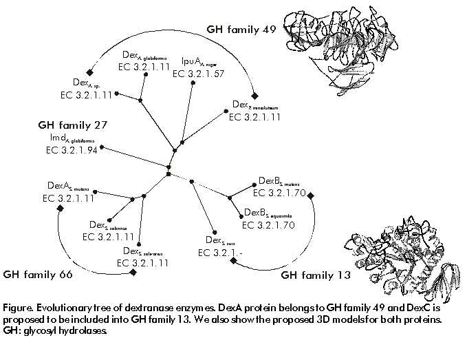
Code Number: BA00059 Introduction The fungus Penicillium minioluteum HI-4 when grown in dextran-containing medium produces enzymes, which participate in dextran assimilation. cDNAs clones corresponding to genes expressed in dextran-induced cultures were identified by differential hybridization. Southern hybridization and restriction mapping analysis of selected clones revealed four non-homologous cDNAs. The four cDNAs were transcriptionally expressed in dextran-containing medium, but not in glucose. One of these cDNAs (dexA) codifies for an endo-dextranase (a-1,6-glucan-6-glucanohydrolase, EC 3.2.1.11) [1], which hydrolyzes the a-1,6-glucosidic linkages within the dextran polymer and between branch points to release smaller oligosaccharides. Here we describe the sequence of the other three genes (dexB, dexC and dexD) that were induced. The protein sequences deduced from the cDNA nucleotide sequences were compared with the protein sequences in GeneBank CDS translations, SWISSPROT, PIR and PDB databases. The search of sequence similarity revealed that DexB shows similarity to glucose permease proteins, DexC to a-glucosidase enzymes and DexD to a-amylases. The three-dimensional (3D) structure of Bacillus cereus oligo-1,6-glucosidase [2] and the 3D structure of different a-amylases [3] have been previously published. Based on common sequence pattern in the structural core region, a plausible 3D model of the core of any protein can be built in the specific protein family. Using this model, structural and functional information can be transferred from one member of the family to the others, even when sequence similarity is low. This can be extremely useful for the identification of residues that participate in active sites or in the interpretation of mutant phenotypes. Materials and Methods cDNA nucleotide sequences were translated into protein sequences using the GeneRunner program [4]. The protein sequences were compared against GeneBank CDS translations, SWISSPROT, PIR and PDB databases using the WWW PSI-BLAST service (http://www.ncbi.nlm.nih.gov/cgi-bin/BLAST/nph-psi_blast). Multiple alignments and phylogenetic trees were done by CLUSTALW [5] and TREETOOL [6] programs. The 3D models were created by WHAT IF [7] program. Results and Discussion The sequence analysis [1] and structure prediction [8] of dexA gene has been published. Here, we reported the sequence analysis of dexB, dexC and dexD genes revealing that they seem to belong to at least three distinct non-homologous sequence families. The first is a sugar transporter family, which contains eukaryotic maltose permease, high- and low-affinity glucose transporter and several others with different specificities (turanose, trehalose, melezitose, palatinose). The second is the a-glucosidase family, of known 3D structure, and comprises prokaryotic dextran-glucosidases (EC 3.2.1.70), oligo-1,6-glucosidases (EC 3.2.1.10), trehalose-6-phosphate hydrolase (EC 3.2.1.93), and prokaryotic and eukaryotic a-glucosidases or maltases (EC 3.2.1.20). The third family, also of known 3D structure, contains several a-amylases (EC 3.2.1.1). Each of the three protein families appears to have a distinct 3D fold. Yet dexA and dexC catalyzes chemically equivalent reactions on similar or identical substrates. The enzymic function of dextran hydrolysis appears to have evolved independently on at least two structural frameworks (Figure). Figure. Evolutionary tree of dextranase enzymes. DexA protein belongs to GH family 49 and DexC is proposed to be included into GH family 13. We also show the proposed 3D models for both proteins. GH: glycosyl hydrolases. The flexible combination of active sites and 3D folds observed in nature can be exploited by protein engineers in designing and optimizing enzymic function. In our case, a successful modification of dextranase enzymic properties led to a more efficient application in the sugarcane factories. In conclusion, dexB (1.6 kb), dexC (1.8 kb) and dexD (1 kb) genes codified for a putative glucose permease, dextran-glucosidase and a-amylase proteins, respectively. In addition, based on the sequence analysis reported here, we propose to include DexC and DexD into glycosyl hydrolases family 13. References 1. García B, Margolles E, Roca H, Mateu D, Raíces M, González ME, et al. FEMS Microbiol Lett 1996;143:175–83. 2. Watanabe K, Hata Y, Kizaki H, Katsube Y, Zuzuki Y. J Mol Biol 1997;269:142–53. 3. Swift HJ, Brady L, Derewenda ZS, Dodson EJ, Dodson GG, Turkenburg JP, et al. Acta Crystallogr 1991;47:535. 4. GeneRunner ver. 3.02 Hastings Software lnc., 1994. 5. Thompson JD, Higgins DG, Gibson TJ. Nucleic Acids Res. 1994;22:4673–80. 6. Maciukenas M. Treetool ver. 1.0, University of Illinois, 1991. 7. Vriend, G. J Mol Graphics 1990;8:52–6. 8. Pons T, Chinea G, Olmea O, Beldarraín A, Roca H, Padrón G, Valencia A Proteins 1998;31:345–54. Copyright Elfos Scientiae 2000
The following images related to this document are available:Photo images[ba00059a.jpg] |
| |||||||||
