 |
| About Bioline | All Journals | Testimonials | Membership | News |
|
||||||
|
||||||
Biotecnología Aplicada, Volume 17, July-September 2000, p. 197 Bispecific Antibody-based Ultrasensitive Immunoassays MR Suresh, D Xu, F Liu, R Vij Faculty of Pharmacy and Pharmaceutical Sciences, University of Alberta, Edmonton, Canada T6G 2N8 From a selection of papers from Biotecnología Habana`99 Congress.
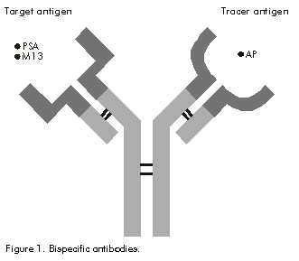
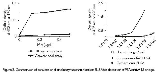
Code Number: BA00061 Bispecific MAbs have non-identical paratopes unlike traditional monospecific MAbs and can be generated chemically, by somatic fusion of two hybridomas or by genetic engineering methods [1]. Bispecific antibodies allow the generation of the highest specific activity immunoprobes, wherein every antibody molecule can be labeled uniformly with the enzyme, as a label. In the present study we have developed bsMAbs against prostate specific antigen (PSA) and M13 phage as a model virus (Figure 1). The second arm in each case was specific to alkaline phosphatase (AP). Figure 1. Bispecific antibodies. The major advances in immunoassays have focused on specificity, sensitivity, speed and convenience. New diagnostic markers have allowed earlier therapeutic intervention than was previously possible. The ability to quantify the analyte concentration in the ultrasensitive range has potential to identify residual and recurring disease several months and even years prior to their estimation by conventional immunoassay procedures. Among the ultrasensitive methods described, the technique of enzyme amplifications is the most sensitive method [2] which is capable of detecting as little as 0.01 attamole (amol) of AP [3]. The technique of enzyme amplification depends on an enzyme label giving rise to a catalytic intermediate that further amplifies the detectable signal. NADP+ for example as the primary substrate can be cleaved to NAD+ by AP. The dephosphorylated cofactor then enters a highly specific redox cycle, where it is reduced by NAD+ specific alcohol dehydrogenase. The oxidized form is regenerated by diaphorase with the concomitant reduction of p-iodonitrotetrazolium violet (INT-violet) reagent to an intensely purple formazan dye. The oxidized form is continuously cycled with the formation of detectable product with every turn of the cycle [4]. PSA is a 30-kD glycoprotein with chymotrypsin-like protease activity produced primarily by the epithelial cells of the prostate gland [5, 6]. Clinically, estimation of PSA levels allows early detection of prostate cancer and provides a way to monitor the treatment response and predict disease recurrence [7]. A number of clinical studies have shown that patients with radical prostatectomy have PSA concentrations < 0.1 mg/L and in most cases < 0.02 mg/L. These levels are undetectable by convencional assays which have a lower limit of detection of 0.1–0.4 mg/L [8]. Availability of ultrasensitive PSA assays may enhance our ability, detect the emergence of micrometastasis which are below the threshold of current assays. Detection of virarl antigens and detection of the viral load present in body fluids is a desirable diagnostic or monitoring tool for many infectious diseases. In case of HIV infection, an HIV antigen assay could be used for measuring viral load to determine the prognosis of the infected individual and to monitor the effectiveness of the antiviral therapy [9]. In an effort to develop highly sensitive ELISA as a routine viral screening assay or viral load assay, phage M13 was chosen as a model virus to demonstrate feasibility since they were relatively safe and are conveniently available. This report describes the development of bispecific antibody based immunoassays for PSA and M13 phage coupled with enzyme amplified ELISA to achieve ultrasensitive detection of analytes. We optimized various parameters of the bispecific assay to achieve the lowest detection limit. A sandwich assay format was used wherein a monospecific solid phase antibody was used to capture the antigen (PSA or M13). Subsequently, the corresponding bsMAb was added along with excess AP. Parallel assays were performed using either p-nitrophenylphosphate for conventional assay or an NADP based cyclic amplification reaction [4]. The results show that in the two examples investigated, we were able to demonstrate ultrasensitive detection of the two antigens which correspond to 250fin PSA/mL and 100 M13 particles or 2.3fg of coat protein (Figure 2). Figure 2. Comparison of conventional and enzyme amplification ELISA for detection of PSA and M13 phage. References 1. Suresh, et al. Bioconjugate Chem 1998; 9(6):635–44. 2. Colin H. J of immunol Methods 1985; 76:389–93. 3. Ellis, et al. J of lmmunol Methods 1985; 87:7–11. 4. Bates, L. TIBTECK 1987;5:204–9. 5. Hara M, et al. J lab Clin Med 1989; 83:3166–70. 6. Wang MC, et al. lnvest Urol 1979;17: 159–63. 7. Kuriyama, et al. Cancer research 1981; 41:465–862. 8. Javad M, et al. C@cal Biochemistry 1995;28(4):407–14. 9. Hanai, et al. Acta Pathol.hficrobiol. Immunol.Scand 1998;suppl2:1–20. Copyright Elfos Scientiae 2000 The following images related to this document are available:Photo images[ba00061a.jpg] [ba00061b.jpg] |
| |||||||||
