 |
| About Bioline | All Journals | Testimonials | Membership | News |
|
||||||
|
||||||
Biotecnología Aplicada 2000;17:235-240 Transfection of Intact and Wounded Skin with a DNA/Polyethylenimine Complex
@ Jorge Berlanga,1 Vivian Sáez,2
Elaine Santana,1 Eileen Riego,1
1Mammalian Cells Genetic Division, 2Pharmaceutical Formulations
Development Laboratory. Received in January, 2000.
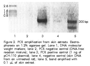
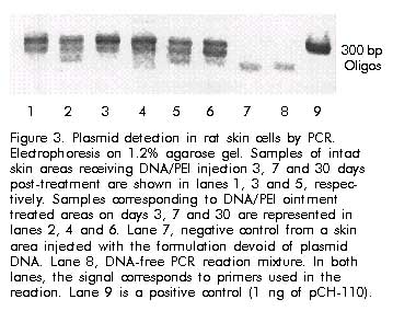
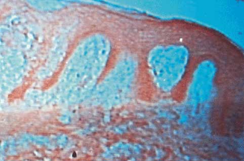
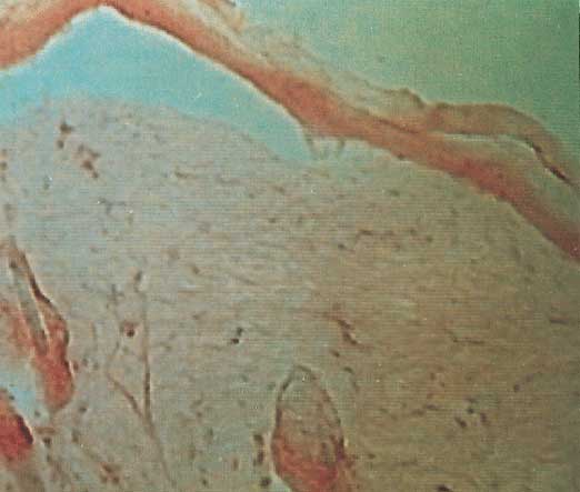
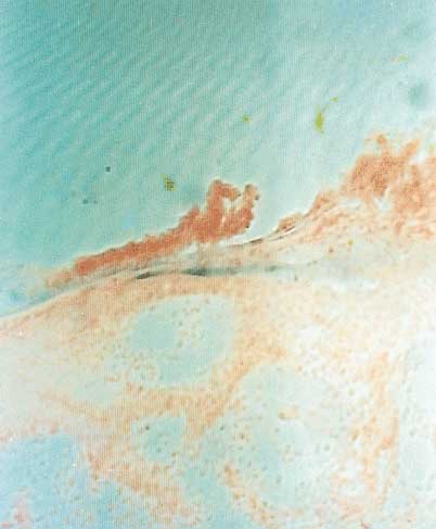
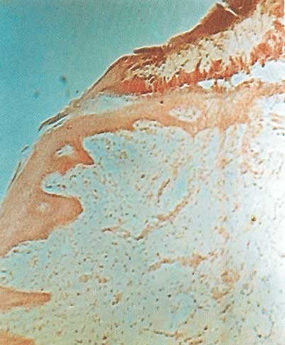
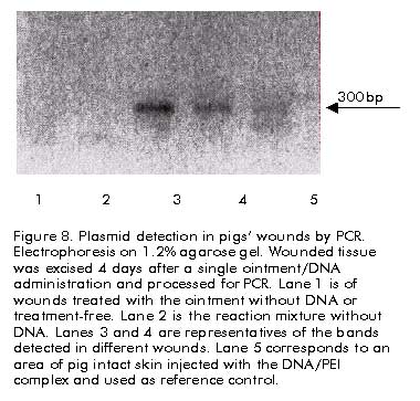
Code Number: ba00072 Abstract Cutaneous cells offer unique advantages to correct local or systemic disorders via in vivo or ex vivo gene transfer techniques. Polyethyleneimine (PEI) is emerging as a highly efficient vector for DNA delivery both in vitro and in vivo. We investigated whether a single intracutaneous injection of a DNA reporter plasmid in complex with PEI, allowed the transfection of intact skin in rats and pigs. The ability of an experimental ointment to deliver to and transfect intact or wounded skin areas, was also examined. We demonstrated that a single intracutaneous injection of the complex allowed the transfection of epidermal cells in both species. A single topic administration of the ointment showed to transfect intact skin areas and keratinocytes at the edges of controlled wounds in pigs. Though integration is not excluded, plasmid dose and its expression intensity appeared progressively reduced in rats. skin, after 30 days of injection or topic administration. These data are the core for additional studies to elucidate the feasibility of using a growth factor-based gene therapy to stimulate wound healing. Keywords: DNA, PEI, skin, transfection Resumen Transfección de piel intacta y lesionada mediante el uso de un complejo ADN/polietilenimina. El fácil acceso al tejido cutáneo permite su fácil manipulación genética y la corrección de trastornos locales o sistémicos mediante la transferencia exógena del material genético apropiado. Aunque de forma limitada, la polietilenimina (PEI) ha sido ensayada exitosamente como vector de ADN en modelos in vitro e in vivo. En este trabajo se estudió la transfección potencial de células de piel, tras una inyección intracutánea del plasmidio reportero combinado con PEI en ratas y cerdos. Se estudió, además, el efecto de la administración tópica de un ungüento experimental que sirve de vehículo al complejo ADN/PEI en la transfección de células cutáneas, sobre tejido intacto y herido. Una sola inyección intracutánea permitió la transfección de la células epidérmicas en ambas especies, y de estructuras de la dermis profunda en ratas. La aplicación del ungüento facilitó la transfección de la piel intacta y de los queratinocitos de los bordes de las heridas en los cerdos. Aunque no se descarta la integración cromosómica, la presencia del plasmidio y su expresión en la piel de las ratas decrecen progresivamente a los 30 días de su inyección o aplicación tópica. Se ejecutarán otros experimentos para definir la factibilidad de acelerar la cicatrización mediante la transfección cutánea con factores de crecimiento. Palabras claves: ADN, PEI, piel, transfección Introduction The current limitations encountered in the use of viral vectors have stimulated an impetus in the development of alternative non-viral methods of DNA delivery. The main goal of this approach is to imitate desirable features of viruses, while avoiding their limitations. This novel therapeutic modality is further appreciated as far as the technique to introduce genes in skin cells is non-laborious, inexpensive, safe, and non-invasive for the patient [1]. The accessibility of the skin and the proliferative capacity of the epidermal cells make keratinocytes ideal candidates for genetic manipulation and gene therapy [2]. Thus, genetically engineered skin has long been proposed as a potential vehicle to correct the deficit of a local or systemic factor [3]. Epidermal cells have been successfully transfected in vivo via particle-mediated DNA delivery [4]. More recent experiments demonstrated that epidermal cells can transiently express plasmid DNA following direct intracutaneous injection [5]. In an attempt to further simplify DNA delivery and enhance transfection efficiency on the skin, some papers examined the feasibility of topically administering the desired gene using different approaches. These have included the direct topical application of DNA-cationic liposome complexes [6], hemagglutinating virus of Japan DNA. liposome complex [7], adenoviral vectors containing a desired gene [8]. Topical transfection has been also accomplished by means of locally generated electric pulses in order to make breaches through the stratum corneum [9]. Polyethylenimine (PEI) is an organic macromolecule with a high cationic-charge-density potential, and has shown to be a highly efficient vector for plasmid delivery both in vitro and in vivo [10]. In general, PEI remains scarcely used for either in vitro or in vivo DNA delivery. In this experiment we studied the effects of a DNA/PEI conjugate local injection on the skin of two animal species. We also examined the ability of a simple ointment-like formulation, vehiculizing the conjugate, to transfect intact or wounded skin edges following a single topical administration. To our knowledge, this is the first in vivo demonstration of the feasibility of using the DNA/PEI complex, formulated as a topical ointment, to transfect intact or wounded skin. Materials and Methods Animals White fur, Yorkshire female piglets with a body weight of 15. 17 kg from the Genetic Porcine Center of Havana were used in this study. Pigs were individually allocated in stainless steel cages (90 x 90 x 90 cm) provided with a waste-disposal system. Adult male Sprague Dawley rats (220. 250 g, CENPALAB, Havana, Cuba), were included in the study, and individually allocated in wire-bottomed cages. Both pigs and rats were housed in certified rooms with controlled indoor environmental conditions, such as light cycle 12/12 -h, room temperature of 17. 20 ºC, controlled ventilation cycles, and relative humidity of 60. 70%. Animals were acclimated for about a week before the experiments, and handled in compliance with current guidelines of the Animal Ethics Committee of the Center for Genetic Engineering and Biotechnology in Havana. Plasmid The expression plasmid pCH-110 (Pharmacia, Uppsala, Sweden), containing a functional lacZ gene driven by the SV40 early promoter was used in this experiment. Plasmid was propagated in Escherichia coli XL-1-Blue MR, and purified by a modified alkaline lysis method [11]. Formulations DNA was combined to PEI (MW 50 kD, Sigma, St. Louis, MO, USA) as described [10], in a ratio of 1 µg of DNA per 3 µL of a 10 mM PEI stock solution in 150 mM NaCl. These proportions result in a ratio of 3 nitrogen atoms of the PEI molecule per phosphate group of DNA. A stock solution DNA/PEI was prepared (20 µg/µL) and used for both the injections and the topic ointment. For the injection, the stock solution was further diluted in 150 mM saline solution to achieve a concentration of 100 ng/µL. The ointment formulation was made from pharmaceutically approved ingredients, usually used for topic administration in the skin: 40% polyethylene glycol (MW 4000, Merck, Germany); 35% propylene glycol (MW 6000, Sigma, St. Louis, MO, USA); 5% glycerol (Riedel-de-Haen, Germany); 10% stearyl alcohol (Sigma, St. Louis, MO, USA); 10% aqueous phase with DNA plasmid. Controlled portions of this mixture were step-wise added to the DNA solution [10], evenly homogenized, and adjusted to a concentration of 1 µg of DNA per milligram of vehicle. Intracutaneous injection Animals were anaesthetised with sodium pentobarbital (Roig-Farma, Barcelona, Spain). Rats received 40 mg/kg by intraperitoneal route, and pigs 30 mg/kg intravenously via a peripheral ear vein. The whole dorsal region was clipped, depilated with a commercial cream (Nair, J&J, USA), and cleansed with a mixture of hibitane 2% and 70% ethanol. Plasmid/PEI injections were carried out in a final volume of 50 µL, using a 1-mL insulin syringe coupled to a 27 ½ needle. The solution was slowly delivered once the needle had been inserted in an angle of approximately 30° in relation to the epidermis. It remained inserted for another 2 min in order to prevent fluid-retrograde diffusion. Control injections included 150 mM saline solution and PEI with no DNA. All the injection sites (ca. 1 cm2) were carefully identified to facilitate further sampling. The body surface area used for the injections was approximately 8.6 and 3.2% for rats and pigs, respectively. Topical administration Before the administration of the ointment containing plasmid onto an intact skin area, the epidermis was pre-treated for about 20. 30 min with propylene glycol in order to enhance its permeability. A thin and uniform layer of the ointment was dispensed onto the skin, which remained undisturbed for the time the animals were anaesthetised (ca. 2 h). In rats, ointment application involved an approximate area of 30 cm2 of the dorsal region. For the pigs, ointment was applied in a well-delimited patched fashion to facilitate further biopsy harvesting. Four patches of 4 x 4 cm each were equidistantly and evenly distributed on each side of the dorsum to complete an area of approximately 128 cm2. In order to prove if the wound edge cells are able to take up and express a foreign DNA, controlled full-thickness wounds (9 mm in diameter) were created on the dorsum of pigs using disposable biotomes (Acuderm Inc., Fl., USA). The ointment was dispensed into each ulcer and its edges with a syringe once haemostasia was attained. No further administrations were conducted with the formulations assayed. Control groups received the same formulations, including PEI, but without DNA. Rats. biopsies for histostaining were collected from treated areas in intact skin and matched controls on days 3, 7, 21 and 30. Samples corresponding to days 3, 7 and 30 were used for PCR analyses (see below). Wounded tissue specimens (in pigs) were resected only on the fourth day post-injury for both histochemistry and PCR. A minimum of 4 wounds was harvested from each animal. DNA extraction and PCR For DNA extraction, skin fragments of 9. 25 mm2 were washed three times in sterile water and placed in a buffer solution containing 5% Chelex (Sigma, St. Louis, MO, USA) 7 µL of 1 M DTT, 5 µL of 10 mg/mL proteinase K in a final volume of 1 mL sterile water. Digestion was left overnight at 56 ºC, vortexed and boiled for 8 min, vortexed again for 10 s and centrifuged at high speed. The supernatant was collected and kept frozen until use. The PCR mixture included the amount of DNA contained in 0.1 µL of the extracted DNA, 18.5 µL of water, 10 pmol of each primers (a and b), 2.5 µL PCR buffer 10x (Heber Biotec, Havana, Cuba) (500 mM KCL, 100 mM Tris-HCl [pH 9.0], 15 mM MgCl2, 1% Triton X-100, 2 mM each dNTP), 1 µL Taq polymerase 5 U (HeberBiotec, Havana, Cuba), to complete 25 µL of total volume. Internal controls included 1 ng of pCH-110 DNA and 1 µg of rat chromosomal DNA as positive controls, and the negative was DNA-free. PCR cycling conditions were: initial denaturation at 96 ºC for 2 min, followed by 1 min at 94 ºC; annealing at 56 ºC for 1 min; extension at 72 ºC for 1 min, to complete 36 cycles; and final extension at 72 ºC for 5 min. In order to ensure the specificity of the PCR, primers amplifying 280 bp of SV-40 early promoter of the pCH-110 plasmid were designed:
(a) sense 5. -GCCTGGCCTCTGCATAAATA-3. (b) antisense 5. -CAGCTGTGGAATGTGTGT CA-3. 20 mer (MT = 50 ºC)
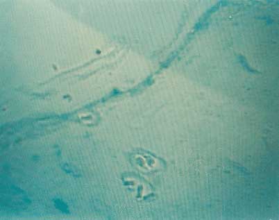
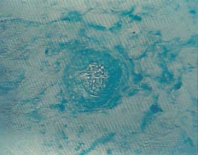
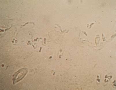
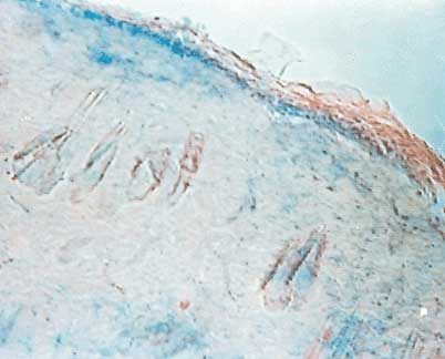
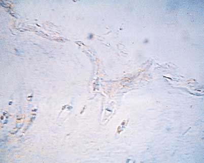
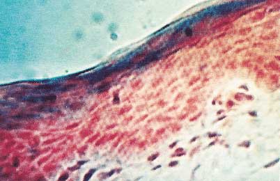
Detection of b-galactosidase (b-gal) activity The skin fragments were processed as previously described [12]. Briefly, fragments were washed twice with PBS and fixed with 2% paraformaldehyde, and 0.2% glutaraldehyde, 0.1% sodium deoxycholate, 0.02% NP-40 detergent at 4 ºC for 20 min. After washing twice with PBS, tissue samples were incubated with Bluo-gal (Sigma, St. Louis, MO, USA) staining solution (100 mM sodium phosphate [pH 7.3], 1.3 mM MgCl2, 3 mM K3 Fe[CN]6, 3 mM K4Fe[CN]6, 1 mg/mL Bluo-gal in N-N-dimethylformamide) at room temperature for about 12 h. Specimens were rinsed with 3% DMSO in PBS, and three times with 70% ethanol, paraffin embedded, sectioned at 5. 6 µm, and fixed for 20 min with 2% paraformaldehyde. Slides were often counterstained with nuclear fast red as described [13] and routinely mounted. PEI cytotoxicity study Slides with 5-µm-sectioned specimens from each sample and at each time point were used for H&E staining. Thus, injected or topically treated, and control areas were examined to reveal evidences of potential PEI-associated cytotoxicity. Parameters included cellular ballooning, karyorrhexis, karyopiknosis, necrosis, apoptotic-like nuclear changes, interstitial edema, and inflammatory infiltrate. Results Intracutaneous injection The histochemical development of intact skin fragments collected 72 h after the injections, showed that b-gal plasmid was expressed by keratinocytes, epithelial appendages, dermal cells, and deep-dermal structures like blood vessels in rats (Figure 1A and 1B). Control areas were negative to histochemical reaction (Figure 1C). Injection in pigs, however, showed that b-gal expression was mostly circumscribed to the epidermal keratinocytes. Some fibroblasts located in the upper dermis were often found expressing b-gal (data not shown). In general, the histostaining pattern was more uniform and even in rats as compared to pigs, which showed a diffuse and irregular pattern on the epidermal layer. The analysis of H&E-stained specimens collected from injected areas of both species, showed that skin was normal, with no evidence of cytotoxicity attributable to PEI. The presence of plasmid DNA after injections could be easily detected by PCR in 0.1 µL of skin extracts (Figure 2). The products of DNA amplification using the above mentioned primers were observed at 72 h, and even at 30 days after intracutaneous injection (Figure 3). Figure 2. A) Representative image of a rat skin area injected with DNA/PEI complex in normal saline. X-gal development demonstrated the expression of the reporter plasmid by epidermal cells. The outer sheets of hair follicles close to the epidermis are also stained. The expression was detected 72 h after injection. Not counterstained 5-mm paraffin section; magnification x10. B) Deep dermal structures like blood vessel walls and peripheral fibroblasts were positively stained with X-gal following DNA/PEI complex intracutaneous injection. Blue background is seen in dermal collagen fibres. The expression was detected 72 h after injection. Not counterstained 5-mm section; magnification x25. C) Representative image of a matched-control skin area injected with PEI and without plasmid DNA. Not counterstained 5-mm embedded section; magnification x10. Figure 2. PCR amplification from skin extracts. Electrophoresis on 1.2% agarose gel. Lane 1, DNA molecular weight markers; lane 2, PCR negative control (DNA-free reaction mixture); lane 3, PCR positive control (1 ng of pCH-110 plasmid); lane 4, negative control (skin DNA from an untreated rat); lane 5, band amplified with 0.1 mL of skin extract. Figure 3. Plasmid detection in rat skin cells by PCR. Electrophoresis on 1.2% agarose gel. Samples of intact skin areas receiving DNA/PEI injection 3, 7 and 30 days post-treatment are shown in lanes 1, 3 and 5, respectively. Samples corresponding to DNA/PEI ointment treated areas on days 3, 7 and 30 are represented in lanes 2, 4 and 6. Lane 7, negative control from a skin area injected with the formulation devoid of plasmid DNA. Lane 8, DNA-free PCR reaction mixture. In both lanes, the signal corresponds to primers used in the reaction. Lane 9 is a positive control (1 ng of pCH-110). Topical administration The administration of the DNA/PEI ointment onto intact skin of both rats and pigs, demonstrated the feasibility of transfecting cutaneous cells by exposing an area of keratinized but pre-conditioned epidermis to the formulation. The histochemical analysis of the skin from both animal species showed that epidermal cells expressed b-gal. In rats, the uppermost fibroblasts expressed the reporter gene (Figure 4A). Hair follicles expression was considered doubtful, though. Skin areas which received DNA-free ointment were negative to b-gal expression (Figure 4B). Expression in pigs was only restricted to suprabasal epidermal cells (Figure 5A). Ocasionally, staining was detected in some fibroblast of papillary dermis. Control skin areas did not show expression (Figure 5B). Plasmid residence in topically treated skin was demonstrated by PCR (Figure 3). These findings are derived from biopsies collected 72 h after the topical administration in rats and pigs. Figure 4. A) Representative specimen corresponding to a rat skin area which was topically exposed to DNA/PEI-containing ointment. Epidermal cells consistently express b-gal. Patches of dermal cells nearby the dermis were also positive. 5-mm section counterstained with nuclear fast red; magnification x10. Figure 4. B) A matched control of a rat skin area treated with the ointment without DNA. Some unspecific background is seen. Not counterstained 5-mm section; magnification x10. Figure 5. A) Fragment of pig skin topically treated with the DNA/PEI-containing ointment. Expression is clearly seen by suprabasal keratinocytes on day 3 post-treatment. 5-mm section counterstained with nuclear fast red. Magnification x40. Figure 5. B) Matched control of a skin area exposed to the ointment without plasmid DNA. 5-mm section counterstained with nuclear fast red; magnification x25. Persistence of the plasmid and its expression In order to evaluate the persistence of the DNA plasmid and its encoded product in the cells of injected and topically treated skin areas, a time course experiment was conducted. For practical reasons this experiment only included rats. Histochemical reactions were conducted on biopsies collected on days 3, 7, 21 and 30 post-treatment. The PCR data indicated that the intensity of the bands was reduced along the time in both injected and topically treated skin areas. Surprisingly, bands were still clearly noticeable 30 days after the treatment (Figure 3). In relation to the expression profile, an intense staining was identified in areas topically exposed or injected with the DNA conjugate on days 3 and 7. A drastic reduction of the expression was seen on day 21 for both administration procedures, whereas no expression was detected on day 30 (Figure 6). Here again, H&E-stained specimens showed no evidence of any PEI-associated cytotoxic effect. Figure 6. No expression was detected in rats’ skin 30 days after DNA/PEI administration. 5-mm section counterstained with nuclear fast red; magnification x25. Transfection of wound edges An independent experiment was carried out to address if epidermal edges of full-thickness wounds in pigs could be transfected by a single administration of the DNA/PEI-containing ointment. To answer this question, clean and standard biopsy ulcers were induced and immediately exposed to the formulation as a single DNA dosification. Bleeding was always controlled before treatments. Matched wounds in different pigs, treated with the ointment without DNA or treatment-free, were included as controls of the experiment. Fragments of intact untreated skin were also included as negative controls. Ulcers were harvested and processed for PCR, and histochemical analysis 4 days after being created and treated. The microscopic analysis of 5-µm sections obtained from the wound edges demonstrated that cells on the ingrowing epidermis expressed b-gal with an acceptable intensity (Figure 7A). By using a calibrated eyepiece we estimated that the expression involved cells in a length of about 1.5 mm outwards the edges. No b-gal expressing cells were detected in the incipient granulation tissue. Control wounds did not show expression at the edges or in granulation tissue (Figure 7B). The presence of plasmid DNA in the wounds was confirmed by PCR at that time (Figure 8). Figure 7. A) Keratinocytes expressing b-gal in one of the wound edges 4 days after treatment with the DNA/PEI ointment. Double paraffin embedding to prevent tissue distortion during sectioning. 5-mm section counterstained with nuclear fast red; magnification x10. Figure 7. B) Epidermal leading edges were negative to b-gal expression in control wounds. 5-mm section counterstained with nuclear fast red; magnification x10. Figure 8. Plasmid detection in pigs’ wounds by PCR. Electrophoresis on 1.2% agarose gel. Wounded tissue was excised 4 days after a single ointment/DNA administration and processed for PCR. Lane 1 is of wounds treated with the ointment without DNA or treatment-free. Lane 2 is the reaction mixture without DNA. Lanes 3 and 4 are representatives of the bands detected in different wounds. Lane 5 corresponds to an area of pig intact skin injected with the DNA/PEI complex and used as reference control. Discussion We have confirmed the feasibility of the intracutaneous injection as a DNA delivery system to transfect skin cells. Other studies had previously shown that skin puncture in pigs [13] and mice [5] allowed an efficient DNA transfer, turning the skin cells in a local or systemic bioreactor. Our results of intracutaneous injection in rats. skin extend these observations to other species. As aforementioned, injection of DNA/PEI complex in rats. skin resulted in an in-block transfection of different cutaneous structures, whereas in pigs b-gal expression was mostly seen in the epithelium. We speculate that this difference may be first explained by the characteristics of each species skin and its influence on the DNA instillation conditions. The rat is a lose skin animal; thus, a cutaneous fold could be easily held for puncture and instillation; rats. skin offered resistance during liquid instillation; and the pressure created within the papule could possibly facilitate a larger and in-depth DNA diffusion and uptake. On the contrary, the pig is a tight skin animal, with thicker dermis, and folds could not be held; therefore, puncture remained roughly in an epithelial or subepithelial position, which did not offer resistance to liquid instillation. Nevertheless, differences among species, tissues, and cell sensitivities to foreign DNA uptake have been described [14] and can not be ruled out in this experiment. Our results show a simple experimental formulation. made up with pharmaceutically approved ingredients. that carries the DNA/PEI complex, and allows the direct introduction and expression of the foreign DNA in skin cells in vivo upon its topical administration. Although the amount of secreted protein per unit of DNA/PEI complex is not addressed in this work, it is noteworthy that a single administration of the ointment was sufficient to consistently transfect epidermal cells in both intact and wounded skin sites. The mechanisms whereby keratinocytes uptake DNA in vivo are unknown [15], however, epidermal cells seem to be an active processing machinery, so that as early as in 24 h keratinocytes express significant amounts of mature IL-8 following intracutaneous injection of its coding DNA in a naked form [13]. In the models of intact skin the cornified layers were not removed. as histologically confirmed. The pre-treatment of the skin with propylene glycol, and its incorporation into the ointment may explain the DNA penetrability and diffusion across the epidermal cells junctions [16]. This assumption may support our somewhat surprising finding given by the presence of upper dermal fibroblasts expressing b-gal following a single ointment application. Although rat epidermis is thiner than that of pigs, it is difficult to elucidate how the DNA/PEI complex was sorted across the stratified epidermis, penetrated down to dermis and was eventually uptaken from the extracellular space by resident fibroblasts. An speculative explanation for this observation is sustained by two facts: 1) dermal connective tissue contains a high proportion of acidic positively charged glycosaminoglycans [17] and 2) PEI confers a high cationic charge density to DNA/PEI complex; thus, repulsive electrostatic forces would be locally generated, which may favor complex diffusion and endocytosis by a negatively charged cell membrane [10]. Our preliminary results of epidermal edges transfection and expression of b-gal warrant future more refined experiments to definitively demonstrate the effectiveness of this procedure in enhancing wound healing by introducing growth factor coding genes [18]. Sun et al. [19] showed that fibroblast growth factor coding genes enhance wound healing in diabetic mice when wound cells were transfected by direct application of the plasmid or assisted with liposomes. In pigs, wound-healing time was significantly shortened via particle bombardment with the epidermal growth factor coding gene [20]. Although an outstanding morphological similitude between human and pig skin makes conceivably to manipulate human epidermal cells in cutaneous injuries, caution must be observed though, as these models are based on controlled acute ulcers, which largely differ from human chronic and deep burn wounds. Thus, designing topic formulations for gene therapy-mediated wound healing must envision DNA protection against in situ extracellular degradation by serum or invading bacterial DNases [21]. Recent studies suggest that PEI may circumvent both extracellular and intracellular DNA degradation, as PEI acting as a proton sponge can ensnare DNA, structuring a mature intracomplex that may assist in the multistage process of transfection [22]. In the time course of our experiment, plasmid DNA band intenstity and b-gal staining gradually declined over time. Yet, it remains to be elucidated whether an eventual chromosomal integration ocurred, even when other findings based on intracutaneous injection of a b-gal reporter gene [13] are similar to ours. Albeit we know by previous observations (unpublished data) that the regenerated epidermis in incisional wounds is negative to b-gal expression, the demonstration of lack of integration should alleviate safety concerns about possible long-term side effects. This is particularly relevant in the case of wound healing and a growth factor with tumor promoter activity. Finally, PEI proved to be a safe agent as no evidence of local irritance or cytotoxicity was obtained in any case. This observation contributes to justify the use of PEI as an efficient and economic DNA vector for incoming cutaneous transfection experiments. Further in vivo models based on the topical delivery of growth factor coding genes in complex with PEI will be completed, with the aim of examining the feasibility of this approach to enhance cutaneous wound closure. References
Copyright Elfos Scientiae 2000
The following images related to this document are available:Photo images[ba00072i.jpg] [ba00072c.jpg] [ba00072b.jpg] [ba00072f.jpg] [ba00072k.jpg] [ba00072h.jpg] [ba00072g.jpg] [ba00072d.jpg] [ba00072l.jpg] [ba00072e.jpg] [ba00072a.jpg] [ba00072j.jpg] [ba00072m.jpg] |
| |||||||||
