 |
| About Bioline | All Journals | Testimonials | Membership | News |
|
||||||
|
||||||
ARTICULO ORIGINAL COMPLETO / FULL ORIGINAL PAPER
HUMORAL IMMUNOLOGICAL RESPONSE OF PATIENTS WITH EPIDEMIC NEUROPATHY AGAINST A VIRUS ISOLATED FROM THE CEREBROSPINAL FLUID OF A CLINICAL CASE Alexis Musacchio, Luis Herrera, Pilar Rodriguez, Reinaldo Alvarez, Martha Gonzalez-Griego, Eduardo Penton and Jose de la Fuente. Group for the Study of the Epidemic Neuropathy. Centro de Ingenieria Genetica y Biotecnologia. P.O. Box 6162, La Habana, Cuba.
Code Number: BA95051
Sizes of Files:
    Text: 38K
    Graphics: Photos (jpg) 344K
Recibido en enero de 1995. Aprobado en julio de 1995. Keywords: coxsackie, epidemic neuropathy, immunology, virus.
SUMMARY Serum specimens from patients with epidemic neuropathy (EN) were tested in immunoblots for the presence of IgG, IgM and IgA antibodies against the structural proteins of: a virus isolated from the cerebrospinal fluid of a patient (isolate C-47), coxsackie B2 and poliovirus. Sera from blood bank donors, healthy workers from the CIGB and healthy children were used as controls. All the sera had IgG antibodies against the C-47 VP0 protein, but only sera from patients with EN had IgG and/or IgM/IgA antibodies against the C-47 VP1 protein. Immunological response to poliovirus was as expected for a vaccinated population. Practically all the sera recognized the viral VP1 structural protein, as the most important protein for protection against this virus. In the case of coxsackie virus B2 (CVB2), most of the sera recognized VP0, VP1 and VP2 structural proteins, thus indicating the previous circulation of this virus among the population. Antibodies against the C-47 isolate were detected in the CSF of the patients but not in healthy controls. The data obtained from the analysis of areas with low and high incidence of the disease showed that the virus associated with the EN circulated among the population. These results supported our hypothesis that the causal agent of the disease could be the virus in combination with nutritional deficiencies and stress that made the population more susceptible to the appearance of clinical symptoms because of the infection with the virus, although it could play the main role in the neurological damage.
RESUMEN Muestras de suero de pacientes con neuropatia epidemica (EN) se analizaron en inmunoblots para la presencia de anticuerpos tipo IgG, IgM e IgA contra las proteinas estructurales de un virus aislado del liquido cefalorraquideo (LCR) de un paciente (aislamiento C-47), virus coxsackie B2 (CVB2) y polio. Se emplearon como control sueros provenientes de donantes de sangre, trabajadores sanos del CIGB y ninos sanos. Todos los sueros mostraron reaccion frente a la proteina VP0 del C-47, pero solo los provenientes de pacientes con EN tenian IgG y/o IgM/IgA contra la proteina VP1. La respuesta inmunologica frente al virus polio fue la esperada para una poblacion vacunada. Practicamente todos los sueros reconocian la VP1 como la proteina mas importante para la proteccion frente al virus polio. Para el virus CVB2, la mayoria de los sueros reconocieron las proteinas VP0, VP1 y VP2, indicando la previa circulacion de este virus en la poblacion. Se detectaron anticuerpos tipo IgG contra el aislamiento C-47 en el LCR de pacientes y no en controles sanos. Los datos obtenidos de regiones con baja y alta incidencia de la enfermedad mostraron que el virus asociado a la EN circulo en la poblacion. Estos resultados apoyaron nuestra hipotesis de que el agente causal de la EN puede ser el virus en combinacion con deficiencias nutricionales y stress que hacen a la poblacion mas susceptible a la aparicion de sintomas clinicos producto de la infeccion con el virus, aunque este pueda jugar el papel fundamental en el dano neurologico.
INTRODUCTION An epidemic of neuropathy producing central and peripheral symptoms appeared in Cuba between 1991 and 1993. The clinical and epidemiological characterization of the disease has been published elsewhere (Llanos et al., 1993). The disease was termed epidemic neuropathy (EN) and three clinical forms were identified: purely ocular (optic neuropathy), peripheric neuropathy and mixed cases. About 50 000 cases were diagnosed until October 1993. Different hypothesis about the etiology of the disease were suggested.woxic-nutritional and biological factors were argued as being involved in the pathogenesis of the epidemic. The toxic-nutritional hypothesis was supported by laboratory evidences and may play a role as a background conditioning requirement for the disease. Two types of viral cytopathic effects (CPE) were observed when the cerebrospinal fluid (CSF) from patients was inoculated into a Vero cell monolayer. Most of the samples showed a weak, slowly progressing and delayed CPE (Rodriguez et al., 1994) and some other samples showed an enterovirus-like CPE that was characterized as Coxsackievirus A9 (CoxA9) by a neutralization test with the Limb-Beyesh-Melnick (LBM) pool of sera and by partial determination of the virus genome sequence (Mas et al., 1993; Roca et al., 1994; Riego et al., 1994). Coxsackievirus, which include 23 group A serotypes (CVA-1 to CVA- 22 and CVA-24) and six group B serotypes (CVB-1 to CVB-6), are common infectious agents that cause a wide spectrum of diseases, ranging from flu-like symptoms to severe infections of the central nervous system (More, 1982). They belong to the enterovirus genus of the Picornaviridae family. Like the other members of the family, the coxsackievirus are non enveloped icosahedral viruses of approximately 28 nm (300 A). Each virion contains one copy of a single-stranded messenger-sense RNA genome of approximately 7 500 nucleotides, enclosed in a roughly spherical capsid. A single polypeptide is post-translationally processed through two precursors (VP4 + VP2 = VP0 (~ 37.4 kDa) and VP1 + VP3) to originate the four coat protein subunits. The capsid is composed of 60 copies of each of the four coat protein subunits VP1 (~ 33 kDa), VP2 (~ 30 kDa), VP3 (~ 26 kDa), VP4 (~ 7.4 kDa). The development of the infection produced by the enteroviruses is characterized by a significant antibody response. During infection, antibodies to the denatured viral antigens appear before antibodies to the native antigens, and subsequently the levels of antibodies to the denatured antigens are the first to fall (Melnick, 1990). The immune response to enterovirus infection consists mainly in the production of neutralizing antibodies as demonstrated by the severity of these infections in agammaglobulinemic patients (MacKinney et al., 1987). The detection of serum antibodies to enterovirus is widely used for the diagnosis as a complement to virus isolation and more recently to virus detection by hybridization. Although most of the CSF samples from patients with EN showed a CPE not typical for enteroviruses, these isolates presented some similarities with this genus in the size and morphology of the virus particles (Rodriguez et al., 1994), in the crossreactivity of viral epitopes (Castro et al., 1994), and, at least for some of them, in the genome sequence as shown by PCR analysis (Alvarez et al., 1995). This fact suggested that we could be dealing with a mutant enterovirus or a new unidentified virus. However, due to the difficulties in growing these viruses and considering the existence of some degree of homology between these viruses and the coxsackievirus, the characterization of the immunological response of patients with EN was performed against a virus isolate classified as coxsackievirus A9 obtained from the CSF of a patient (isolate C-47/IPK). Here it was shown that patients with EN had a characteristic immunological response against C-47 viral proteins and that the virus circulated in the population. The relevance of these findings for the multicausal hypothesis of the etiology of the EN is discussed.
MATERIALS AND METHODS Virus preparation The C-47/IPK (C-47) isolate (classified as coxsackievirus A9) was obtained at the Tropical Medicine Institute "Pedro Kouri" (IPK) from the CSF of a female adult patient with EN showing essentially optical symptoms. The coxsackievirus B 2 (CVB2) and polio 1 virus strains were employed as references and were obtained from the IPK collection. Viruses were propagated twice in Vero cells prior to analysis. After propagation in Vero cells, virus were partially purified (Falcon et al., 1994). Samples of the CSF and sera from patients and controls Samples were aseptically collected from the hospitals and transported in ice to the CIGB. At the center, samples received a code number and were aliquoted and stored at -20 C until use. Characteristics of the individuals from whom the samples were collected are summarized in tables 1 and 2. Immunobloting SDS:polyacrylamide gel electrophoresis (SDS-PAGE) was performed according to the procedure described by Laemmli (1970). For electrophoresis, 400 ug of the virus preparation were diluted 1:2 in 2X electrophoresis sample buffer (1x: 0.0625 M Tris-HCl, pH 6.8, 2% SDS, 10% glycerol, 5% beta-mercaptoethanol, 0.01% bromophenol blue), placed in a boiling water bath for 20 min at 100øC and loaded into a 10 cm long slot. SDS-PAGE were run for 45-60 minutes with a 20 mA constant current at room temperature on a 0.75 mm thick 10% polyacrylamide gel (pH 8.8) with a 3% acrylamide concentration gel (pH 6.8). After electrophoresis, the antigens were transferred from the gel to 0.2 um pore size nitrocellulose filters (Schleicher & Schuell, Germany) in an electroblotting buffer containing 0.025 M Tris-HCl, 0.15 M glycine, 20% methanol, pH 8.3, for 1h at 40V. After blotting, the membrane was stained with Ponceau S-red to demonstrate successful transfer of proteins to the nitrocellulose membrane. The filters were washed 3 times for 10 min in PBS and incubated for 1h at 37 C in a blocking solution containing 5% skim milk (Oxoid, England) in PBS. Later, the membrane was cut in 3 mm wide strips. Serum samples were added (human serum 1:20) and the strips were incubated for 2h at room temperature with continuous shaking. The strips were washed 3 times, for 10 min each in PBS and horseradish-peroxidase-conjugated goat antihuman IgG, IgA or IgM antibodies were added in 5% milk in PBS. The incubation was for 1h at room temperature with shaking. The strips were then washed once for 10 min in PBS, once for 5 min in 0.02 % NP-40 in PBS and once for 5 min in PBS, and placed in a color developing solution (5 mL 4-Cl-I-naphtol dissolved in 1 mL methanol, 15 uL hydrogen peroxide and 25 mL PBS). The reaction was stopped after 15 min by dipping the blots in distilled water.
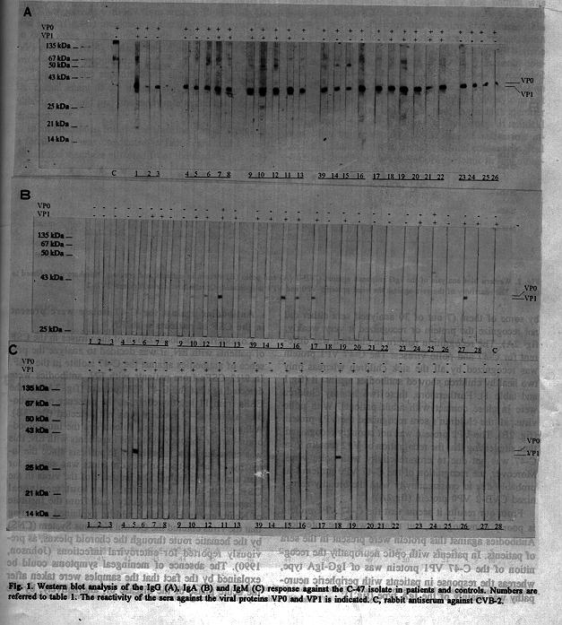
RESULTS AND DISCUSSION The C-47 virus isolate elicited a characteristic differential response in patients with EN In an attempt to find out if the immunological response against the C-47 isolate was masked by the presence in the sera of the population of antibodies against enteroviruses that have previously circulated in the country (P. Mas, personal communication) or by the response against the poliovirus vaccine, immunoblots were performed with the sera of patients and controls against C-47, CVB2 and polio viruses. As shown in figure 1A, all the sera examined had IgG against the C-47 VP0 protein because of the crossreactivity of this protein among the enteroviruses.
Table 1 Data from patients and controls included in these studies.
-------------------------------------------------------------
No.   Patients and controls Specimen Sex   Clinical    Region
      (Number of the                       diagnosis^b
      clinical history)^a
-------------------------------------------------------------
1     4478, from a blood  Serum     M   Healthy   C. Habana
      donor bank
2     4479, from a blood  Serum     F   Healthy   C. Habana
      donor bank
3     4451, from a blood  Serum     M   Healthy   C. Habana
4     1-T, from CIGB      Serum     M   Healthy   C. Habana
5     2-T, from CIGB      Serum     F   Healthy   C. Habana
6     3-T, from CIGB      Serum     M   Healthy   C. Habana
7     7-T, from CIGB      Serum     M   Healthy   C. Habana
8     11-T, from CIGB     Serum     F   Healthy   C. Habana
9     Q1082               Serum     M   ON       P. del Rio
10    Q1095               Serum     M   ON       P. del Rio
11    Q1096               Serum     F   ON       P. del Rio
12    54790               Serum     M   ON       P. del Rio
13    Q1657               Serum     M   ON       P. del Rio
14    90072               Serum     M   PN       P. del Rio
15    Q1183               Serum     M   PN       P. del Rio
16    Q1180               Serum     F   PN       P. del Rio
17    4NP                 Serum     NR  PN    Stgo. de Cuba
18    5NP                 Serum     NR  PN    Stgo. de Cuba
19    6NP                 Serum     NR  PN    Stgo. de Cuba
20    7NP                 Serum     F   PN    Stgo. de Cuba
21    8NP                 Serum     M   PN    Stgo. de Cuba
22    9NP                 Serum     F   PN    Stgo. de Cuba
23    161-N, child        Serum     M   Healthy   C. Habana
24    O-1-B-N, child      Serum     F   Healthy   C. Habana
25    N-17-N, child       Serum     M   Healthy   C. Habana
26    OHB8-N, child       Serum     M   Healthy   C. Habana
27    E-1, child          Serum     F   ON        C. Habana
28    E-2, child          Serum     F   ON        C. Habana
29    Q1207           CFS and Serum M   ON       P. del Rio
30    Q1204           CFS and Serum F   ON       P. del Rio
31    51094           CFS and Serum F   ON       P. del Rio
32    N0131           CFS and Serum F   ON       P. del Rio
33    Q1216           CFS and Serum F   ON       P. del Rio
34    Q1222           CFS and Serum M   ON       P. del Rio
35    Q1044           CFS and Serum M   ON       P. del Rio
36    Q1178           CFS and Serum F   PN       P. del Rio
37    Q184            CFS and Serum F   PN       P. del Rio
38    Q1173           CFS and Serum M   PN       P. del Rio
39    Q1174           CFS and Serum M   PN       P. del Rio
40    N0143           CFS and Serum M   ON       P. del Rio
41    FP16            CFS and Serum F   Healthy   C. Habana
42    FP18            CFS and Serum M   Healthy   C. Habana
43    FP19            CFS and Serum F   Healthy   C. Habana
44    FP20            CFS and Serum F   Healthy   C. Habana
45    FP21            CFS and Serum F   Healthy   C. Habana
46    FP22            CFS and Serum M   Healthy   C. Habana
47    FP6             CFS and Serum F   Healthy   C. Habana
48    FP8             CFS and Serum F   Healthy   C. Habana
49    FP9             CFS and Serum M   Healthy   C. Habana
50    FP11            CFS and Serum M   Healthy   C. Habana
51    FP12            CFS and Serum M   Healthy   C. Habana
52    SJ1             Serum         F   Healthy    San Juan
53    SJ2             Serum         F   Healthy    San Juan
54    SJ3             Serum         F   Healthy    San Juan
55    SJ4             Serum         F   Healthy    San Juan
56    SJ5             Serum         M   Healthy    San Juan
57    SJ6             Serum         F   Healthy    San Juan
58    SJ7             Serum         F   Healthy    San Juan
59    SJ8             Serum         F   Healthy    San Juan
60    SJ9             Serum         F   Healthy    San Juan
61    SJ10            Serum         F   Healthy    San Juan
62    SJ11            Serum         F   Healthy    San Juan
63    SJ12            Serum         M   Healthy    San Juan
64    SJ13            Serum         F   Healthy    San Juan
65    SJ14            Serum         F   Healthy    San Juan
66    SJ15            Serum         M   Healthy    San Juan
67    SJ16            Serum         F   Healthy    San Juan
68    SJ17            Serum         F   Healthy    San Juan
69    SJ18            Serum         F   Healthy    San Juan
70    SJ19            Serum         M   Healthy    San Juan
71    SJ20            Serum         F   Healthy    San Juan
72    SJ21            Serum         F   Healthy    San Juan
73    SJ22            Serum         F   Healthy    San Juan
74    SJ23            Serum         M   Healthy    San Juan
75    SJ24            Serum         M   Healthy    San Juan
76    SJ-E-1          Serum         F   PN         San Juan
77    SJ-E-2          Serum         F   PN-ON      San Juan
78    SJ-E-3          Serum         F   ON         San Juan
79    SJ-E-4          Serum         F   ON-PN      San Juan
80    SJ-E-5          Serum         M   PN         San Juan
81    SJ-E-6          Serum         M   PN         San Juan
82    C-1             Serum         M   Healthy   Candelaria
83    C-2             Serum         F   Healthy   Candelaria
84    C-3             Serum         M   Healthy   Candelaria
85    C-4             Serum         F   Healthy   Candelaria
86    C-5             Serum         F   Healthy   Candelaria
87    C-6             Serum         F   Healthy   Candelaria
88    C-7             Serum         F   Healthy   Candelaria
89    C-8             Serum         M   Healthy   Candelaria
90    C-9             Serum         F   Healthy   Candelaria
91    C-10            Serum         M   Healthy   Candelaria
92    C-11            Serum         F   Healthy   Candelaria
93    C-12            Serum         F   Healthy   Candelaria
94    C-13            Serum         M   Healthy   Candelaria
95    C-14            Serum         F   Healthy   Candelaria
96    C-15            Serum         F   Healthy   Candelaria
97    C-16            Serum         F   Healthy   Candelaria
98    C-17            Serum         F   Healthy   Candelaria
99    C-18            Serum         F   Healthy   Candelaria
100   C-19            Serum         M   Healthy   Candelaria
101   C-20            Serum         M   Healthy   Candelaria
102   C-30            Serum         M   Healthy   Candelaria
103   C-E-1           Serum         M   ON        Candelaria
104   C-E-2           Serum         F   ON        Candelaria
105   C-E-3           Serum         F   ON        Candelaria
106   C-E-4           Serum         F   ON        Candelaria
107   C-E-5           Serum         F   ON        Candelaria
108   C-E-6           Serum         F   ON        Candelaria
109   C-E-7           Serum         F   ON        Candelaria
110   C-E-8           Serum         M   ON        Candelaria
111   CI1             Serum         NR  Healthy    Caimanera
112   CI2             Serum         NR  Healthy    Caimanera
113   CI3             Serum         NR  Healthy    Caimanera
114   CI4             Serum         NR  Healthy    Caimanera
115   CI5             Serum         NR  Healthy    Caimanera
116   CI6             Serum         NR  Healthy    Caimanera
117   CI7             Serum         NR  Healthy    Caimanera
118   CI8             Serum         NR  Healthy    Caimanera
119   CI51            Serum         NR  Healthy    Caimanera
120   IM299           Serum         NR  Healthy        Imias
121   IM300           Serum         NR  Healthy        Imias
122   IM301           Serum         NR  Healthy        Imias
123   IM302           Serum         NR  Healthy        Imias
124   IM303           Serum         NR  Healthy        Imias
125   IM304           Serum         NR  Healthy        Imias
126   IM305           Serum         NR  Healthy        Imias
127   IM306           Serum         NR  Healthy        Imias
128   IM307           Serum         NR  Healthy        Imias
-------------------------------------------------------------
^aAdult individuals (> 20 years old) unless otherwise
stated. Individuals included in the focal studies are in Table
2.^bHealthy controls were obtained from blood donors, CIGB workers and surgical patients. Patients with EN were classified according to the predominant symptoms in optic neuropathy (ON) and peripheric neuropathy (PN). NR: not recorded.
Table 2 Data from individuals included in the focal studies
-------------------------------------------------------------
Focus No. Sex Age Living   Relat-  Suspected  Diag-  Observat-

                 together  ionship by         nosis  ions
                          with the interview  confirmed
                   a                b          c         d
--------------------------------------------------------------
I     1   F 36     *   Patient      *          *    ON-PN
      2   M 17     *   Son                          NORMAL
      3   F 9      *   Daughter                     NORMAL
      4   F 36         Neighbor                     NORMAL
      5   F 29         Neighbor                     NORMAL
II    6   F 40     *   Patient      *          *    PN
      7   F 71     *   Mother                       NORMAL
      8   F 14     *   Daughter                     NORMAL
      9   M 6      *   Son                          NORMAL
      10  M 47         Sexual                       NORMAL
      11  F 20         Neighbor                     NORMAL
III   12  F 30     *   Patient      *          *    PN
      13  M 30     *   Husband                      NORMAL
IV    14  F 47     *   Patient      *          *    ON-PN
V     15  F 23     *   Patient      *          *    ON-PN
      16  F NR     *   Mother                       Normal
      17  M 59     *   Father                       Normal
      18  F 26         Friend       *          *    ON-PN
      19  F 47         Coworker                     PN
VI    20  M 73     *   Patient      *          *    PN
VII   21  M 35     *   Patient      *          *    ON
      22  F 31     *   Wife                         NORMAL
VIII  23  F 24     *   Patient      *          *    PN
      24  F 46     *   Mother                       PN
      25  M 52     *   Father       *          *    PN
      26  F 27     *   Brother      *               Normal
      27  M 20         BrotherON-                   PN
      28  F 52         Neighbor     *               PN
      29  F 22         Neighbor     *               PN
IX    30  M 11    *    Patient      *          *    ON-PN
      31  F 2     *    Brother                      Normal
      32  F 30    *    Mother       *               PN
      32  M 36    *    Father       *          *    PN
X     34  F 13    *    Patient      *          *    ON-PN
      35  F 12    *    Patient      *               ON-PN
      36  F 6     *    Brother                      Normal
      37  F 32    *    Mother       *               ON-PN
      38  M 63         Father       *          *    PN
      39  M 14         Brother                      Normal
------------------------------------------------------------
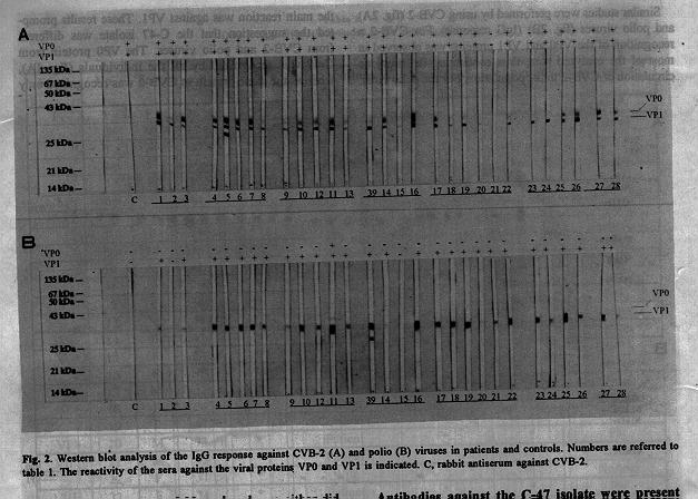
^aLiving in the same house.^bClinical symptoms of EN referred by the patient and observed during the focal study. ^cDiagnosis confirmed according to the clinical specifications of the disease. ^dPatients were classified according to the predominant symptoms in optic neuropathy (ON) and peripheric neuropathy (PN). However, the recognition of the C-47 VP1 protein was achieved only in the sera of patients or in that of people exposed to the virus (fig.1A and table 1). The IgA (fig.1B) and IgM (fig.1C) response against the C-47 isolate was also detected in the sera of adult patients and children, including two healthy children (fig.1A, C and table 1). Similar studies were performed by using CVB-2 (fig. 2A) and polio viruses (fig. 2B) (IgG response). For CVB-2 a recognition of the VP0 and VP1 proteins was observed in most of the analyzed sera, thus confirming the previous circulation of CVB-2 in the population. For the polio virus, the main reaction was against VP1. These results prompted the suggestion that the C-47 isolate was different from CVB-2 and polio viruses. The VP0 protein from C-47 was recognized by all the individuals (fig. 1A), whereas the same protein in CVB-2 was recognized only by some of them (7 out of 30 analyzed sera either did not recognize the protein or recognized it very weakly; fig .2A). The recognition pattern in children was different for C-47 and polio viruses. The C-47 VP1 protein was recognized by all the sick children whereas only two healthy children showed antibodies against it (fig.1 and table 1). Furthermore, these two healthy children were in close contact with adult patients. With polio virus, all the children's sera recognized the VP1 protein (fig. 2B). The VP2 protein was not strongly recognized, thus suggesting that the recognition of VP2 protein from C-47 was not due to crossreactivity with polio virus. Moreover, the recognition of the C-47 VP0 protein was probably due to crossreactivity with the broadly recognized CVB-2 VP0 protein (fig.2A).
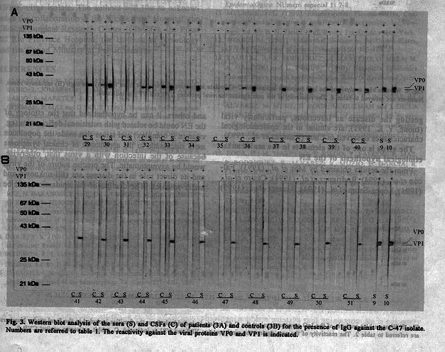
From our results it seems that the C-47 VP1 protein is poorly immunogenic, but related with the infection. Antibodies against this protein were present in the sera of patients. In patients with optic neuropathy the recognition of the C-47 VP1 protein was of IgG-IgA type, whereas the response in patients with peripheric neuropathy was mainly of the IgA type (fig.1). Antibodies against the C-47 isolate were present in the CSF of patients with EN Considering the localization of the viruses in the CSF of patients with EN, it was decided to analyze the presence of antibodies against the C-47 isolate in the CSF. In 9/12 CSFs from patients with EN, antibodies were detected against the C-47 VP0 protein (fig. 3A). In the CSFs from 11 healthy controls, the presence of antibodies against C-47 proteins was not detected (fig. 3B). Although it was impossible to confirm the intra-blood-brain-barrier antibody synthesis in patients with EN, this was considered the most likely hypothesis since the intensity of the signals in western blots was similar for sera and CSF samples. The presence of the virus in the CSF (200-300 particles/mL) without meningeal symptoms and with acellularity (data from the Institute of Neurology in Havana) could be explained assuming that the virus entered the Central Nervous System (CNS) by the hematic route through the choroid plexus, as previously reported for enteroviral infections (Johnson, 1990). The absence of meningeal symptoms could be explained by the fact that the samples were taken after the acute phase of the disease or, alternatively, by the absence of tropism of these viruses for the CNS. However, in an attempt to explain the role of the virus in the EN, this is one of the most contradictory facts.
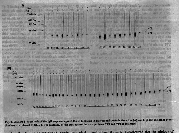
The virus circulated within the population with no correlation with the incidence of the disease A study was conducted in high and low incidence zones after the onset of the epidemic. The analysis of sera from patients with EN from areas with low (fig.4A) and high (fig.4B) incidence of the disease, showed that in both areas all the individuals recognized the C-47 VP1 protein (fig.4), indicating the circulation of the virus within the population.
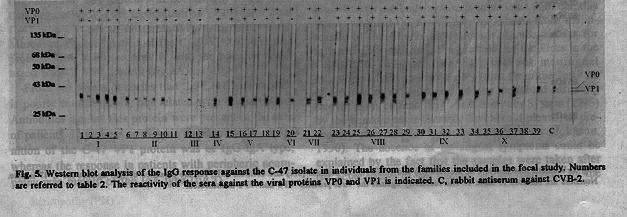
This evidence supported our initial hypothesis that the causal agent of the disease was not only the virus, although it could play the main role in the neurological damage. In an attempt to follow the transmission of the virus, a focal study was conducted in families where at least one of the members suffered from the disease (table 2). In 7 out of 8 focus studied, contacts recognized C-47 VP0, VP1 and VP2 proteins (fig.5), showing the same recognition pattern as the patients. The only focus that did not show this recognition pattern (focus III in table 2) corresponded to a patient whose diagnosis was not confirmed. Focus IV and VI (table 2) were not considered in the analysis because only one patient was included in each. These results are in accordance with those previously showing that the virus circulated within the population and no obvious pattern of transmission can be followed since, as previously stated, the disease was most probably multicausal. However, from 28 contacts included in the study, 10 (35.7%) were recorded as suffering from the disease (table 2). The fact that an obvious pattern of contagion and transmitability was not found, could be explained considering a high number of infected persons with a low frequency of clinical symptoms, as for example in the case of enterovirus infections (Melnick, 1990). Finally, several epidemiological findings found during the SMON epidemic in Japan suggesting the disease as infectious, particularly viral (Inoue, Y. K. 1991), are similar to the epidemiological characteristics observed in Cuba (Ramirez et al., 1993).
The virus is one of the elements in the multicausal etiological hypothesis of the EN From the results obtained by Mas et al., (1993); Falcon et al., (1994); Rodriguez et al., (1994); Castro et al., (1994); Lopez-Saura et al., (1994); Alvarez et al., (1995) and others, it can be hypothetized that the etiology of the EN could be related with nutritional deficiencies (eg. vitamin B complex) and stress that made the population more susceptible to the appearance of clinical symptoms because of the infection with a virus that, otherwise, would be asymptomatic. The precise nature of the virus and its direct role in the disease are still unknown and under research. The decline of the epidemic could be explained by the elimination of the predisposing element, by the elimination of susceptible persons in the population or by a combination of both factors. The vitamin administration to the population would prevent the appearance of the clinical symptoms but not spread of the virus. Thus, the seroconvertion in different groups and regions will not be related with the disease and should be essentially the same. The recombinant human interferon-alpha2b (HeberonR, Heber Biotec S.A., Havana, Cuba) therapy, that resulted useful for the treatment of the peripheral neuritis associated to the EN (Lopez-Saura et al., 1994), will act on the virus without eliminating the predisposing element. Furthermore, in some cases, people that eventually recover from the disease could relapse again with clinical symptoms. However, in a population with a good nutritional balance, the risk of the appearance of the disease should be low.
ACKNOWLEDGEMENTS We wish to thank Dr. P. Mas for providing the C-47/IPK virus isolate. The authors will like to acknowledge the collaboration of the "Frank Pais" Hospital, the Clinical-Surgical Research Center (CIMEQ), the Pinar del Rio Province Hospital, the Institute of Neurology and the Cuban Ministry of Public Health.
REFERENCES 1. ALVAREZ, M.; V. MUZIO; M. MARRERO; M. BARRO; D. ROSARIO; L. JOMARRON; A. MARTIN; C. SARIOL; M. CANDELARIO; J. MAESTRE; J. DE LA FUENTE; L. HERRERA; M. GUZMAN; P. MAS and G.KOURI (1995). Detection by Polymerase Chain Reaction of enterovirus-like sequences in patients with epidemic neuropathy in Cuba. BiotecnologIa Aplicada 12: 46-51. 2. CASTRO, F. O.; J. BERLANGA; C. ALFONSO; P. RODRIGUEZ; M. HECHEVARRIA; O. HAYES; R. PEREZ; D. PICHARDO; M. PEREZ; I. DORTA; E. LOPEZ; M. C. LOPEZ; L. HERRERA and J. DE LA FUENTE (1994). Evaluation in animal models of the neurovirulence of isolates from patients with epidemic neuropathy. BiotecnologIa Aplicada 11: 138-144. 3. FALCON, V.; J. REYES; O. ANCHETA; P. RODRIGUEZ; R. ALVAREZ; N. BARANOSKY; M. C. DE LA ROSA; P. MAS; G. PADRON; G. KOURI; L. HERRERA and J. DE LA FUENTE (1994). Study by transmission electron microscopy of virus strains isolated from the cerebrospinal fluid of patients with epidemic neuropathy. BiotecnologIa Aplicada 11: 151-159. 4. INOUE, Y. K. (1991). Inoue-Melnick virus and associationed diseases in man: Recent Advances. Prog. Med. Virol. 38: 167-179. 5. JOHNSON, R. T. (1990). Viral Infections of the Nerval System. In: Virology, Ed. by B. N. Fields et al. Raven Press, Ltd., New York. 6. LAEMMLI, U.K. (1970). Cleavage of structural proteins during assembly of the head of the bacteriophage T4. Nature 227: 680- 685. 7. LLANOS, G. C.; D. ASCHER and D. C. BROWN (1993). Epidemic neuropathy in Cuba. Epidemiological Bulletin PAHO 14: 1-4. 8. LOPEZ-SAURA, P.; F. HERNANDEZ; V. LABARTA and the Group for the Study of the Epidemic Neuropathy (1994). Interferon alpha-2b in Epidemic Neuropathy. J. of Interferon Res. 14 (Suppl.1): S120. 9. MACKINNEY, R. E.; S. L. KATZ and C. M. WILFERT (1987). Chronic enteroviral meningoencephalitis in agammaglobulinemic patients. Rev. Infect. Dis. 9: 334-354. 10. MAS, P.; M. P. RODRIGUEZ; M. G. GUZMAN; M. ALVAREZ; V. MUZIO; O. ANCHETA; A. GOYENECHEA; H. ROCA; L. MORIER; R. ALVAREZ; M. MARRERO; A. MUSACCHIO; A. CASTILLO; V. FALCON; J. L. PELEGRINO; L. LUACES; A. BALMASEDA; M. BARRO; L. RODRIGUEZ; D. GARCIA DEL BARCO; M. SOLER; B. GARCIA; M. A. RIVAS; R. LLEONART; S. RESIK; G. PADRON; L. JOMARRON; M. CANDELARIO; M. MUNE; A. MARTIN; D. ROSARIO; M. G. MORALES; J. LAFERTE; J. REYES; C. SARIOL; R. RICARDO; J. L. MAESTRE; A. RODRIGUEZ; N. BARANOSKY; M. GONZALEZ GRIEGO; P. LOPEZ; E. PENTON; M. LIMONTA; J. DE LA FUENTE; L. HERRERA; A. LLOP and G. KOURI (1993). Resultados preliminares de laboratorio virologico en estudios de casos de Neuropatia Epidemica Cubana. Boletin Epidemiologico. Numero especial 1: 7-8. 11. MELNICK, J. L. (1990). Enteroviruses: Picornaviruses, Coxsackieviruses, Echoviruses and Newer Enteroviruses. In: Virology, Ed. by B. N. Fields et al. Raven Press, Ltd., New York. 12. MOORE, M. (1982). Enteroviral Disease in the United States, 1970-1979. Journal of Infect. Diseases 146: 103-108. 13. RAMIREZ, A.; R. RODRIGUEZ; A. MARRERO; G. MESA; M. A. GALINDO and L. INIGUEZ (1993). Neropatia Epidemica Cubana: Breve resena epidemiologica. Boletin Epidemiologico Numero especial 1: 1-5. 14. RIEGO, E.; M. MARRERO; E. CALVO; R. BRINGAS; M. ALVAREZ; M. MUNE; H. ROCA; M. GUZMAN and J. DE LA FUENTE (1994). Sequence analysis of coxsackie A9 viruses isolated during 1990-1994 in Cuba from patients with meningitis, myocarditis and epidemic neuropathy. Biotecnologia Aplicada 11: 145-150. 15. ROCA, H.; M. MARRERO; E. RIEGO; V. FALCON; P. MAS; P. RODRIGUEZ; R. ALVAREZ; V. MUZIO; B. GARCIA; R. LLEONART; G. KOURI; L. HERRERA and J. DE LA FUENTE (1994). Molecular characterization of a virus associated with the outbreak of a polyneurophathy epidemic in Cuba. Miami Short Reports 4: 89. 16. RODRIGUEZ, M. P.; R. ALVAREZ; D. GARCIA DEL BARCO; R. LLEONART; V. FALCON; J. REYES; F. O. CASTRO; J. BERLANGA; A. MUSSACHIO; P. MAS; L. HERRERA and J. DE LA FUENTE (1994). Caracterizacion de las cepas virales obtenidas a partir de aislamientos de los liquidos cefalorraquideos de pacientes con cuadros de Neuropatia Epidemica. Advances in Modern Biotechnology 2: 4.
Copyright 1995 Sociedad Iberolatinamericana de Biotecnologia Aplicada a la Salud
The following images related to this document are available:Photo images[ba95051b.jpg] [ba95051c.jpg] [ba95051a.jpg] [ba95051e.jpg] [ba95051d.jpg] |
| |||||||||
