 |
| About Bioline | All Journals | Testimonials | Membership | News |
|
||||||
|
||||||
Production Of Hepatitis B Surface Antigen Provokes Morphological Changes In The Endoplasmic Reticulum Of Kluyveromyces lactis Eduardo Martinez, Viviana Falcon, Juan Morales-Grillo and Gerardo Ferbeyre Center for Genetic Engineering and Biotechnology, P.O. Box. 6162, Cubanacan, Playa, Havana City, Cuba.
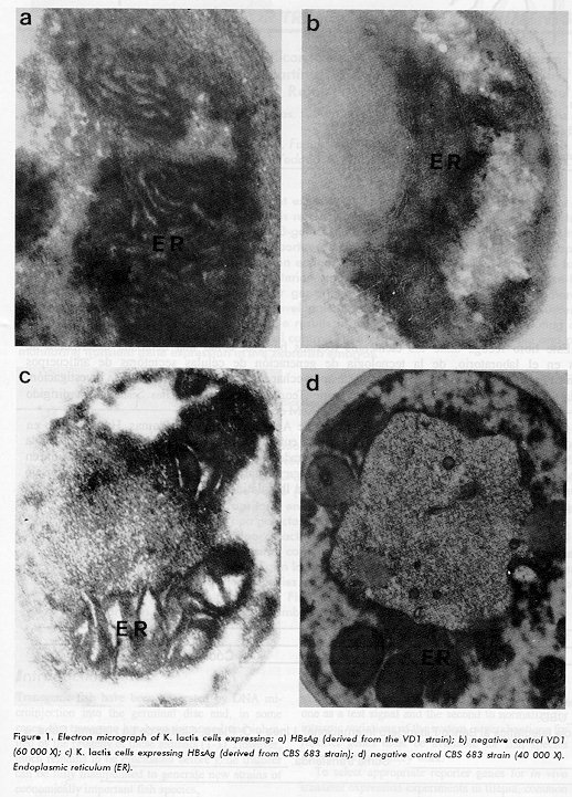
ABSTRACT Electron microscopy studies of Kluyveromyces lactis cells expressing hepatitis B surface antigen show enlargement and dilation of the endoplasmic reticulum. Key words: HBsAg, electron microscopy RESUMEN Estudios de microscopia electronica de celulas de Kluyveromyces lactis que expresan el antigeno de superficie de la hepatitis B muestran alargamiento y dilatacion del reticulo endoplasmatico. Palabras claves: HBsAg, microscopia electronica Introduction Kluyveromyces lactis has been developed as a host for the production of heterologous proteins. Several results show a high secretory capacity of this yeast, using signal sequences from several different origins (1, 2). The hepatitis B surface antigen (HBsAg) consists of a membrane structure into which a major viral protein (S), and two related minor viral proteins, middle (M) and large (L), are embedded (3). HBsAg proteins have been expressed in yeasts. The secretion of the M-protein into the culture medium was reported in Hansenula polymorpha (4), and the entrance of the L-protein into the secretory pathway of Saccharomyces cerevisiae has also been described (5). Recently, the expression of the S-protein in K. lactis was reported (6). Here it is reported how the production of the S-protein induces morphological changes in the endoplasmic reticulum (ER) of this yeast. Experimental procedure K. lactis strains expressing HBsAg (S-protein), fungal rennin and VD1 as the negative control were cultured as described by Martinez E, et al and Ferbeyre G, et al (6, 7). Samples of cells from these cultures were fixed in glutaraldehyde (2.4 %) and post-fixed in osmium tetroxide (1 %), then dehydrated in acetone and embedded in spurr resin (8). The preparation was examined under a transmission electronic microscope JEOL-JEM-2000 EX. Results and Discussion The electron microscope viewing of cells expressing HBsAg revealed enlargement and dilation of the ER (Figure 1a). This phenomenon was not observed in cells from the control strain VD1 (Figure 1b). To ensure that it is not a feature of the strain VD1, another strain derived from CBS 683 and capable of producing S-HBsAg was studied too, and similar results were obtained (Figure 1c). Figure 1d shows a CBS 683 strain (negative control). These morphological changes have not been observed when other heterologous proteins were expressed in K. lactis (7). Similar morphological alterations in the ER have been found during the expression of the L-protein in S. cerevisiae (5). However, the same results were not found for the S- protein. The morphological changes observed, could result from the accumulation of the HBsAg within the ER, suggesting that this protein enters the secretory pathway of K. lactis. In this sense, our group has found S-protein particles at low levels in the culture media and in the periplasmic space (8). The HBsAg particles are secreted in mammalian cells, but the particles are accumulated within large dilated areas of the endoplasmic reticulum and remain within these structures prior to secretion from the cells (9). It seems that, due to the complexity of the HBsAg antigen, such particles encounter a rate-limiting step somewhere between the endoplasmic reticulum and the Golgi of the secretory apparatus of K. lactis, which has also been suggested for other foreign proteins secreted from S. cerevisiae (10). This result could be used to isolate host cell mutants that show an enhanced secretion phenotype (11).
References 1. Van den Berg JA, Van der Laken KJ, Van Ooyen AJJ, Renniers TCHM, Rietveld K, Schaap A, et al. Kluyveromyces as a host for heterologous gene expression: Expression and secretion of Prochymosin. Bio/Technology 1990;8:135-139. 2. Fleer R, Yeh P, Amellal N, Maury I, Fournier A, Bacchetta F, et al. Stable multicopy vectors for high-level secretion of recombinant human serum albumin by Kluyvero- myces yeasts. Bio/Technology 1991;9:968-973. 3. Heermann KH, Goldmann U, Schwartz W, Seyffarth T, Baum- garten H, Gerlich WH. Large surface proteins of hepatitis B virus containing the pre-S sequence. J Virol 1984;52:396- 402 4. Shen SH, Bastien L, Nguyen T, Fung M, Slilaty S. Synthesis and secretion of hepatitis B middle surface antigen by the methylothophic yeast Hansenula polymorpha. Gene 1989;84:303-309. 5. Biemans R, Thines D, Rutgers T, Wilde M, Cabezon T. The large surface protein of hepatitis B virus is retained in the yeast endoplasmic Reticulum and provokes its unique enlargement. DNA and Cells Biology 1991;10:191-200. 6. Martinez E, Morales J, Aguiar J, Pineda Y, Izquierdo M, Ferbeyre G. Cloning and expression of hepatitis B Surface Antigen in the yeast Kluyveromyces lactis Biotechnology Letter 1992; 14:83-86. 7. Ferbeyre G, Martinez E, Torrens I, Aguiar J, Villareal A, Gonzalez T, et al. Secretion of recombinant fungal renin in Kluyveromyces lactis. Biotecnologia Aplicada 1991;8:335-334. Copyright 1996 Elfos Scientiae
The following images related to this document are available:Photo images[ba96105a.jpg] |
| |||||||||
