 |
| About Bioline | All Journals | Testimonials | Membership | News |
|
||||||
|
||||||
THE EFFECT OF THE EPIDERMAL GROWTH FACTOR TREATMENT SCHEDULE ON THE HEALING OF FULL-THICKNESS WOUNDS IN PIGS Jorge Berlanga,^1 Jorge Lodos,^1 Violeta Labarta,^1 Nelson Merino,^2 Tania Gonzalez,^1 Orlando Hayes,^1 Pedro Puentes,^1 Julio Mulet^1 and Pedro Lopez-Saura^1
^1 Center for Genetic Engineering and Biotechnology. PO Box 6162, Havana,
Cuba. Received in December, 1996. Accepted for publication in February, 1997.
Code Number:BA97038
Size of Files:
    Text: 34.4K
    Graphics: Line drawings (gif) -10.4K
ABSTRACT The pharmacodynamic effect of different epidermal growth factor (EGF) application frequencies on the healing of full thickness skin wound biopsies (8 mm diameter) in pigs is studied. A total of 100 wounds were created, grouped and medicated as: I, II, III treated with an EGF- containing formulation (EGF 10 ug/g plus 1 % silver sulfadiazine) every 24, 48 and 72 h, respectively. Medicated-control wounds (IV) were treated every 24 h with 1 % silver sulfadiazine, and group V was the non-medicated control. Dressings were changed every day and wounds were cleansed and treated accordingly. Wounded tissue was harvested on day 8, and used for histologic examination or computerized-assisted planimetry (10/10 wounds). Percentage of total re-epithelized area, medium radius, circularity factor, wound perimeter, epithelial linear ingrowth, dermal reconstitution, epithelial migration, and wound contraction were considered. EGF-daily treated wounds had a total re-epithelization of 80 %. It was significantly stimulated as compared to the others (p < 0.05). Similarly, group I had the largest rate of epithelial linear ingrowth and the lowest values of medium radius and wound perimeter (p < 0.05). Epithelial migration was significantly stimulated (p < 0.05) in the EGF-treated groups irrespective of the treatment frequency as compared to group IV. The lowest value in the circularity factor was detected in group I (p < 0.05). Dermis reconstitution was significantly improved in groups I and II in relation to group IV (p < 0.05), but wound contraction was not modified. We showed that EGF promotes wound repair on the basis of frequently repeated exposures to the target tissue.
Key words: EGF, re-epithelization, dermal reconstitution RESUMEN Se estudio la respuesta reparativa de lesiones de grosor total en cerdos ante variaciones en la frecuencia de aplicacion de una formulacion con factor de crecimiento epidermico (FCE, 10 ug/g) y sulfadiacina de plata 1 %. Se indujeron 100 lesiones de 8 mm que se agruparon y trataron como: I, II, III, tratados con FCE cada 24, 48 y 72 h, respectivamente. Los grupos IV y V fueron controles medicado y espontaneo. Los animales se sacrificaron al octavo dia y se colectaron las ulceras para estudio histologico o planimetrico (10/10). Se estudiaron los siguientes parametros: porciento de area total reepitelizada, radio medio, factor de circularidad, perimetro, crecimiento lineal del epitelio, reconstitucion dermica, migracion epitelial, e indice de contraccion. Las heridas del grupo I mostraron un 80 % de reepitelizacion total, la que fue significativamente superior al resto de los grupos (p < 0,05). Estas mostraron el mayor nivel de crecimiento lineal y los menores valores de radio medio y perimetro con respecto a los otros grupos (p < 0,05). Se constato un incremento significativo de la migracion en todos los grupos tratados con FCE con relacion al grupo IV, (p < 0,05). La circularidad fue significativamente menor en el grupo I con relacion a los otros grupos (p < 0,05). La reconstitucion de la dermis fue significativamente superior en los grupos I y II con respecto al IV (p < 0,05). No se demostraron diferencias en el nivel de contraccion de las heridas. Este experimento demostro que el FCE estimula la reparacion cutanea de forma frecuencia de tratamiento-dependiente.
Palabras claves: FCE, reepitelizacion, reconstitucion dermica Introduction Although wound healing is a complex biological process and its molecular and cellular regulation is not fully understood (1), data indicate that peptide growth factors and their receptors play key roles in it. Five major growth factor families seem to contribute significantly to wound healing: EGF, TGF-beta, IGF-I, PDGF and FGF (2).
The epidermal growth factor (EGF) stimulates mitosis of epidermal keratinocytes (3) and dermal fibroblasts (4), and promotes angiogenesis (5). We have shown that a topical treatment with EGF enhances the healing of full-thickness wounds at specific concentrations, using a model that involves both epidermal resurfacing and fibrovascular reconstitution (6). While no physiological role for growth factors has been definitively established in normal wound healing, it is probable that they may accelerate the healing events in healthy individuals due to earlier delivery or increased levels of growth factors that occur naturally (7).
No data are available documenting the extent, nor the effect of the local bioavailability of EGF in semisolid formulations once it is applied to an injured site. However it has been shown that prolonged/continuous exposures, or repeated applications of EGF are necessary to accelerate or improve healing. Thus, some in vitro experiments have shown that cells must be continuously exposed to EGF for 8-12 h before they start to divide (8, 9). Results from in vivo models provide additional evidence supporting the importance of prolonged and continuous exposure of EGF to stimulate wound healing (10, 11). It may be inferred that the pharmacodynamic effect of locally applied EGF is largely dependent on: the application frequency, the use of effective concentrations and appropriate formulation vehicles to ensure a sustained exposure of the peptide onto the damaged skin.
This paper shows the effect of different treatment schedules of EGF on the healing of controlled full-thickness skin wounds in pigs. Materials and Methods Animals Five male Yorkshire pigs weighing 15-17 kg were obtained from the National Swine Genetic Center (Havana, Cuba). Animals were clinically examined and adapted for about 10 days to a new management and living conditions before the experiment. Pigs were individually housed in stainless steel cages with a waste disposal system (INPUD, Cuba). Indoor environmental conditions such as, air exchange cycles (25-27/h), room temperature (18-21 C) relative humidity (40-60 %) and light cycle (12/12 h) were controlled throughout the experiment. Animals were fed 3 times per day with commercial pelleted feed (CENPALAB, Cuba) and ad libitum access to water. Wound model Animals received intramuscular diazepam (1.5 mg/kg) and atropine sulfate (0.07 mg/kg) as pre-anesthetic sedative medications. Anesthesia was induced with sodium pentobarbital (30 mg/kg) administered through a marginal ear vein.
The dorsal thoraciclumbar region of each animal was clipped, depilated and prepped with a povidine-iodine solution. The surgical area was delineated, and 20 full-thickness-8 mm diameter punch-wounds were done on each animal by blunt excision, using disposable cutaneous biotomes (Acu-Punch, Acuderm inc. USA). A total of 100 ulcer injuries were used in this experiment; 20 were assigned to each experimental group. Each group was represented in every pig with 4 wounds. Ten wounds were induced on each side of the dorsum, grouped in 5 hemilateral blocks of 2 matched wounds, symmetrically and equidistantly distributed. Each matched pair of wounds received the same treatment. Wound margins were labeled with india ink to facilitate further processing; wounds were dressed with 2 x 2 cm non-adherent material (Johnson and Johnson, New Brunswick, NJ) and covered with gauze secured with Vet-Wrap (3M, St. Paul, MN). Dressings were changed daily, and wounds were cleansed with 70 % alcohol prior to cream application. Animals were slaughtered on day 8 post-surgery by anesthesia overdose (pentobarbital 60 mg/kg), and ulcers were harvested for processing. All the procedures of this experiment were approved by the Animal Welfare Committee of the Center for Genetic Engineering and Biotechnology (Havana, Cuba). Formulation Recombinant human EGF was produced in Saccharomyces cerevisiae at the Center for Genetic Engineering and Biotechnology (Havana, Cuba). The product was obtained with more than 95 % purity (12). Hebermin (HeberBiotec S.A., Cuba) was formulated with 10 ug of recombinant human EGF/g and 1 % silver sulfadiazine (SSD) in a hydrophilic vehicle. The biological activity of EGF was assessed by a receptor-binding assay (13), and its concentration in the cream was determined by an ELISA system (14). Experimental groups Four treatment schedules were tested: (I) Hebermin every 24 h; (II) Hebermin every 48 h; (III) Hebermin every 72 h; (IV) 1 % silver sulfadiazine every 24 h and group V was a control for spontaneous healing, treated only with daily local desinfection. The corresponding first treatments were applied immediately after wounding and followed until the 7^th day post-injury. Sample harvesting and healing analysis
Ulcers and a surrounding area of intact skin (> 5 mm) were collected including the panniculus carnosus in block to stabilize wound tissue, and maintain the normal geometry of the samples.
Twenty wounds were studied in each experimental group. Ten samples were used for histologic analysis, and the other 10 were used for computer- assisted planimetry measurements.
Samples were fixed in 10 % buffered formalin for 72 h, and bisected along the diameter for histologic evaluations. The halves were embedded in paraffin, and 5 um-thick sections were cut. Then, they were deparaffinized, cleared, rehydrated, stained with hematoxylin/eosin, Masson's trichrome and van Giesson's dyes.
Morphometric assessment was conducted on mid-wound histologic sections. To ensure consistency and non-biased evaluations, slides were coded by an external observer, and evaluated blindly. Tissue specimen-images were captured with a CCD color video camera (Sony DXC-107) mounted on a microscope and displayed on a high-resolution monitor. The camera was coupled to an IBM-compatible computer through a framegrabber card (Video Blaster SE-100). An image processing package (Digipat System, EICISOFT. 428, 24 St. Vedado, Havana, Cuba) was used for all measurements.
For the computer planimetry study, epithelial layers were harvested and processed as referred (15). Briefly, excised whole skin-blocks were incubated in a 0.5 M sodium bromide solution for 24 h at 37 C. Epidermis was carefully detached from the dermal layer, placed and dried onto a paper sheet. Then each image was scanned with a Hewlett-Packard Scanjet II. Scanned images were processed using the Digipat image processing package and the non-epithelized area was determined. The following calculations were further made:
b) Wound medium radius (expressed in mm).
c) Circularity factor. It was defined as:
             Min{Rm'sigma(Rm)}
    C = 1 - -------------------
                   Rm
where Rm is the medium radius, sigma Rm is the median
quadratic dispersion of Rm and the Min function returns the
minimal of its arguments. C: varies from 1 (perfect circle) to 0.
d) Wound perimeter (expressed in mm).
e) Epithelial linear ingrowth. This parameter was calculated by dividing the healed area (in mm^2) of each wound by the average value of the perimeter per experimental group. It is expressed in millimeters and represents the linear growth of the epithelial layer onto the repaired area (16). Histologic determinations
a) Dermal reconstitution was qualitatively graded as (6):
1: some collagen fibrils should be present, resembling a primitive degree of organization, focally distributed, without horizontal alignment along the wound bed. Neovascularization should be noted as an immature event, with a limited number of primitive vessels lacking blood. A moderate inflammatory infiltrate is observed, as well as focal positivity to van Giesson's stain.
2: a general image of extracellular matrix reconstitution, with mature and organized granulation tissue containing horizontally deposited collagen fibrils. Affinity to van Giesson's staining is observed with varied intensity across the area. Some active, dilated and blood-containing vessels should be noted. Swelling is minimal.
3: complete extracellular matrix reconstitution, with mature and organized collagen fibrils, horizontally deposited in the neodermis. Even affinity to van Giesson's staining should appear. Some vessels might contain blood or might be found collapsed by surrounding collagen fibrils.
b) Epithelial migration rate: was determined by adding the lengths of both migrating epithelial tongues from the wound edges onto the neodermis of each hemisection. This is a linear parameter expressed in millimeters.
c) Wound contraction: was assessed by subtracting the distance in millimeters between the unwounded margins at a mid-dermal level to the original wound diameter (16).
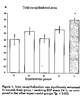
All measurements are expressed as means (standard errors). Total re- epithelized area, linear ingrowth, wound contraction, epithelial migration rate, wound perimeter, medium radius, circularity factor and dermal reconstitution were compared using the Mann-Whitney U test. A significance value of p < 0.05 was used. All tests were 2-tailed. Results Percentage of total wound re-epithelization Wounds from group I had a total re-epithelized area of 80 % at day 8 post- surgery. This was significantly larger than the percent of re- epithelization calculated for the other experimental groups (p < 0.05). Thus it seemed that the EGF-daily treatment had a greater stimulating effect on the healing.
Group II (EGF every 48 h) did not show a significant enhancement of the total re-epithelization in relation to groups IV (p = 0.052) and V (p = 0.061). Total re-epithelization of group III wounds was not improved with respect to both treated (p = 0.07) and untreated (p = 0.068) control groups (Figure 1).
The rate of linear ingrowth in group I wounds had the largest value of linearized epidermal healing in relation to the other experimental groups (p < 0.05). The other wound-geometry parameters associated to the rate of epithelial response, such as wound perimeter and medium radius, demonstrated that epithelial healing was significantly stimulated (p < 0.05) by a daily-EGF treatment program (Table 1). Table 1. Values are the mean and standard error of each epithelial- related parameter. A significant epithelial response is shown by wounds treated with EGF every 24 h. The epithelial migration rate was significantly hindered in wounds treated with 1 % silver sulfadiazine every 24 h. * (p < 0.05) Mann-Whitney U test.
---------------------------------------------------------------------------
Experimental Linear    Epithelial  Circularity   Medium        Wound
groups       ingrowth  migration   factor        radius        perimeter
             (mm)      (mm)        (arbirtary    (mm)          (mm)
                                     units)
---------------------------------------------------------------------------
  I      2.67 ñ 0.3*  6.74+/-2.2   71.2+/-11.6*  1.69+/-0.6*   14.19+/-6.3*
 II      1.69+/-0.3   6.56+/-2.6   77.1+/-7.6    2.21+/-0.6    18.70+/-6.7
III      1.30+/-0.3   6.43+/-3.6   86.8+/-4.2    2.59+/-0.4    19.53+/-3.0
 IV      1.80+/-0.4   4.03+/-2.5*  78.1+/-9.8    2.10+/-0.8    17.16+/-7.2
  V      1.22+/-0.5   6.66+/-2.5   83.0+/-7.3    2.66+/-0.6    19.18+/-4.8
---------------------------------------------------------------------------
Epithelial migration
Histomorphometric evaluation of the re-epithelized space along the hemisection diameter of the wound indicates the migratory response of the epidermal layer. An enhancement of epithelial migration onto the wound- hemisection diameters was observed in all groups treated with EGF, when they were compared to group IV (SSD). None of the EGF-treated groups differed significantly from group V of spontaneous healing (Table 1).
Comparison of data from group IV reveals that the 1 % silver sulfadiazine treatment hindered epithelial migration. This group showed the lowest mean value of epithelial migration, which was significantly lower than group V of spontaneous evolution. Circularity factor As shown in Table 1, the lowest mean value in the circularity factor was found in group I. This was significantly lower than the values calculated for the other groups (p < 0.05). Dermal reconstitution Changes in the macroscopic features of wounds from group I were observed at day 3 after injury. Wounds showed a visible reddening and a more abundant leakage, possibly associated with neovascularization. An unexpected finding was the evolution of the wounds treated with 1 % silver sulfadiazine. The margins of these wounds were swollen from day 2 to day 6 in most of the cases.
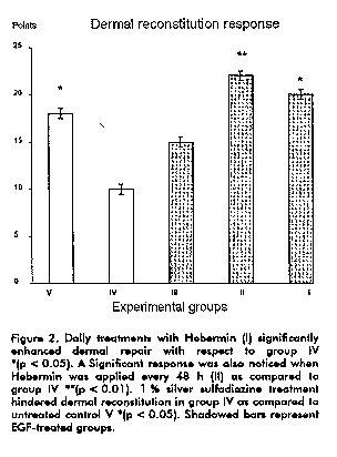
Histologic evaluation of the wounds showed that dermal reconstitution was importantly stimulated in groups receiving the most frequent exposures to EGF. A negative effect was found in group IV treated with silver sulfadiazine alone as compared to group V (spontaneous healing).
Group I showed a significantly more pronounced reconstitution of the dermal layer as compared to group IV (p < 0.05). Wounds exposed to daily EGF treatments showed a mature or transitional granulation tissue, with horizontally aligned collagen fibrils in a mesh woven fashion. Fibrils showed affinity to van Giesson and Masson s trichrome stains. Most of the wounds studied in this group had abundant active vessels. Inflammatory infiltrate was moderate and limited to the lower dermis and it was characterized by the presence of mononuclear cells. Wounds exposed to EGF every other day (II) also had an important enhancement of dermis reconstitution as compared to group IV (treated with 1 % silver sulfadiazine alone, p < 0.05). Similar histologic findings to those described in group I were observed in wounds derived from group II. Briefly, thin collagen fibrils appeared densely packed in a woven fashion, horizontally and with parallel orientation with respect to the epidermis.
A modest, not statistically significant effect, was found in wounds treated with EGF every 72 h, relative to group IV. A qualitatively superior response and a higher score value was observed in relation to group IV wounds (Figure 2).
Treatment with 1 % silver sulfadiazine did not stimulate granulation tissue organization or maturation. The slides from this group revealed a poor deposition of collagen material; the collagen fibrils detected appeared short, thickened and unaligned, usually with a vertical orientation pattern. These wounds were characterized by an intense inflammatory infiltrate of mononuclear cells involving the whole scar tissue. Furthermore, a limited neovascularization was observed in these wounds. The general histological appearance of the matrix of the dermal area in such wounds reminded the typical image of "productive or substrate phase". Wound contraction The rate of wound contraction was also studied in this experiment as this event is largely dependent upon the presence of infiltrating fibroblastic- like cells into the neodermis. In this parameter, there was no statistical differences among the experimental groups.
Discussion Although a definitive physiological role of endogenous EGF in animals remains unknown (17), several experimental models have demonstrated that treatments with exogenous EGF, may stimulate wound healing and tissue repair. Wound models designed to assess the role of EGF exogenous treatments in healing, provided evidences on the critical requirements to be met in order to achieve a significant EGF-pharmacodynamic performance, and thus, a clinically measurable tissue response (18).
This experiment shows that wounded tissue response is at least partly dependent on the frequency to which the injured site is exposed to topical EGF administrations. Our results agree with previous data indicating that the sustained release of appropriate concentrations of EGF is necessary to stimulate wound healing (19). Repeated topical treatments of partial- thickness burns with EGF creams accelerated epidermal regeneration (20). Similarly, frequent applications of EGF induced hyperplastic regeneration of tympanic membrane perforations and increased tensile strength of corneal incisions in cats (11).
According to this experiment, the two most important events of the healing of full-thickness wounds, dermal reconstitution and epidermal regeneration, respond to EGF in an exposure frequency-dependent fashion. Thus, the EGF- daily treatment promoted a significant stimulation of total wound healing, which was associated to a significant enhancement in the rate of epithelial-linear resurfacing of the damaged area. Accordingly, these wounds, after receiving seven consecutive treatments, exhibited significant reductions of the inner perimeter, as well as of the medium radius.
The fact that only the wounds exposed to daily treatments with EGF were significantly healed, leads us to infer that wound healing is largely dependent on cellular proliferation, a late event triggered after the initial 48-72 h post-wounding (21). This assumption is supported by observations of Rehinwald and Green (22), which documented the recruitment of a higher percentage of cells to leave the resting state (G0), and enter and remain in the mitotic cycle when challenged with EGF. According to Brown (21) this cellular proliferative recruitment is only observed in vivo if the wounded tissue is continuously or repeatedly exposed to EGF. In our experiment these postulates couple with a daily regime of EGF treatments. Thus, the more frequent the exposures to EGF, the larger the mitotic stimulation of residual epidermal cells.
Epithelial migration is a critical event in re-epithelization (23). We observed that EGF was able to stimulate epithelial migration, even in wounds treated every 72 h, as compared to wounds treated every day with 1 % SSD. Although no statistical difference was noticed between EGF-treated groups and the rate of epithelial migration in the spontaneous control; groups receiving EGF showed an epithelial migratory response that exceeded 50 % of the initial wound diameter (8 mm).
The ability of EGF to promote growth-independent keratinocyte locomotion contributes to the mechanism by which topically applied EGF can promote cutaneous wound healing (24). It has been recently observed in in vitro experiments that connective tissue components act synergistically with EGF to direct keratinocyte migration. Collagen was shown to be a preferential migration-substrate, inasmuch as EGF enhances the expression of integrin, a well known collagen receptor (24). These results suggest that the deposition of organized collagen fibrils onto the upper neodermis is a key element to support epithelial cell migration, and therefore wound healing.
The assessment of wound geometry variables such as the wound circularity factor was also useful to evaluate the effect of EGF on healing. As re- epithelization does not necessarily take place symmetrically around the wound contour, our hypothesis was that wounds exposed to EGF, with a larger re-epithelized area, would show a substantial reduction in their circularity factor.
Indeed, wounds from group I revealed a significantly reduced value in the circularity factor. Variations in the circularity factor seemed a characteristic feature of the geometry of re-epithelization in circular full-thickness wounds, while its intensity is dependent on the extent of the epithelial growth.
Dermal matrix reconstitution was significantly stimulated by EGF every 24 and 48 h when compared to treatment with 1 % SSD alone. In addition, the positive impact of EGF, even when it was infrequently applied, was qualitatively reflected on the intensity of dermis repair in wounds derived from group III as compared to 1 % SSD-treated wounds. Although the most frequently EGF-treated wounds were not statistically different to untreated control wounds, the latter were below 20 points in the score system, which was supported by coarse qualitative differences in collagen bundle deposition and organization.
Wounds receiving the 1 % silver sulfadiazine treatment showed a torpid evolution, and exhibited a poorly organized granulation tissue in relation to untreated wounds. An opportunistic contamination of these wounds was ruled out as judged by the microscopic findings, which was supported by the well known anti-microbial effect of silver sulfadiazine. Since the 1 % silver sulfadiazine cream was the excipient used for EGF formulations, all comparisons throughout the experiment were done with treatment IV.
Even when the two most frequent EGF treatments enhanced dermal repair, this effect was statistically irrelevant in relation to the response found in untreated control wounds. It is difficult, at the moment, to hypothesize in this respect since to our knowledge, the combination of rh-EGF with 1 % silver sulfadiazine has only been assayed in epidermal regeneration of partial-thickness wounds (20, 23) and chronic ulcers (25), which are quiet different experimental systems to ours.
The overall analysis of our data indicates that EGF induced fibroblast chemotaxis and stimulated collagen deposit. However it is likely that the combination with 1 % silver sulfadiazine or the formulation vehicle itself, somehow eclipsed the EGF biological activity.
We had previously demonstrated (6) that daily EGF-topical applications, increase the rate of wound contraction in a similar full-thickness wound model in rats. However in this experiment, contraction was not significantly modified by EGF. Taking into account that full-thickness wounds heal predominantly by contracture, with epidermal regeneration playing a minor role (26); we may suggest that the improvement in wounds (healed area, linear ingrowth, and circularity reduction), were mediated by EGF stimulation to epithelial regrowth.
In this experiment we have shown that EGF treatment plays an important role in promoting wound re-epithelization on the basis of frequent or repeated exposures to the target tissue. Further studies should be conducted to obtain alternative topic formulations that ensure a prolonged EGF biological stability and a continuous release of the peptide without negative interactions with its native biological properties. References 1. Curtsinger L, Pietsch J, Brown G, von Fraunhofer A, Ackerman D, Polk H, Schultz G. Reversal of adriamicin-impaired wound healing by transforming growth factor-beta. Surgery, Gynecology and Obstetrics 1989;168:517-522. 2. Bennett N, Schultz G. Growth factors and wound healing: biochemical properties of growth factors and their receptors. The American Journal of Surgery 1993;165:728-737. 3. Rheinwald JG, Green H. Epidermal growth factor and the multiplication of human epidermal keratinocytes. Nature 1977;265:421-424. 4. Lembach KJ. Induction of human fibroblasts proliferation by epidermal growth factor (EGF): Enhancement by an EGF-binding arginine esterase and by ascorbate. Proc Natl Acad Sci USA 1976;73:183-187. 5. Schreiber AB, Winkler ME, Derynck R. Transforming growth factor - alpha: A more angiogenic mediator than epidermal growth factor. Science 1986;232:1250-1253. 6. Berlanga J, Moreira E, Perez L, Boix E, Gonzalez T, Lopez-Saura P. Wound healing promotion in rats treated with EGF is dose dependent. Biotecnologia Aplicada 1996;13:181-185. 7. Bennett N, Schultz G. Growth factors and wound healing: Part II. Role in normal and chronic wound healing. The American Journal of Surgery 1993;166: 74-81. 8. Aharonov A, Pruss RM, Herschman HR. Epidermal growth factor: Relationship between receptor regulation and mitogenesis in 3T3 cells. J Biol Chem 1978;253:3970-3977. 9. Haigler HT, Carpenter G. Production and characterization of antibodies blocking epidermal growth factor: Receptor interactions. Biochim Biophys Acta 1980;598:314-325. 10. Brightwell JR, Riddle SL, Eiferman RA et al. Biosynthetic human EGF accelerates healing of neodecadron-treated primate corneas. Invest Ophtalmol Vis Sci 1985;26:105-110.
11. Woost PG, Brightwell JR, Eiferman RA, Schultz GS. Effect of growth factors with dexamethasone on healing of rabbit corneal stromal incisions. Exp Eye Res 1985;40:47-60. 12. Cinza AM, Quintana M, Lombardero J, Poutou R, Perez E, Perez Tosar L et al. A batch process for production of human epidermal growth factor in yeast. Product characterization. Biotecnologia Aplicada 1991;8:166-173. 13. Macias A, Perez R, Lage A. Estudios sobre el factor de crecimiento epidermico. II. Desarrollo de un radioreceptor analisis para la determinacion de cantidades picomolares. Interferon y Biotecnologia 1985;2:115-127. 14. Vazquez J, Freyre M, Duarte C, Ferra E, Lopez I, Perez E, Gavilondo J. Radio and enzyme immunoassay for human epidermal growth factor with mouse monoclonal antibodies. Biotecnologia Aplicada 1990;7:42-51. 15. Tredget E. Personal communication 1995. Plastic Surgery Research Laboratory, Department of Surgery, University of Alberta, Edmonton, Canada. 16. Mellin T, Casheen D, Ronan JJ, Murphy B, Di Salvo J, Thomas K. Acidic fibroblast growth factor accelerates dermal wound healing in diabetic mice. The Journal of Investigative Dermatology 1995;104:850-855. 17. Calabro A, Milani S, Paladini I, Orsini B, Salvadori G, Surrenti C. Role of epidermal growth factor in peptic ulcer healing. Review Article. Digestive Diseases and Sciences 1995;40:2497-2504. 18. Schultz G, Rotatori D, Clark W. EGF and TGF-alpha in wound healing and repair. J Cell Biochem 1991;45:346-352. 19. Buckley A, Davidson JM, Kamerath CD et al. Sustained release of epidermal growth factor accelerates wound repair. Proc Natl Acad Sci USA 1985;82:7340-7344. 20. Brown GL, Curtsinger LJ, Brightwell JR et al. Enhancement of epidermal regeneration by epidermal growth factor. J Exp Med 1986;163:1319- 1324. 21. Stenn KS, Depalma L. In: The molecular and cellular biology of wound repair 1988. (Clark REF and Hensen PM, eds), pp. 321-325, Plenum Press, New York. 22. Rheiwald JG, Green H. Epidermal growth factor and the multiplication of cultured human epidermal keratinocytes. Nature (London) 1977;265:421- 423. 23. Brown GL, Nanney LB, Griffen J, Cramer AB, Yancey JM, Curtsinger LJ et al. Enhancement of wound healing by topical treatment with epidermal growth factor. N Engl J Med 1989;321:76-79. 24. Chen JD, Kim JP, Zhang K, Sarret Y et al. Epidermal growth factor (EGF) promotes human keratinocyte locomotion on collagen by increasing the alpha2 integrin subunit. Exp Cell Res 1993; 209:1-8. 25. Brown GL, Curtsinger L, Jurkiewicz MJ, Nahai F, Schultz G. Stimulation of healing of chronic wounds by epidermal growth factor. Plast Reconstr Surg 1991;88:189-194. 26. Gross J, Farinelli W, Sadow P, Anderson R, Bruns R. On the mechanism of skin wound "contraction": A granulation tissue "knockout" with a normal phenotype. Proc Natl Acad Sci USA 1995;92:5982-5986. Copyright 1997 Elfos Scientiae The following images related to this document are available:Line drawing images[ba97038a.gif] [ba97038b.gif] |
| |||||||||
