 |
| About Bioline | All Journals | Testimonials | Membership | News |
|
||||||
|
||||||
Biotecnología Aplicada 1998;15:83-87 EPIDERMAL GROWTH FACTOR STIMULATED RE-EPITHELIALIZATION IN PIGS. THE POSSIBLE ROLE OF ACUTE-WOUND PROTEASES Jorge Berlanga,1 Jorge Lodos,1 Osvaldo Reyes,1 Juan F Infante,2 Enrique Caballero1 and Pedro López-Saura1 1Center for Genetic Engineering and Biotechnology. PO Box 6162, Havana 10600, Cuba. E-mail: jorge.berlanga@cigb.edu.cu Received in June, 1997. Accepted for publication September, 1997.
Code Number: BA98012
ABSTRACT The epithelial response to a daily topical treatment regime with three different epidermal growth factor (EGF) concentrations was evaluated in five pigs. Animals were kept under controlled environmental conditions, and 30 partial-thickness wounds were inflicted on the dorsum. These were assigned to treatments with 50, 10 and 2 µg of human reacombinant EGF (hr-EGF/g). Control wounds received cream vehicle or were untreated. Wounds were cleansed, inspected and treated every day accordingly. The wound-derived exudate was collected from the untreated wounds during the first 24 h post-injury, and filter-sterilized to qualitatively estimate the presence of protease activity on a fluorescent synthetic peptide by electrophoresis. After 7 EGF treatments, pigs were sacrificed on day 8 and the wounds were harvested and processed to determine the percentage of total re-epithelialization, average radius, and wound perimeter, as the linear ingrowth. The computerized morphometric study proved that only the largest dose significantly stimulated re-epithelialization, whereas the other doses were ineffective. It was also shown that the acute phase-wounds exudate exhibits proteolytic activity which completely degraded the fluorescent substrate. This proteolytic activity was similarly inhibited by 0.5 and 5 µg of a Kunitz-type protease inhibitor. Our results lead us to speculate that the bioavailability of topically applied EGF to accelerate wound healing may be reduced by proteases derived from local acute wounds. Key words: wound healing, EGF, re-epithelialization RESUMEN Se estudió el efecto de la aplicación tópica de tres concentraciones de factor de crecimiento epidérmico (FCE) sobre la respuesta epitelial en cinco cerdos. Los animales se mantuvieron en condiciones controladas, induciéndose 30 heridas de grosor parcial en la región del dorso. Un número similar de heridas en cada animal fue asignado a tratamiento con 50, 10 o 2 µg de FCE humano recombinante (FCE-hr/g) de vehículo. Los controles consistieron en heridas tratadas con el vehículo o libres de tratamiento. Las heridas fueron inspeccionadas, higienizadas, y medicadas diariamente según correspondió, sin practicar escarectomía. El exudado de las heridas control no tratadas fue cuidadosamente colectado durante las primeras 24 h post-lesión y esterilizado para determinar la presencia de proteasas sobre un sustrato fluorescente mediante ensayo cualitativo. Luego de siete tratamientos los animales se sacrificaron, y se colectaron las heridas para determinación morfométrica de: porciento del área epitelizada, radio medio, perímetro y crecimiento lineal. Sorpresivamente se constató que sólo la dosis de 50 µg estimuló significativamente la regeneración epitelial, en tanto que las otras resultaron inefectivas. Paralelamente observamos la presencia de actividad de proteinasas en el exudado estéril de heridas en fase aguda, la que fue inhibida por 0,5 y 5 µg del inhibidor ensayado. Estos resultados sugieren indirectamente que el FCE aplicado sobre lesiones no contaminadas en fase aguda puede ser potencialmente degradado, lo que puede limitar su potencia biológica, biodisponibilidad y efectividad clínica. Palabras claves: cicatrización, FCE, reepitelización Introduction Re-epithelialization is an important and critical event by which the wound is resurfaced by epithelial cells, involving both cellular locomotion and proliferation (1). Several studies using partial thickness-skin injuries in animals and humans demonstrated that the spontaneous healing rate is not optimum, since the addition of exogenous growth factors like the epidermal growth factor (EGF) accelerated healing (reviewed in 2, 3). A continuous and prolonged release of EGF on the wound is required to achieve a consistent pharmacodynamic response to treatment. Its bioavailability is thus optimized within a proper therapeutic window (4) by the control of the cytokine concentration and its application on the healing site (5). Brown and coworkers (6) were the first to demonstrate that EGF at 10 µg/g formulated as a cream with 1 % silver sulfadiazine, stimulated re-epithelialization of partial-thickness skin wounds in pigs. Other experiments also involved the use of EGF formulated with 1 % silver sulfadiazine in the healing process of partial and full-thickness skin wounds (7-9). We assessed the effect of three different EGF concentrations (10, 5, and 2 µg/g) in a semisolid hydrophilic cream vehicle devoid of silver sulfadiazine on the healing of a full-thickness skin wound model in rats (8), and demonstrated that healing was stimulated at 10 and 5 µg/g. Nevertheless a more pronounced effect was found with the largest EGF dose level in relation to re-epithelialization. The experiment suggested the existence of different sensitivities to EGF concentrations between the dermis and the epidermis. Using a similar formulation we recently observed that EGF at 10 µg/g stimulated the healing of full-thickness wounds in pigs on the basis of frequent treatment applications (manuscript in preparation). A topical EGF formulation in a hydrophilic vehicle devoid of silver sulfadiazine has not been apparently developed, and to our knowledge it has never been experimentally or clinically evaluated apart from our own experience. This experiment was therefore undertaken to evaluate the epithelial response to three different EGF concentrations in a simple hydrophilic formulation. The findings referring that re-epithelialization was stimulated only when EGF was formulated at 50 µg/g, and that wound exudates exhibit proteolytic activity on a synthetic substrate, led us to suggest that local EGF bioavailability may be potentially reduced by wound-derived proteases. Materials and Methods Animals Five Yorkshire gilts with body weight between 16-18 kg were obtained from the National Swine Genetic Center (Havana, Cuba). Animals were clinically examined, individually caged, and adapted to the technical staff and manipulation for 10 days before the experiment. Pigs were housed in stainless steel cages provided with a mechanical restriction system and waste disposal plate underneath (INPUD, Cuba). Indoor environmental conditions such as air exchange cycles (25-27/h), room temperature (18-21 ºC), relative humidity (40-60 %), and light cycles (12/12 h) were controlled during the experiment. Commercial pelleted swine feed (CENPALAB, Cuba) was offered three times a day and access to water was unrestricted. Wound model Animals received intramuscular administration of diazepam (1.5 mg/kg) and atropine sulfate (0.07 mg/kg) as preanesthetic sedative medication. Anesthesia was induced with intravenous sodium pentobarbital (30 mg/kg) through the marginal vein of the ear. The dorsum of each animal was clipped, depilated and prepared with povidine-iodine solution. The surgical area was delineated and 30 partial-thickness wounds of 6 mm of diameter were induced on each animal. Wounds were created by a split circular incision involving epidermis and the upper dermis with disposable cutaneous biotomes (Acu-Punch, Acuderm inc. USA). The incised fragment was clamped with forceps and carefully resected with microsurgical scissors. The reproducibility of the model was confirmed by histological assessment of the excised fragments. Thirty wounds were assigned to each experimental group, and each group was represented in every pig with 6 wounds. Fifteen wounds were induced on each side of the dorsum, grouped in five hemilateral blocks of 3 matched pairs, symmetrically distributed at equal distances. Wounds were assigned to the following groups: treated with (I) 50, (II) 10 and, (III) 2 µg of human recombinant EGF (hr-EGF/g) of hydrophilic vehicle. Group IV was treated with the hydrophilic vehicle alone and group V was used as a spontaneous evolution control. After brief hemostasia, wounds received EGF cream or placebo applications daily for 7 days. Wounds were dressed with 2 x 2 cm nonadherent dressings (Johnson and Johnson, New Brunswick, NJ), and covered with gauze secured with Vet-Wrap (3M, St. Paul, MN). Dressings were changed daily for 4 days. All the wounds were cleansed with 70 % alcohol and either medicated or not according to treatment. From day 4 onward wounds were not dressed and the scabs were not disturbed. Animals were sacrificed on day 8 post-surgery by anesthesia overdose, and ulcers were harvested for processing. All the procedures of this experiment were in accordance to the guidelines of the Animal Welfare Committee of the Animal Facility at the Center for Genetic Engineering and Biotechnology. Formulation Recombinant human EGF was produced in Saccharomyces cerevisiae at the Center for Genetic Engineering and Biotechnology, Havana. It was obtained with more than 95 % purity (10). The biological activity of EGF was assessed by a receptor-binding assay (11), and its concentration was determined by an ELISA system (12). According to this analysis, the actual concentration of EGF in each formulation was 53.4, 12.3, and 2.5 µg/g. The polypeptide was formulated in a hydrophilic vehicle. Sample harvesting and healing analysis Ulcer area and a surrounding margin of intact skin (> 5 mm) were collected by a full-thickness excision including the panniculus carnosus in block to stabilize wound tissue, and to maintain the normal geometry of the samples. Planimetry measurements For the computerized planimetry study, epithelial layers were harvested and processed as previously reported (13). Wounds were incubated in 0.5 M NaBr, 0.025 % trypsin for 24 h at 37 ºC. The epidermis was carefully detached from the dermal layer and attached onto a paper sheet. Epithelial images from each group were scanned with a Hewlett-Packard Scanjet II. Scanned images were processed using the Digipat (Digipat System, EICISOFT, Havana) software package, and the non-epithelized area was measured. The following parameters were further calculated: (a) percentage of total re-epithelized area, (b) average wound radius, (c) wound perimeter, and (d) epithelial linear ingrowth. This parameter was calculated by dividing the healed area (in mm2) of each wound by the average value of the perimeter per experimental group. It is expressed in millimeters and represents the linear growth of the epithelial layer onto the wounded area (14). Qualitative proteases assay Wound fluid (20-40 µL) was collected from the spontaneous evolution group on the first and second days post-surgery during dressing changes. The harvested samples were pooled, and its protein concentration determined as 23 mg/mL by the Lowry method (15). The material was diluted 1:1 in physiological saline solution, and sterile filtered through a 0.22 µm membrane. Five microliters aliquots were stored at -70 ºC. The proteolytic activity of the wound fluid was qualitatively assessed using the fluorescent-synthetic peptide method (16). Briefly, 3 µg of the fluorescent-peptide Ac-K(-flu) PLSRTLSVAAK-NH2 (17) were incubated in 3 µL of the diluted fluid for either 12 or 24 h at room temperature. A parallel assay was conducted under the above experimental conditions including two concentrations (0.5 and 5 µg) of the ShPI. It is a Kunitz type protease inhibitor isolated from a sea anemone S. helinathus. This protease inhibitor proved to be active against serine, cysteine and aspartic proteases (18). Aliquots of the wound fluid were preincubated with ShPI for 30 min at room temperature. Afterwards 3 µg of the synthetic substrate were added and the mixture incubated for 12 h. The dose-inhibitory activity of the protease inhibitor was checked against 3 µg of trypsin (Sigma, St. Louis, USA). The analysis was performed by 0.8 % agarose gel electrophoresis, at 100 V for 5 min in Tris-HCL buffer pH 7.2. The electrophoretic bands were detected under UV light at 265 nm wave length. Gel images were captured by a CCD color video camera (Sony DXC-107, Japan) coupled to an IBM-compatible computer through a framegrabber card (Video Blaster SE-100). Data processing All measurements are expressed as mean SD. Re-epithelialization and related data were compared using the non-parametric Mann-Whitney U test. All tests were 2-tailed and a significance of p < 0.05 was established. Results Epithelial resurfacing In general all the wounds exhibited similar morphologic features and no complications were observed throughout the study. Most of the wounds leaked a clean plasma-like exudate, which was more abundant on the first day post-injury. The computerized-assisted morphometric measurements demonstrated that total wound epithelial resurfacing was only significantly enhanced by the treatment with the largest EGF concentration. The wounds treated with EGF at 50 µg/g exhibited a significant reduction (p 0.05) of the non-epithelized area at the injured zone. Accordingly, these wounds had a significantly larger re-epithelialization rate (p 0.05) and thus the largest percent of total re-epithelized area (Figure 1). Figure 1. A significant response was only induced by EGF at 50 µg/g. The other doses were ineffective in stimulating wound re-epithelization. *p < 0.05 Mann-Whitney U test. Medium radius and perimeter values were significantly reduced (p < 0.05) in the wounds treated with 50 µg/g of EGF (Table 1). Table 1. Wound-geometry associated parameters. A significant reduction in perimeter and medium radius values was detected in wounds treated with EGF-50 µg/g. Both parameters are expressed in millimeters.
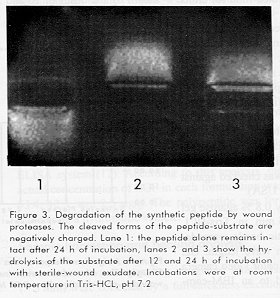
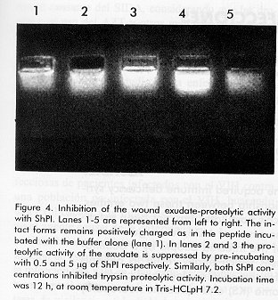
*p < 0.05 Mann-Whitney U test. Similarly, the largest EGF dose consistently stimulated the epithelial linear ingrowth rate. This group of wounds exhibited a significantly larger epithelial response (p < 0.05) when compared either to the other EGF doses or to control groups. However no evidence of a pharmacodynamic effect was shown by the doses of 10 and 2 µg of EGF used in this experiment (Figure 2). Figure 2. After seven days of evolution, the largest epithelial resurfacing was observed in wounds treated with EGF-50 g/g. Other doses were unable to promote epithelial restoration. *p < 0.05 Mann-Whitney U test. Fluid proteolytic activity The wound fluid was able to hydrolyze the peptide-substrate at neutral pH after 12 or 24 h of incubation. However the synthetic peptide alone remained intact after 24 h of incubation in Tris-HCL (pH 7.2) at room temperature (Figure 3). Figure 3. Degradation of the synthetic peptide by wound proteases. The cleaved forms of the peptide-substrate are negatively charged. Lane 1: the peptide alone remains intact after 24 h of incubation, lanes 2 and 3 show the hydrolysis of the substrate after 12 and 24 h of incubation with sterile-wound exudate. Incubations were at room temperature in Tris-HCL, pH 7.2 In other experiments the protease inhibitory activity of ShPI was evaluated against the wound fluid. The concentrations of 0.5 and 5 µg of the protease inhibitor completely suppressed the proteolytic activity of the wound fluid. These concentrations also protected the substrate against the proteolytic activity of trypsin (Figure 4). Figure 4. Inhibition of the wound exudate-proteolytic activity with ShPI. Lanes 1-5 are represented from left to right. The intact forms remains positively charged as in the peptide incubated with the buffer alone (lane 1). In lanes 2 and 3 the proteolytic activity of the exudate is suppressed by pre-incubating with 0.5 and 5 ?g of ShPI respectively. Similarly, both ShPI concentrations inhibited trypsin proteolytic activity. Incubation time was 12 h, at room temperature in Tris-HCLpH 7.2. Discussion Several issues could contribute to the current limitations in the widespread use of recombinant growth factors in wound healing; these include short half-lives, wound proteases, toxicity at high doses, and lack of bioavailability of these proteins when formulated in saline or cream vehicles (19). This experiment focused on the pharmacodynamic profile of EGF concentration in the process of re-epithelialization, and provided indirect evidence suggesting that polypeptide bioavailability may be shortened by wound-derived proteases. EGF promoted wound re-epithelialization only at a concentration of 50 µg/g. The fact that re-epithelialization was not enhanced by EGF at 10 µg/g, is controversial with respect to previous findings including our own (6-9, 20). In those experiments, this concentration effectively stimulated re-epithelialization of partial (6, 7) and full-thickness (8, 9, 20) wound models. Methodological and experimental differences between previous experiments and the present one may explain this contradiction: (I) differences in the composition of the EGF-topical formulations, (II) type and origin of the wounds, (III) site and extension of the wounds, (IV) individual idiosyncrasy, and (v) subject species. Besides, animal and human trials with EGF-topical treatments have yielded differences in terms of efficacy, so that EGF-receptor-mediated therapies remain experimental after more than thirty years of the initial discovery (19-23). Throughout the wound re-epithelialization process, the earliest event is cell migration whereas cellular division does not occur until 48-72 h later (1). It has been demonstrated that hr-EGF significantly promotes keratinocyte migration on an extracellular matrix in a dose-dependent fashion, and this process is mediated through a consistent up-regulation of 21 integrin (24). This experiment also provided data indicating that the quantity and composition of the matrix are limiting factors for the rh-EGF-induced keratinocyte migration. Thus connective tissue components may act synergistically with soluble EGF concentrations to direct epithelial migration. These findings support previous in vivo observations by our group, indicating that re-epithelialization is preferentially stimulated by larger EGF concentrations in a full-thickness wound model (8). Proteolytic activity in acute and especially in chronic cutaneous lesions is significantly increased over normal tissue (25). Other data have shown that wound proteases may degrade exogenously applied growth factors and even their cell membrane receptors (26). In addition to this, the extracellular matrix might be denatured by an imbalanced wound protease secretion, accounting for a poor epithelial cell attachment and thus a reduced migratory response at the injured area (26, 27). These results have supported the experimental assessment of certain classes of protease inhibitors to prevent EGF degradation by wound-derived proteases, which seems to enhance its stability and therefore its effectiveness in animal models (28). Although we are aware that the system used in this experiment offers qualitative information and precludes the possibility of identifying a specific class of protease, it allowed to determine the proteolytic effect of the acute phase wound derived fluid. Because the wounds were aseptically handled and the fluid was filter-sterilized with the substrate prior incubation, bacterial contamination may be reasonably ruled out. Thus, the proteolytic effect observed may be attributed to wound-derived proteases. This fact provides an indirect evidence on a potential local degradation of EGF at the wound area. We observed that an incubation period of 12 h was enough to degrade the synthetic substrate, but whether degradation occurs during shorter incubation periods is currently investigated. Classic papers have documented that cells have to be exposed to EGF for 8-10 h before they start to divide (29, 30). Thus, if EGF is eventually degraded by wound proteases during the first 12 h after treatment a negligible clinical response is expected if any. The findings indicate that epithelial regeneration was solely stimulated by the largest EGF dose, and demonstrate the intrinsic proteolytic activity of the wound fluid. This may explain the pharmacodynamic inefficacy showed by the other EGF concentrations used in this experiment, on the basis of a reduced bioavailability. Further studies are warranted to eventually demonstrate if the exudate of acute aseptic wounds may degrade EGF. References 1. Stenn KS, Depalma L. In: The molecular and cellular biology of wound repair 1988. (Clark REF and Hensen PM, eds), pp. 321-325, Plenum Press, New York. 2. Schultz G, Rotatori D, Clark W. EGF and TGF- in wound healing and repair. J Cell Biochem 1991;45:346-352. 3. Lawrence WT, Diegelmann RF. Growth factors in wound healing. Clin Dermatol 1994;12:157-169. 4. Buckley A, Davidson JM, Kamerath CD et al. Sustained release of epidermal growth factor accelerates wound repair. Proc Natl Acad Sci USA 1985;82:7340-7344. 5. Sporn M, Roberts A. Autocrine secretion-10 years later. Ann Int Med 1992; 117:408-414. 6. Brown G, Curtsinger L, Brightwell J, Ackerman D, Tobin G, Polk H et al. Enhancement of epidermal regeneration by biosynthetic epidermal growth factor. J Exp Med 1986;163: 1319-1324. 7. Brown GL, Nanney LB, Griffen J, Cramer AB, Yancey JM, Curtsinger LJ et al. Enhancement of wound healing by topical treatment with epidermal growth factor. N Engl J Med 1989;321:76-79. 8. Berlanga J, Moreira M, Pérez LC, Boix E, González T, López-Saura P. Wound healing promotion in rats treated with EGF is dose dependent. Biotecnología Aplicada 1996;13:181-185. 9. Brown GL, Curtsinger L, Jurkiewicz MJ, Nahai F, Schultz G. Stimulation of healing of chronic wounds by epidermal growth factor. Plast Reconstr Surg 1991; 88:189-194. 10. Cinza AM, Quintana M, Lombardero J, Poutou R, Pérez E, Pérez Tosar L et al. A batch process for production of human epidermal growth factor in yeast. Product characterization. Biotecnología Aplicada 1991;8:166-173. 11. Macías A, Pérez R, Lage A. Estudios sobre el factor de crecimiento epidérmico. II. Desarrollo de un radioreceptor análisis para la determinación de cantidades picomolares. Interferón y Biotecnología 1985;2:115-127. 12. Vázquez J, Freyre M, Duarte C, Ferra E, López I, Pérez E et al. Radio and enzyme immunoassay for human epidermal growth factor with mouse monoclonal antibodies. Biotecnología Aplicada 1990;7:42-51. 13. Montagna W. In: Swine Biomedical Research 1966, eds Bustad LK, and Mc Clettan, RO. Frayn, Seattle, pp. 285-296. 14. Mellin T, Casheen D, Ronan JJ, Murphy B, Di Salvo J, Thomas K. Acidic Fibroblast Growth Factor accelerates dermal wound healing in diabetic mice. The Journal of Investigative Dermatology 1995;104:850-855. 15. Lowry OH, Rosenbrough NJ, Farr AL, Randall RJ. Protein measurement with the folin phenol reagent. J Biol Chem 1951;193:265-275. 16. Reyes O, Figueroa I, Garay H, González L, Cruz JL. Síntesis en fase sólida de un péptido fluoresceinado para la determinación cualitativa de proteasas. Biotecnología Aplicada 1996;13: 101-104. 17. Promega. Technical Bulletin No. 185. Instructions for use of product V5880 1994. 18. Delfín J, González Y, Díaz J, Chávez M. Protease inhibitor from Stichodactyla helianthus: Purification, characterization and immobilization. Archives of Medical Research 1994;95:199-204. 19. Chen JD, Kim JP, Zhang K, Sarret Y et al. Epidermal growth factor (EGF) promotes human keratinocyte locomotion on collagen by increasing the 2 Integrin subunit. Exp Cell Res 1993;209: 1-8. 20. Okumura K, Kiyohara Y, Komada F, Iwakawa S, Hirai M, Fuwa T. Improvement in wound healing by epidermal growth factor (EGF) ointment. (I). Effect of nafamostat, gabexate or gelatin on stabilization and efficacy of EGF. Pharmaceutical Research 1990;7:1289-1293. 21. Schultz GS. Epidermal growth factor in wound healing. In: Grotendorst GR, Hjelmeland LM, Gills JP, editors. Advances in applied biotechnology: biological response modifiers for ophtalmic tissue repair. Woodlands, Texas: Portfolio Publishing 1990;8:171-182. 22. Mast B, Schultz G. Interactions of cytokines, growth factors, and proteases in acute and chronic wounds. Wound Repair and Regeneration 1997;4:411-420. 23. Kiyohara Y, Komada F, Iwakawa S, Hirai M, Fuwa T, Okumura K. Improvement in wound healing by epidermal growth factor (EGF) ointment (II). Effect of protease inhibitor nafamostat on stabilization and efficacy of EGF in burn. J Pharmacobiodyn 1991;14:47-52. 24. Aharonov A, Pruss RM, Herschman HR. Epidermal growth factor: Relationship between receptor regulation and mitogenesis in 3T3 cells. J Biol Chem 1978;253:3970-3977. 25. Haigler HT, Carpenter G. Production and characterization of antibodies blocking epidermal growth factor: Receptor interactions. Biochim Biophys Acta 1980;598:314-325. 26. Schultz GS. Epidermal growth factor in wound healing. In: Grotendorst GR, Hjelmeland LM, Gills JP, editors. Advances in applied biotechnology: biological response modifiers for ophtalmic tissue repair. Woodlands, Texas: Portfolio Publishing 1990;8:171-182. 27. Mast B, Schultz G. Interactions of cytokines, growth factors, and proteases in acute and chronic wounds. Wound Repair and Regeneration 1997;4:411-420. 28. Kiyohara Y, Komada F, Iwakawa S, Hirai M, Fuwa T, Okumura K. Improvement in wound healing by epidermal growth factor (EGF) ointment (II). Effect of protease inhibitor nafamostat on stabilization and efficacy of EGF in burn. J Pharmacobiodyn 1991;14:47-52. 29. Aharonov A, Pruss RM, Herschman HR. Epidermal growth factor: Relationship between receptor regulation and mitogenesis in 3T3 cells. J Biol Chem 1978;253:3970-3977. 30. Haigler HT, Carpenter G. Production and characterization of antibodies blocking epidermal growth factor: Receptor interactions. Biochim Biophys Acta 1980;598:314-325. Copyright 1998 Elfos Scientiae The following images related to this document are available:Photo images[ba98012d.jpg] [ba98012c.jpg]Line drawing images[ba98012a.gif] [ba98012b.gif] | |||||||||||||||||||||||||||||||||||||
| |||||||||
