 |
| About Bioline | All Journals | Testimonials | Membership | News |
|
||||||
|
||||||
Biotecnología Aplicada 1998;15:173-175 A SINGLE-STEP SCREENING PROCEDURE FOR Pichia pastoris CLONES, BY PCR Camilo Ayra-Pardo,1 *Ciro García Martínez2 and Gustavo A de la Riva1 1Plant Division and 2Vaccine Division, Center for Genetic Engineering and Biotechnology. PO Box 6162, Havana 10600, Cuba. Phone: (53-7) 21 8164, 21 8466 ext. 1424;Fax: (53-7) 21 8070, 33 6008; E-mail: Juan.Morales@cigb.edu.cu Received in July, 1997. Accepted for publication in November, 1997.
Code Number: BA98027
ABSTRACT The screening of Pichia pastoris transformants is always necessary to avoid any possible selection of "false" transformants resulting from a gene conversion event between the transforming vector his4 gene and the mutant his4 locus in the Pichia genome. True transformants can be identified by conventional Southern blot or by polymerase chain reaction (PCR) analysis of purified genomic DNA, although both methods have the disadvantage of being time-consuming and the number of clones that can be simultaneously screened is limited. In this report, we describe a faster method for the screening of P. pastoris clones, using a single-step PCR procedure from fresh-intact colonies. Key words: PCR, Pichia pastoris, methylotrophic yeast, AOX1 RESUMEN El análisis de transformantes de Pichia pastoris es siempre necesario para eliminar la selección de "falsos" positivos resultantes del evento de conversión génica entre el gen his4 del vector de transformación y el locus his4 en el genoma de Pichia. Los transformantes positivos pueden ser identificados por técnicas convencionales como Southern blot o reacción en cadena de la polimerasa (RCP), a partir de ADN genómico purificado. No obstante, ambos métodos tienen la desventaja de ser procedimientos de larga duración y de permitir el análisis de un número limitado de clones simultáneamente. En este reporte describimos un método de pesquisaje en un solo paso de clones de P. pastoris mediante RCP de colonias frescas intactas. Palabras claves: RCP, Pichia pastoris, levadura metilotrófica, AOX1 Introduction The methylotrophic yeast Pichia pastoris has been widely used as a suitable host for the expression of foreign genes. In the last few years, a highly efficient expression system (available as a kit from Invitrogen; San Diego, USA) was designed using the methanol-inducible P. pastoris alcohol oxidase1 (AOX1) promoter and vectors which, carrying a cloned sequence of interest, are inserted by recombination into the Pichia genome with replacement (or not) of the resident AOX1 locus (1). When the AOX1 locus is not affected by recombination, the clones are able to use methanol efficiently as a carbon source (Mut+ phenotype). On the contrary, when the AOX1 locus is replaced, only the AOX2 gene remains active and methanol is then used very slowly (Muts phenotype). Since the commercially available P. pastoris strains are mutated at the his4 locus and the vectors used carry a functional his4 gene, after transformation the clone selection is currently accomplished by growing in a histidine-deficient media. However, about 10 % to 20 % of the transformants are the result of a gene conversion event between the his4 gene of the transforming vector and the mutant his4 locus in the Pichia genome, making a subsequent screening step of true transformants necessary. True transformants can be identified by conventional Southern blot or by polymerase chain reaction (PCR) analysis of purified genomic DNA, though both methods have the disadvantage of being time-consuming and the number of clones that can be screened simultaneously is limited. Recently, Linder et al. (2) proposed a method for the PCR screening of P. pastoris clones from colonies previously treated with lyticase. In this report, we describe a faster, single-step PCR screening procedure that uses intact yeast colonies, being the only limiting factor the thermal-cycler space. Materials and Methods The P. pastoris strain GS115 (Invitrogen; San Diego, USA) was used in our experiments after transforming it with a pPIC-3K vector (3) carrying a 2.13 kb DNA fragment coding for the toxic region of the Cry1Ab protein of Bacillus thuringiensis var. kurstaki HD-1. The resulting clones were called PPK19. Fresh colonies were taken from YNB (Difco, USA) glucose plates and used as a template in the PCR reaction mixture. PCR of PPK19 clones was performed in a HYBAID thermal-cycler (UK) using AOX1 primers (Table 1). Table 1. Protocol for a single-tube PCR screening procedure.
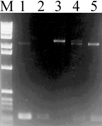
The values in bold letters correspond to the quantities per sample. Results and Discussion The protocol for the single-step PCR screening procedure is depicted in Table 1. The present method was designed for both the detection of Pichia integrants and the determination of the clone Mut phenotype by PCR using 5´ and 3´ AOX1 primers. For the appropriate interpretation of the PCR results when pHIL and pPIC plasmids are used, the distances in base pairs between the AOX1 primers should be added to the size of the cloned insert (e.g.; pHIL-D2: + 0.159 kb, pPIC-3K: + 0.228 kb). From Mut+ clones two bands were generated and detected, one corresponding to the insert (2.35 kb) and the other to the AOX1 gene (approx. 2.2 kb), while for Muts clones only the band corresponding to the insert (2.35 kb) was detected (Figure 1). Figure 1. Analysis of PCR products obtained from intact P. pastoris colonies. Volumes of 10 m L of the PCR reactions were run on a 1.2 % agarose gel and stained with ethidium bromide. Lane 1: a Mut+ clone carrying the pPIC-3K vector without insert (size of the resulting bands: 2.2 kb and 0.228 kb); lane 2: no yeast colony as a negative control; lane 3: a Muts PPK19 clone (size of the resulting band: 2.35 kb); lane 4: a Mut+ PPK19 clone (size of the resulting bands: 2.2 kb and 2.35 kb); lane 5: wild-type yeast strain showing the characteristic band of the AOX1 gene (2.2 kb) and lane M: DNA ladder (1 kb ladder, BRL). In an attempt to obtain a higher PCR product yield, we tried the incubation of yeast cells in a common microwave oven or alternatively in a water bath at 100 ºC for disruption of the cell wall and membranes prior to amplification. However, no improvement was attained (data not shown). It should be mentioned that inconsistent PCR results were obtained after keeping plates stored at 4 ºC for several weeks. The reasons accounting for this observation can not be formulated. A possible explanation could be that the strength of the cell wall of certain methylotrophic yeasts increases with age (4), making the rupture inefficient by this published method. Given this, it follows that the efficient amplification of any genomic sequence by our PCR procedure should necessarily require the use of intact fresh yeast cells (preferably not older than two weeks) as a source of template. In addition, a proper design of the primer set and optimization of PCR conditions should be accomplished in order to obtain a reliable DNA amplification. For Pichia clone screening we recommend an extension step of at least 2 min to ensure amplification of the 2.2 kb AOX1 locus in Mut+ clones. In summary, this new screening method has the advantages that it is time-saving and cost-effective since it does not require expensive reagents (cell-wall degradative enzymes) or specialized equipment for cell disruption. Consequently, it is feasible to obtain a ready-to-amplify sample by just introducing a pinpoint-size portion of a fresh yeast colony directly into the reaction tube, thus avoiding other rather complicated steps and reducing handling. Finally, misclassification of Mut+ clones as Muts was not observed. This procedure, together with our previous report (5) on the genetic transformation of P. pastoris via colony electroporation would greatly facilitate routine genetic manipulation of yeast and speed up future work on this topic. Acknowledgment This work was supported by grant 3031-243 from the Cuban Council of State. We thank Orlando Luis Pardo and Ariel F. Martínez for revision of the manuscript. References
Copyright 1998 Elfos Scientiae The following images related to this document are available:Line drawing images[ba98027a.gif] |
| |||||||||
