 |
| About Bioline | All Journals | Testimonials | Membership | News |
|
||||||
|
||||||
Biotecnología Aplicada 1999;16:222-225 Confirmation of Unusual Epidemiological Behavior of the Human Respiratory Syncytial Virus During Two Consecutive Epidemics (Seasons 94/95 and 95/96) in Havana City, Cuba Ángel Valdivia,1 Danay Chacón,1 Clara Savón,1 Odalys Valdés,1 Grehete González,1 Reynel Cancio,1 Julio C Sánchez,2 Germán Rogés,1 Blanca García-Barreno,3 José A Melero,3 Ángel Goyenechea1 1 Deparment of Virology. Institute of Tropical Medicine "Pedro Kourí". PO Box 601, Marianao 13, Havana, Cuba. Fax: (53-7) 24 6051, 22 0633; E-mail: a.valdivia@ipk.sld.cu 2 Center for Genetic Engineering and Biotechnology. PO Box 6162. Havana 6, Cuba. 3 National Center of Fundamental Biology, Majadahonda 28220 Madrid, Spain. Received in January, 1999. Accepted for publication in March, 1999. Code Number:BA99035 Abstract Twenty-seven strains of the human respiratory syncytial virus, isolated from three consecutive outbreaks, were sequenced at the National Center of Fundamental Biology (Madrid, Spain). The nucleotide sequence showed two unique characteristics related to what had been previously reported in the literature: (i) all of them were identical, and (ii) all showed a great homology with the Spanish Long reference strain (with only five changes). Upon this unusual finding and with the main objective of excluding a probable contamination with another Long strain from Sweden, selected regions of both reference strains and other laboratory strains were sequenced. The Spanish Long strain was only used as a confirmatory control of the results obtained in laboratories from Spain. The results obtained by the authors discarded any possible contamination when the percentages of homology between the sequenced regions of the two foreign strains and the Cuban isolates were analyzed and compared. The results also corroborated that the Swedish Long strain was even more separated from the Cuban isolates than the Spanish strain. This report presents a theory to explain the unusual epidemiological behavior of this virus in Cuba. Keywords: HRSV, molecular epidemiology, respiratory syncytial virus Resumen Confirmación de un comportamiento epidemiológico inusual del virus sincitial respiratorio humano durante dos epidemias consecutivas (temporadas 94/95 y 95/96) en Ciudad de La Habana, Cuba. Veintisiete cepas del virus sincitial respiratorio, aisladas durante tres brotes consecutivos, fueron secuenciadas en el Centro Nacional de Biología Fundamental de Madrid, España. La secuencia nucleotídica mostró dos características distintivas en relación con lo que se había reportado anteriormente en la literatura: (i) todas ellas eran idénticas y (ii) todas mostraron una gran homología con la cepa de referencia Long proveniente de España (con sólo cinco cambios). A partir de este hallazgo inusual y con el principal objetivo de excluir una posible contaminación con otra cepa Long de origen sueco, se secuenciaron regiones selectas de las dos cepas de referencia y de las existentes en el laboratorio. La cepa Long española se utilizó sólo como control para confirmar los resultados obtenidos en los laboratorios españoles. Los resultados obtenidos descartaron cualquier posible contaminación cuando se analizaron y compararon los porcentages de homología entre las regiones secuenciadas de las dos cepas foráneas y los aislamientos cubanos. Los resultados también corroboraron que la cepa Long de origen sueco se aleja de los aislamientos cubanos aún más que la cepa española. Este reporte presenta una teoría para explicar el comportamiento epidemiológico inusual de este virus en Cuba. Palabras claves: epidemiología molecular, virus sincitial respiratorio, VSR Introduction Human respiratory syncytial virus (HRSV) is the leading cause of lower respiratory infections in young children and infants [1]. HRSV belongs to the genus Pneumovirus, family Paramyxoviridae. Its genome is a single negative strand RNA molecule of about 15,000 nucleotides [2], which is transcribed into polyadenylated mRNAs from a single promoter located at the 3’ end [3]. It has two surface glycoproteins (G and F), which are the main targets of neutralizing and protective antibodies. G glycoprotein is responsible for virus binding to the cell surface receptor [4]. F glycoprotein mediates the fusion of viral and cell membranes [5]. Variability of HRSV isolates was first demonstrated at antigenic level in a neutralization test performed with hyperimmune serum [6]. Different panels of monoclonal antibodies were later used to subdivide HRSV isolates into two antigenic groups, A and B [7, 8], that correlate with genetically distinct viruses [9]. Further studies of HRSV variability have focused mainly on the G protein for two reasons: (i) the G protein shows the largest antigenic and genetic differences between viruses of the two antigenic groups [10], and (ii) the G protein is one of the targets of the neutralizing and protective antibody responses. In this work, two HRSV reference strains (Long) were characterized by PCR and nucleotide sequencing, and compared to 26 HRSV isolates obtained during two consecutive outbreaks in Havana City, Cuba. Materials and Methods Viruses, cells and Long reference strains The two HRSV Long strains used in this study were kindly provided by Dr. Monica Grandien (Swedish Center of Disease Control) and by Dr. JA Melero (National Center of Fundamental Biology [NCFB], Madrid, Spain) in 1992 and 1997, respectively. On arrival at the laboratory, the strains were multiplied in cell cultures until great volumes were obtained, which were later aliquoted and stored at -70 ºC for further use, in order to avoid a high number of passages. Both Long strains were cultured in Hep-2 cells in plastic flasks (Costar, USA) with a surface area of 75 cm2 containing 15 mL of minimum essential medium (MEM, with glutamine, penicillin, streptomycin, and 5% fetal calf serum). When an extensive cytopathogenic effect (CPE) was present, the cells were detached into the tissue culture medium by shaking with sterile glass beads and 0.5 mL of these suspensions were dispensed and stored at -70 ºC until further use for PCR analysis. Nucleic acid extraction, reverse transcription and PCR were performed as previously described by Cane and Pringle [11–13]. RNA extraction All small-scale RNA extractions were carried out in 1.5 mL microfuge tubes without any attempt to separate RNA from DNA. Briefly, 0.5 mL from infected cell suspensions was centrifuged during 5 min at 10,000 rpm. The cell pellet was resuspended in 0.5 mL of 3.5 M urea, 200 mM NaCl, 10 mM Tris-HCL pH 7.8, 5 mM EDTA, 0.75 mM MgCl2, 0.5% SDS, and 0.35% NP-40 detergent. Half a milliliter of phenol-chloroform (1:1) (equilibrated with 150 mM NaCl, 10 mM Tris-HCL pH 7.8, 1 mM EDTA) was added. The mixture was then vortexed for about 5 s and centrifuged for 10 min at 10,000 rpm. The aqueous phase was separated and treated again with phenol-chloroform. One milliliter of ethanol was added, and the solution was incubated at -20 ºC for 20 h, the precipitate was pelleted by centrifugation, washed with 0.5 mL 70% ethanol and dissolved in distilled water. Amplification of HRSV RNA Preparation of cDNA was performed with about 20 mg of total spectrophotometrically quantified RNA in a 20 mL volume containing 100 ng of each primer. First, the template RNA was mixed with both primers and placed at 65 ºC. After 15 min, the solution was placed on ice and completed to 20 mL with 100 mM Tris-HCl pH 8.3; 500 mM KCl; 25 mM MgCl2; 25 mM of each dNTP; 20 U Rnasin (Boehringer Mannheim GmbH, Germany) and 5 U of AMV reverse transcriptase (Boehringer Mannheim, Germany). The reaction was carried out at 42 ºC for 30 min. Finally, the reaction mix was placed at 95 ºC for 5 min and kept on ice until PCR was developed. PCR mix was set up to a volume of 100 mL, which contained 100 mM Tris-HCL pH 8.3, 500 mM KCl, 25 mM MgCl2, distilled water, and 2.5 U Taq DNA polymerase (Boehringer Mannheim, Germany). The amplification was developed for 30 cycles in a Perkin Elmer thermal cycler (USA). Each cycle consisted of denaturation at 93 ºC for 1.5 min, annealing of the primers at 55 ºC for 1.5 min, and chain elongation at 72 ºC for 1.5 min. After the last cycle of amplification, 10 mL of the reaction solution were analyzed by electrophoresis on 2% agarose gels with Tris-borate buffer. Controls Distilled water, mixed buffer solutions, full-time open vial with the final buffer mixture, and RNA from Hep-2 cells were included as negative controls [14–16]. The reaction of cDNA synthesis and the PCR were developed using a strict protocol, taking precautions to prevent contamination [17]. Primers The primers used for the G gene were supplied by Dr. JA Melero. These primers were designed to amplify between nucleotides 1 and 316 (316 bp), and between nucleotides 533 and 13 (F gen) (400 bp). Primer sequences and position in the G gen are shown in the Table. Table. Primer sequences and position in the G gen.
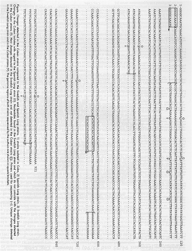
Sequencing of the PCR products PCR products of the G gene from both Long reference strains were electrophoresed using 1% agarose (low melting point) in TNE buffer, and visualized using ethidium bromide. DNA was isolated using a Wizard column DNA kit (Promega, USA). DNA sequences were determined by the dideoxy chain termination method [19] and using a Sequenase sequencing kit (USB, USA). These kits were employed according to the manufacturer’s protocol. The primers used to sequence the G gene were the same used for amplification (Table). In a typical experiment, 100 ng of purified PCR product (5 mL or 200 ng) were mixed with 2 mL or 10 pmol of primer, heated at 95 ºC for 3 min, quickly chilled on ice, and used for sequencing. The reaction was stopped by adding formamide containing bromophenol blue and xylene cyanol dye, heated at 85 ºC for 3 min, and chilled prior to electrophoresis in 6% polyacrylamide gels containing 6 M urea. The gels were fixed in 10% acetic acid, wrapped in Saran Wrap®, and autoradiographed at room temperature without intensifying screens. In all cases, five units were used to obtain sequence data. Results The five changes detected in Cuban isolates are shown in the Figure, being compared to the Spanish and Swedish Long strains. The changes observed in these isolates do not appear in the Swedish Long strain and some changes observed in the latter do not appear in the Cuban isolates. Figure. Changes detected in the Cuban strains compared to the Swedish and Spanish Long strains. 1) strains isolated in Cuba, 2) Spanish Long strain, 3) Swedish Long strain. Changes in the Cuban isolates with regard to the Spanish Long strain (O). Nucleotides found in the Swedish Long strain and which do not coincide with the changes detected in the Cuban strains (à). New changes detected in the Swedish Long strain and not detected in the Cuban strains ( ). Primers used for sequencing (®). Unique change detected in the Swedish Long strain and in the Cuban strains (Ñ). The genome regions of both strains sequenced by the authors are shown in square brackets. The Figure was constructed on the basis of previously published nucleotide sequences of the Spanish strain [18]. The only change in Cuban strains present in our Swedish Long strain was in nucleotide 79 (T®C), which had been previously reported [10]. Discussion Sequence analysis of numerous HRSV isolates, mostly of the A group, showed extensive variability of the G glycoprotein [11, 18, 20, 21]. Several issues about HRSV evolution emerged from these studies: • Viruses from antigenic group A belong to different lineages that correlate with previously identified genotypes [12, 13, 18]. Most epidemics are produced by viruses classified into more than one genotype. At a local level, replacement of predominant genotypes was observed during consecutive years [22]. • HRSV genotypes have a worldwide distribution, and viruses isolated in distant places and in different years may be more closely related than viruses isolated in the same place on two consecutive days [23]. In 1996, we combined the use of monoclonal antibodies and restriction analysis of PCR products of the selected region of N gene as part of the characterization of two outbreaks of HRSV in Havana City, Cuba. All the isolated strains (21 belonging to an outbreak occurred during 1994 and 1995, and 6 belonging to another consecutive outbreak occurred in 1996), were identified as RS Subgroup A by means of monoclonal antibodies and gave N gene fragment restriction pattern NP4 [24–26]. Previously studied RS virus Long strains A2 and RSS2 belonged to restriction pattern NP4 [11]. In 1997, the strains were sent to the NCFB to be sequenced, in order to carry a more discriminatory method and to perform the analysis of the most variable gene [18]. The nucleotide sequence of the strains isolated in Cuba in two consecutive outbreaks showed two major characteristics distinct from previous reports: • The nucleotide sequences of the isolated strains from two consecutive outbreaks in Havana City were identical. • The nucleotide sequence pattern was very similar to that of the oldest Long strains that circulated in the world and coincided with the N gene fragment restriction mapping NP4 obtained in 1996, as aforementioned. In some cases, the nucleotide sequences obtained from clinical specimens and tissue culture were identical. Only five changes were found in comparison with the Spanish Long strain : nucleotide 58 (A®C), nucleotide 79 (T®C), nucleotide 260 (A®T), nucleotide 640 (A®G) and nucleotide 888 (T®A). With these unusual findings and with the main objective of discarding any possibility of contamination with the Swedish reference strain, the following strategy was conducted: 1. To use RNA from the two reference strains and two sets of oligonucleotides that would generate two PCR products and which would include the regions where the only five changes differentiating the Spanish strain from the Cuban ones were detected. 2. To align the sequenced regions of both reference strains with the Cuban isolates. 3. To define the percentages of homology of each strain with respect to the Cuban isolates. The percentages of homology between the strains and the Cuban isolates had been previously obtained at NCFB: • For the fragment from position 40 to 234 (194 nucleotides), and compared to the Spanish Long strain, homology was 98.5%. • For the fragment from position 588 to 912 (324 nucleotides), homology with the Spanish strain was 99.4%. • For the two fragments together and still comparing the Spanish strain with the Cuban isolates, homology was 99.1%. The percentages of homology between the Cuban isolates and the sequenced regions of the Swedish Long strain were: • For the fragment from position 40 to 234 (194 nucleotides), and compared to the Swedish Long strain, homology was 97.5%. • For the fragment from position 588 to 912 (324 nucleotides), homology with the Swedish strain was 99.4%. • For the two fragments together and still comparing the Swedish strain with the Cuban isolates, homology was 98.7%. The results attained cleared up two concerns raised at the Laboratory in Spain, when the Cuban strains isolated at the Institute of Tropical Medicine "Pedro Kourí" were sequenced: 1. The RSV strains isolated in Cuba are not a product of contamination with the Swedish Long strain. 2. The percentages of homology obtained show that the Swedish Long strain is more distant from the Cuban isolates than the Spanish strain. In one of the most important studies on evolution developed to date [27] involving the characterization of 93 HRSV isolates, it was determined that the most variable regions of the G gene are located between nucleotides 283 and 411, and between nucleotides 799 and 917. The higher number of differences found between our Long Swedish strain and the Cuban isolates are included exactly in the most preserved regions of the gen, which additionally sustains that the variation is not the result of contamination. There are many factors that, in the opinion of the authors, may have contributed to the peculiar behavior of the molecular epidemiology of this virus in Cuba: • There is a possibility that the number of strains that entered in Cuba in the last 40 years has been reduced, due to a series of diverse reasons which included limited traveling, especially in lower age groups. • The geographical condition of an island is also a condition that limits the introduction of strains that could be circulating in neighboring countries, together with the absence, as part of the fauna, of other primate species that could be involved in transmission. • The outburst of the tourist industry and the economic relationships have been in progressive increase since the beginning of the 90’s, but until now it does not seem to be associated with the introduction of new strains in Havana City. A reason that could explain this is that "family" tourism, which includes the usual reservoirs of this virus, young children, is mostly enjoyed in regions out of Havana, some of them even physically separated from the great island, as the keys and peninsulas of the littorals (Varadero, Cayo Largo and Cayo Coco, for example). Further molecular characterization of isolates from outbreaks should continue in order to determine the moment in which variations are introduced, and should not be restricted to the capital city but expanded to other areas. Acknowledgments We thank Dr. Jorge González for critical reading of the manuscript. This work was funded in part by a grant (B1095-2026-E) from Comisión Interministerial de Ciencia y Tecnología for Spanish-Cuban Cooperation. References
Copyright Elfos Scientiae 1999 The following images related to this document are available:Photo images[ba99035a.jpg] |
| |||||||||
