 |
| About Bioline | All Journals | Testimonials | Membership | News |
|
||||||
|
||||||
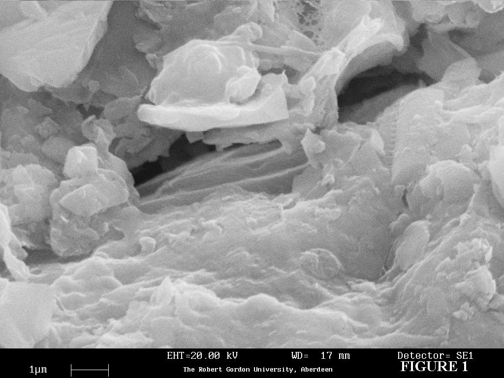
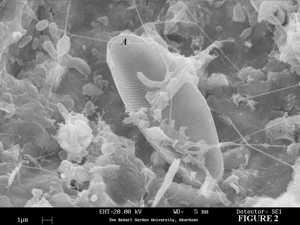
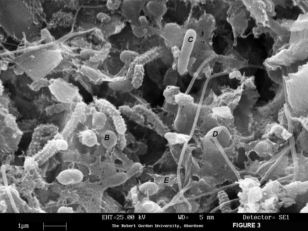
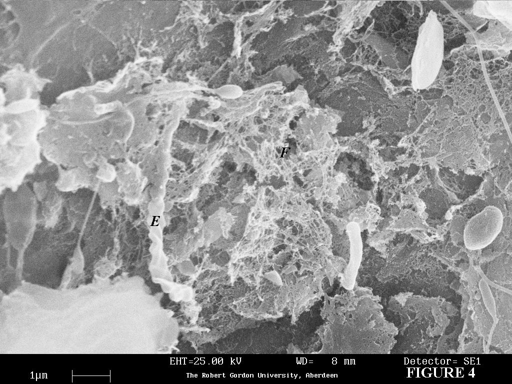
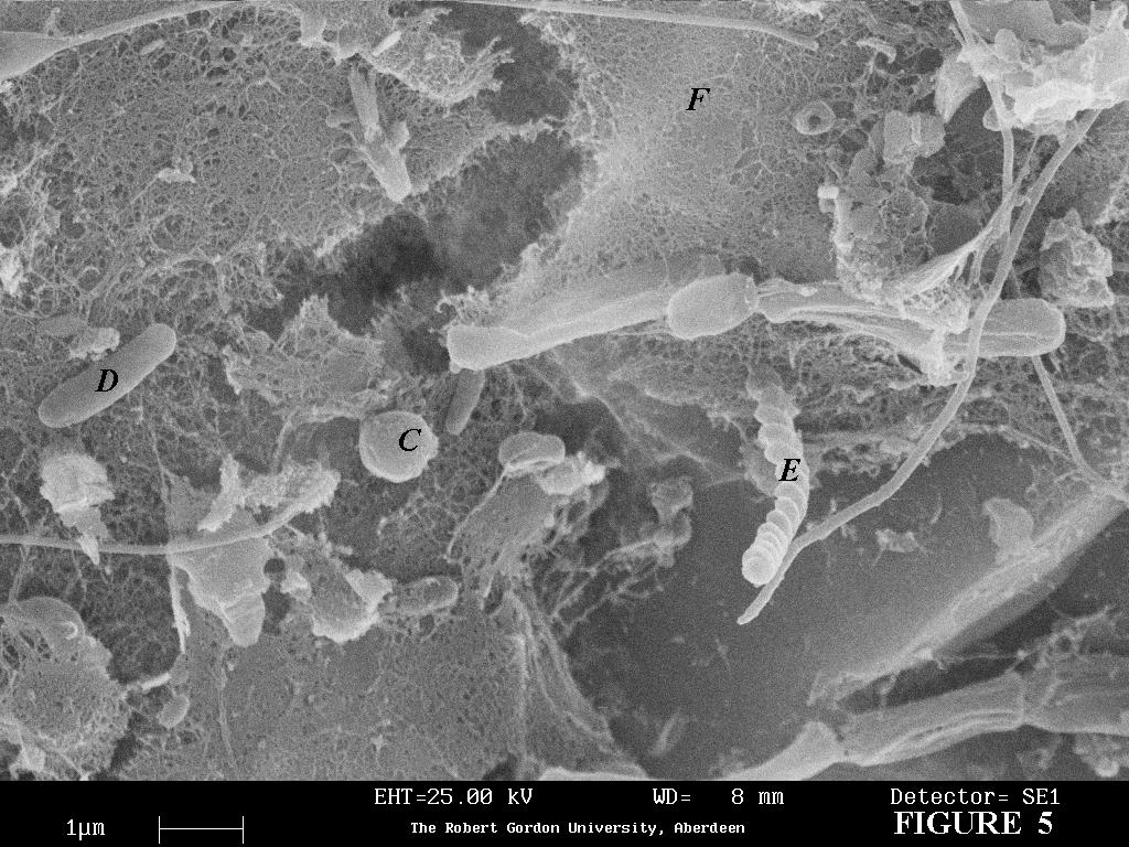
Biofilm, Volume 6, Paper 1 (BF01001) 2001 Visualisation of the establishment of a heterotrophic biofilm within the schmutzdecke of a slow sand filter using scanning electron microscopy Samantha P. Law1*, Maureen M.A.L. Melvin1 and Andrew J. Lamb2 1School of Applied Sciences, The Robert Gordon University, St. Andrew Street, Aberdeen AB25 1HG, UK. Tel: +44 (0) 1224 262842 *Corresponding author - E-mail: s.law@rgu.ac.uk Received: July 10th 2001 Code Number: BF01001 ABSTRACT The unique feature of slow sand filters is the schmutzdecke, a biofilm formed at the sand - water interface. Scanning electron microscopy was used to observe the surface of sand obtained from the schmutzdecke of a newly seeded and a mature slow sand filter. The complex nature of the biofilm formed on the sand was observed. A distinct difference was seen between the freshly seeded schmutzdecke compared to the schmutzdecke from a mature filter. It was found that a larger amount of extracellular matrix was present within the schmutzdecke removed from a mature filter. The images obtained show that sand, at the sand-water interface of slow sand filters, offers an attractive environment for the colonisation of a variety of micro-organisms including diatoms, algae, bacteria and their extracellular products. Keywords: biofilm, schmutzdecke, slow sand filtration, scanning electron microscopy Introduction In slow sand filtration, water percolates slowly through a porous sand bed (Visscher, 1990) allowing the production of potable water by a combination of physical -chemical and biological particulate removal mechanisms (Weber-Shirk and Dick, 1997). The unique feature of slow sand filters is the 'schmutzdecke' that forms at the sand - water interface on the surface of the sandbed, which acts as an interface for biofilm development. It consists of bacteria, algae, protozoa, invertebrates and certain extracellular products. The schmutzdecke removes natural organic matter, transforms synthetic organic compounds and retains pathogens, producing microbiologically safe potable water. In the Grampian region of Scotland, water is extracted from the River Dee, as it is a water of excellent quality (Mellanby, 1990). Like all waters from upland sources, it can become discoloured during heavy rainfall. This is due to the presence of recalcitrant natural organic matter, mainly humic and fulvic substances, which are large molecular weight compounds that cannot readily be biodegraded and are therefore not removed by the schmutzdecke. To produce an aesthetically pleasing drinking water, the discoloration of the river water is removed prior to filtration by using ozone, a powerful oxidant. The high molecular weight, poorly biodegradable humic and fulvic substances present are oxidised into biodegradable, low molecular weight compounds (Melin and Odegaard, 1999). Therefore, ozonation increases the biodegradable organic carbon content of the raw water, encouraging the growth of heterotrophic micro-organisms within the schmutzdecke. The schmutzdecke represents a zone of nutritional affluence in a surrounding sea of relative poverty (Sutherland, 1983) as the micro-organisms present at the sand - water interface receive a constant source of nutrient rich water. Scanning electron microscopy (SEM) is an invaluable method to determine the diversity of micro-organisms attached to the sand surface and was used to observe the physical relationships between the sand surface and schmutzdecke (biofilm) development. The establishment of the heterogeneous biofilm was determined by observing the differences between a freshly seeded and a mature schmutzdecke. This method, developed for the SEM analysis of micro-organisms, is simple, relatively inexpensive and rapid. Importantly this method also avoids the use of the toxic reagents cacodylic acid and osmium tetroxide. Method Schmutzdecke samples were collected from a 1 day and 40 day old filters at Invercannie water treatment works by removing approximately100g of the top layer of sand, one meter from the side of the filter to avoid any side-wall effects (Yordanov et al, 1999). Schmutzdecke samples washed in nitric acid were used as a control in SEM analysis. During the procedure, great care was taken in the preparation of these samples in order to prevent damage to biological material and the formation of artefacts or unrecognisable features. The schmutzdecke samples were fixed in 5% gluteraldehyde (5% v/v 0.1M phosphate buffer, pH 7.4) for 3 hours, rinsed twice in 0.1M phosphate buffer, pH 7.4, then post-fixed in a 2% gluteraldehyde - 3% formaldehyde solution (v/v 0.1M phosphate buffer, pH 7.4) for 1 hour. The combination of these aldehydes provided a good quality of fixation as formaldehyde rapidly penetrates tissues stabilising the structure of biological material whilst gluteraldhyde permanently fixes the sample. The samples were then rinsed twice with 0.1M phosphate buffer, pH 7.4 before being sequentially dehydrated for 20 minutes in 70%, 90% (both v/v 0.1M phosphate buffer, pH7.4) and 100% acetone. A B7010 critical point drier (Agar Scientific, Essex, UK) was used to dry the schmutzdecke samples using acetone and carbon dioxide as the transitional fluid. Critical point drying was used instead of air drying to prevent the disruption of cellular structures, maintaining the stability of the fixed structure. The samples were mounted onto aluminium stubs using carbon tape and sputter coated with gold (Polaron, East Sussex, UK). This prevented the build-up of static charges. The samples were viewed on the scanning electron microscope (Leo, Cambridge, UK). Results and Discussion SEM is advantageous as it allows the examination of surfaces at high magnifications. Observations of schmutzdecke samples revealed details of the sand surface and the associated micro-organisms with it in a pseudo three-dimensional image. A micrograph of the control schmutzdecke sample that was washed with nitric acid shows that the nitric acid treatment strips the sand of any micro-organisms present. (Fig. 1) Micrographs of a freshly seeded (1 day old) schmutzdecke sample show the attachment of a diatom (Fig.2) and of coccoid bacteria, rod-shaped bacteria and algae (Fig.3) to the sand surface . A fine matrix of extracellular mucilage can also be observed. Micrographs of a mature (40 day old) schmutzdecke sample show the presence of a thick extracellular matrix attaching the micro-organisms present to the sand surface (Figs.4 and 5). Figure 1. Micrograph of the control schmutzdecke sample Key to micrographs: A = diatom The technique described here has allowed the observation of the complex nature of the biofilm formed upon the sand surface. From the images obtained, sand, at the sand - water interface of slow sand filters, offers an attractive environment for colonisation with micro-organisms such as bacteria, diatoms and algae. SEM observation revealed micro-organisms firmly anchored to the sand, even on day 1 of a freshly seeded slow sand filter, demonstrating that they were not washed away during sample preparation (Figs 2 and 3). By 40 days a substantial extracellular matrix presumably of polysaccharide, was apparent (Figs 4 and 5). Attachment to the sand surface is therefore likely to be by this secreted adhesive mucilage (Allison and Sutherland, 1987) which enhances the ability of these micro-organisms to become anchored to the sand and hence not be washed away during water filtration. Extracellular mucilage produced by bacteria aids the attachment of micro-organisms to surfaces, leading to biofilm formation. Biofilms are notoriously difficult to preserve for SEM observation as a consequence of their high water content (Chenu and Jaunet, 1992). This is because biofilms are highly hydrated with up to 97% of the biofilm matrix being water (Sutherland, 2001); the dehydration process of SEM preparation will lead to the collapse of the biofilm, which gives the incorrect impression that mature biofilms are confluent. Nonetheless, the mesh like structure of adhesive mucilage, possibly extracellular polysaccharides, surrounding the micro-organisms can be clearly visualised by SEM, as shown in figures 2, 3,4 and 5. Its presence is not an artefact of SEM preparation as it is not present on the surface of the control (Fig.1). As only a small area of the slow sand filters at Invercannie was sampled, the micrographs cannot give a full reflection of what is occurring within the filters. The filters are located outdoors and are exposed to the external environment, so there will also be variations in schmutzdecke development due to fluctuations in environmental conditions. However, the micrographs presented here do demonstrate the heterogeneous nature of the biofilms formed in the schmutzdecke. They also show the production, over time, of an extracellular bio-matrix, which is an integral component of an established schmutzdecke within a functioning slow sand filter. ACKNOWLEDGEMENTS I would like to thank Mr Iain Tough (School of Applied Sciences, The Robert Gordon University, Aberdeen, UK) for his assistance in using the SEM. REFERENCES Allison, D.G. and Sutherland, I.W., 1987. The Role of Exopolysaccharides in Adhesion of Freshwater Bacteria. Journal of General Microbiology 133, 1319 - 1327. Chenu, C. and Jaunet, A.M., 1992. Cryoscanning Electron Microscopy of Microbial Extracellular Polysaccharides and Their Association with Minerals. Scanning, 14, 360 - 364. Melin, E.S. and Odegaard, H., 1999. Biofiltration of Ozonated Humic Water in Expanded Clay Aggregate Filters. Water, Science and Technology, 40 (9), 165-172. Mellanby, J.F., 1990. Ozone and Slow Sand Filtration for Treatment of Coloured Water. Summary Report on Invercannie Water Treatment Works Pilot Plant, Grampian Regional Council, Water Services Department. Sutherland, I.W., 1983. Microbial Exopolysaccharides - Their Role in Microbial Adhesion in Aqueous Systems. Critical Reviews in Microbiology, 10 (2), 173 - 201. Sutherland, I.W., 2001. The Biofilm Matrix - an Immobilised but Dynamic Microbial Environment. Trends in Microbiology, 9 (5), 222 - 227. Visscher, J.T., 1990. Slow Sand Filtration - Design, Operation and Maintenance. Journal of American Water Works Association, 82 (6), 67-71. Weber - Shirk, M.L. and Dick, R.I., 1997. Physical - Chemical Mechanisms in Slow Sand Filters. Journal of American Water Works Association, 89 (1), 87-100. Yordanov, R.V., Melvin, M.A.L., Law S.P., Littlejohn, J. and Lamb, A.J, 1999. Effect of Ozone Pre-Treatment of Coloured Upland Water on Some Biological Parameters of Sand Filters. Ozone Science and Engineering, 21, 615-628.
Copyright remains with the author. Biofilm Editorial Office biofilm@biostrat.demon.co.uk The following images related to this document are available:Photo images[bf01001d.jpg] [bf01001e.jpg] [bf01001b.jpg] [bf01001a.jpg] [bf01001c.jpg] |
| |||||||||
