 |
| About Bioline | All Journals | Testimonials | Membership | News |
|
||||||
|
||||||
Effect of Stem Bark Extracts of Enantia chloranta on Some Clinical isolates Razaq F. ATATA*, Alhassan SANI and Stella M. AJEWOLE Department of biological sciences, University of Ilorin,
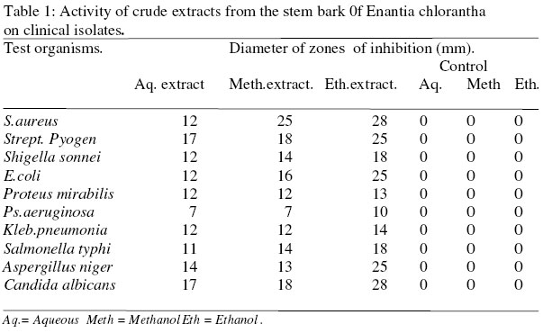
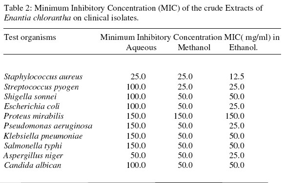
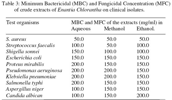
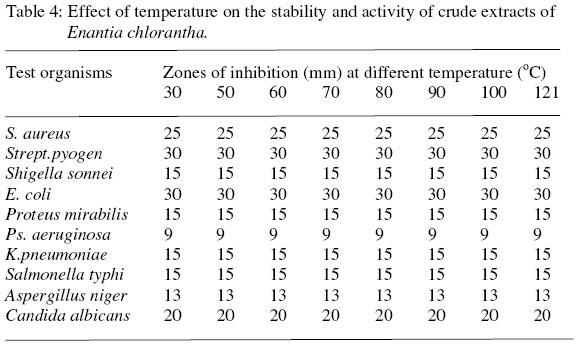
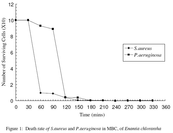
PMB, 1515 Ilorin, Nigeria. Received 10 October 2003 Code Number: bk03024 Abstract Studies on antimicrobial activity of water, methanol and ethanol extracts of the stem bark of Enantia chlorantha, was carried out by the agar ditch diffusion and tube dilution methods. The clinical isolates used include; Staphylococcus aureus, Streptococcus pyogenes, Shigella sonnei, Proteus mirabilis, Klebsiella pneumoniae, Salmonella typhi, Pseudomonas aeruginosa, Aspergillus niger, and Candida albicans. The isolates were obtained from the Department of Medical microbiology and Parasitology of the University of Ilorin Teaching Hospital. Crude extracts of all solvents used inhibited the growth of all the isolates tested, except Pseudomonas aeruginosa . The Minimum Inhibitory Concentration (MIC) ranged between 25mg/ml to 150mg/ml depending on isolate and extracting solvent. Ethanolic extracts showed greater antimicrobial activity than the other two solvents. The killing rate of the Minimum Bactericidal Concentration (MBC) of the ethanolic extract on S. aureus and P.aeruginosa was 21/2hr, and 3hr respectively. Temperature stability study showed that the extracts are stable and active over a temperature range of 30 to 121oC. Key words: Enantia chlorantha, antimicrobial property, plant extract INTRODUCTION It is estimated that there are between 200 000 and 700 000 species of tropical flowering plants that have medicinal properties, this has made traditional medicine relatively cheaper than modern medicine.1 Over the years there have been alarming reports of multiple drug resistant in medically important strains of bacteria and fungi.2-7 The persistent increase in antibiotic resistant strains of organisms has lead to development of more potent synthetic antibiotics such as the 3rd and 4th generations of Cephalosporins by Pharmaceutical companies. These new antibiotics are scarce, costly and not affordable particularly in the developing countries and therefore make compliance difficult. There is therefore need for continuous search for new effective and affordable antimicrobial drugs. Local medicinal plants provide a source of new possible antimicrobial drugs.8 Efforts of Scientists in establishing plants with promising antimicrobial property is yielding fruitful results as a number of plants with high antimicrobial property have been elucidated.9-13 Among plants believed traditionally to have therapeutic effect but which receives little or less scientific research is Enantia chlorantha. Traditionally the bark extract is applied to ulcers and leprous spots for quick healing, decoction is used for washing wounds, bark sap is taken as decoction against diarrhea. This work therefore aims at investigating its suggested antimicrobial activity. MATERIALS AND METHOD Collection and preparation of plant material Stem bark of the plant material (E. chlorantha) used was purchased from herb sellers at Baboko market in Ilorin Kwara State Nigeria. The plant was identified at the the Forestry Research Institute of Nigeria (FRIN) Ibadan. The plant was sun-dried for two weeks until constant weight was obtained. It was ground into powder and stored in sterile glass bottle at 30oC (room temperature) Preparation of extracts Three different solvents viz; distilled water, methanol and ethanol were used for extraction. Fifty gramme of the ground plant material was weighed and suspended in each of the three solvents, the mixtures were then left on shaker at 190 rev/min. for 24hrs at room temperature.14 The extracts from each solvent was decanted, passed through muslin cloth, and then filtered with Whatman No 1 filter paper. The extracts were tested for purity by plating them on nutrient agar and incubated for 24hrs at 37oC and 25oC. The extract from each solvent was then stored in sterile brown bottle kept in refrigerator at 7oC. Concentration of the extracts and preparation of stock solutions The three extracts (water, methanol,and ethanol) were evaporated to semi-solid form on water bath at 100oC and 80oC respectively. The semi-solid yellow extracts obtained were further evaporated in oven at 110oC until constant weight was achieved for each extract. Each solid extract was reconstituted in their respective solvents to obtain a stock solution of 200mg/ml. The stock solutions obtained were then filtered using Millipore membrane filter (0.45um pore size).The sterile extracts obtained were stored in sterile capped bottles. Test organisms and source. The organisms used comprise of two Gram-positive (Staphylococcus aureus and Streptococcus pyogens ), six Gram-negative bacteria ( Escherichia coli, Salmonella typhi, Shigella sonnei, Proteus mirabilis. Pseudomonas aeruginosa, Klebsiella pneumoniae) and two fungi (Aspergillus niger and Candida albicans). The test organisms were collected from the Department of Medical microbiology and Parasitology of the University of Ilorin Teaching Hospital. Standardization of inoculum. Five colonies of each organism used were picked into Nutrient Broth (NB) and incubated at 37oC for 18-24hrs for bacteria while C. albicans,and,the A. niger were incubated at 25oC for 5-7days. Turbidity produced was adjusted to match 0.5 Mc Farland standard (108cfu/ml) which was further adjusted to 105cfu/ml and 103cfu/ml.15 Test for antimicrobial activity of the extracts. Sterile Nutrient Agar (NA) and Potato Dextrose Agar (PDA) plates were prepared. An 18-24hr old standardized culture of bacterial and the C. albicans was separately used to flood the NA and PDA surfaces of each plate respectively16, and excess was drained off. The seeding was done in such away that NA plates contained 105cfu/ml of bacterial isolates while PDA agar contained 103cfu/ml of fungal spores. A sterile cork borer of 5mm diameter was used to make six ditches on each plate. A 0.1ml of the extract from each solvent (equivalent to 20mg of the extract) was dropped into each appropriately labeled ditch. And into the remaining three ditches distilled water, methanol, and ethanol were used as positive controls. The inoculated plates were left on the table for 1hr to allow the extracts to diffuse into the agar.15 The NA plates were incubated aerobically at 37oC and PDA at 25oC for 18-24hrs.The PDA plates containing A.niger was incubated for 7days at 25oC. Zones of inhibition produced after incubation was measured in millimeter (mm). Determination of Minimum Inhibitory Concentration (MIC) of the extracts on the isolates. Broth dilution method15 was used. Varying concentrations of the extracts (200mg/ml, 150mg/ml, 100mg/ml, 50mg/ml, 25mg/ml, and 12.5mg/ml) were prepared. A 0.1ml of each concentration was added to each 9ml of nutrient broth containing 0.1ml of standardized test organism of bacterial cells and fungal spores. The tubes were incubated aerobically at 37oC and 25oC for 24hrs and 7days for bacterial and Fungal isolates respectively. Positive controls were equally set up by using solvents and test organisms without extracts. The tube with least concentration of extract without growth after incubation was taken and recorded as the MIC. Determination of Minimum Bactericidal (MBC) and Fungicidal Concentration (MFC). A 1ml sample from the tubes used in MIC determination which did not show any visible growth after the period of incubation were streaked out on NA and PDA agar to determine the minimum concentration of the extract require to kill the organisms. These concentrations were indicated by the failure of the test organisms to grow on subsequent transfer to NA and PDA plates. The lowest concentration of the extract indicating a bactericidal effect after 24hrs Of aerobic incubation was regarded as the Minimum Bactericidal Concentration (MBC) while the lowest concentration that prevent fungal growth after 7days of aerobic incubation was recorded as the Minimum Fungicidal Concentration (MFC). Determination of death rate of the isolates in the extract Death rate of the most susceptible and the least susceptible bacterial used (Staph. aureus and P. aeruginosa) were carried out using Kelsey and Maurer method.17 This was carried out by mixing 0.5ml of 105cfu/ml of test isolates with 4.5ml of MBC of the ethanolic extract, 0.1ml of the mixture was taken and plated out on sterile nutrient agar at time intervals of 30mins for 300 mins. The plates were incubated at 37oC for 24hrs. The numbers of colonies developed on each plate at the time intervals were counted. Effect of temperature on stability of extracts The extracts were heated in a water bath at 30°c, 600c, 80°c, 100°c, for 30 minutes and in an autoclave at 121°c for 15 minutes. After cooling, the extracts were tested for antimicrobial activity. RESULTS The results showed that the crude extracts have antimicrobial activity against all the isolates tested at 200mg/ml. Ethanolic extract showed greater antimicrobial activity than methanolic and aqueous extracts, as indicated by zones of inhibition ( Table 1) C. albicans was the most susceptible isolate tested, while P. aeruginosa was the least susceptible isolate (Table1). Minimum Inhibitory Concentration (MIC) for each isolate (Table 2) showed that Staphy. aureus, Stre. pyogenes, E. coli, K. pneumonia and A. niger had MIC of 25mg/ml of ethanolic extract while Pr. mirabilis, P. aeruginosa, had MIC of 150mg/ml for all three extracts. MIC of ethanolic and methanolic extracts on S. typhi was 50mg/ml. The Minimum Bactericidal Concentration (MBC) of all isolates ranged between 200mg/ml-50mg/ml (Table 3). The effect of temperature on the stability of the extracts showed that the extracts were stable at temperature range of 30°c-121°c (Table 4). Time course study of ethanolic extract on Staph. aureus, showed that the extract was able to kill all the cells after 31/2hr of exposure (fig.1). The killing effect of the extract on P.aeruginosa, was gradual in the first 2hours of exposure but became drastic 30 mins later and the surviving cells were reduced to zero after 2.5 hours of exposure (fig.1). DISCUSSION The results of this work showed that the bark stem extracts of Enantia chlorantha inhibited the growth of all the bacteria and fungi tested (Table 1). This suggests that the plant extract is broad spectrum in activity and that its mode of action may not be due to inhibition of cell wall synthesis. Similar finding has been reported.1,10. Generally higher antimicrobial activity of the extracts was observed on Staph. aureus, Stre. faecalis, E. coli, K. pneumoniae and A.niger, this is similar to the earlier results obtained.18 when extracts from Aramomum melegueta fruit was used. Whereas moderate antimicrobial activity of the extracts were observed on; S. typhi and C. albican. Ethanolic extracts showed the strongest activity followed by Methanolic extracts and aqueous extracts an indication that ethanol is a better extractant than the two other solvents used in this study. However aqueous extracts showed better antimicrobial effect (with MFC of 100mg/ml) on A. niger and Candida albican than Methanolic and Ethanolic extracts (Table3). The strong activity of the extracts on Staph. aureus, Sh. sonnei and E. coli suggest that it may be used for the treatment of wound infection and diarrhea caused by these organisms. This work also revealed the potential use of extracts of this plant for use in the control of medically important organisms such as; S. typhi (causative agent of typhoid fever), P. aeruginosa, C. albican and Aspergillus niger. Similar results have been obtained with extracts from leaves of Kalanchoe pinnate when tested against S.aureus, E.coli, B.subtilis, Pr.vulgaris, P.aeruginosa and C. albicans19. The killing rate of extracts on Staphy. aureus, showed that the extracts has strong killing effect on the organism, it reduces the cells from 105cfu/mlt to less than 102cfu/ml within 90 minutes of exposure (fig.1). Killing rate of the extracts on P. aeruginosa however took longer time to reduce to less 102cfu/ml; it was observed that after 5hr of exposure to the extract there was sudden increase in numbers of surviving cells (fig1). It could be concluded from this study that the activity of the extracts from bark stem of Enantia chlorantha stem showed activity against the tested isolates and probably justify its local use. Further work on identification and purification of the extracts to find out the active principle responsible for the antimicrobial property of the plant extracts is required. REFERENCES.
© 2003 Nigerian Society for Experimental Biology. The following images related to this document are available:Photo images[bk03024t2.jpg] [bk03024t1.jpg] [bk03024f1.jpg] [bk03024t4.jpg] [bk03024t3.jpg] |
| |||||||||
