 |
| About Bioline | All Journals | Testimonials | Membership | News |
|
||||||
|
||||||
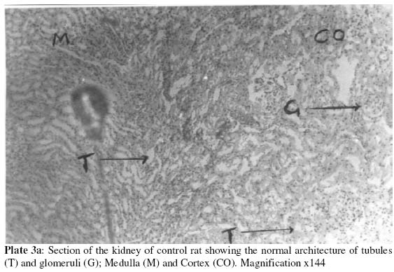
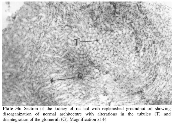
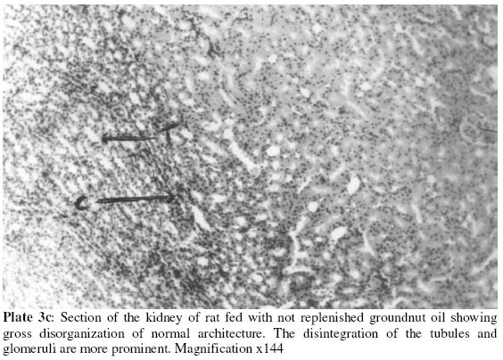
Biokemistri, Vol. 16, No.1, June, 2004, pp.1-10 Histological changes of selected rat tissues following the ingestion of thermally oxidized groundnut oil Florence O. JIMOH 1 and Adewale A. ODUTUGA 2 1Department of Biochemistry, University Ilorin, Ilorin, Nigeria 2Department of Biochemistry, IgbinedionUniversity, Okada, Nigeria 1Author to whom correspondence should be addressed. 1E-mail: asjimoh02@yahoo.com, Tel: 08035822119 Received 17 December 2002 Code Number: bk04001 Abstract The effect of ingestion of dietary oxidized groundnut oil on the histology of wistar rat brain (cerebral cortex), liver, and kidney were examined. Female weanling rats (Rattus norvegicus) were maintained for 12 weeks on intermittently thermoxidzed groundnut oil at 15% dietary level as part of a balanced diet. Fresh (unheated) groundnut oil served as the control. Histological examination of the selected tissues (cerebral cortex, liver, and kidney) showed mild to severe distortion of the normal architecture in animals fed with replenished and not-replenished thermoxidized groundnut oil respectively. Distortion of the layered appearance of the cerebral cortex was observed in addition to the prominence of the pyramidal cells. The liver showed mild to severe distortion of the normal architecture in animals fed with thermoxidized groundnut oil, as well as prominence and widening of the liver sinusoids. The kidney showed alterations in tubules and disintegration of the glomeruli. The results indicate that structural abnormalities had occurred in the animals fed oxidized groundnut oil. Key words:Thermoxidized groundnut oil, replenished and non-replenished, histology, morphology. INTRODUCTION Dietary oil rich in polyunsaturated fatty acids serve as the principal and inexpensive source of essential fatty acids and vitamins (Billek, 1980; Talwar et al, 1989). These oils are however susceptible to oxidative changes during use like frying (Ologan, 2002). This is because the polyunsaturated fatty acids constituents of these oils readily undergo oxidation resulting in the formation of peroxides, aldehydes, ketones, aldehydroesters and ozonides (Frankel, 1980; Kubow, 1992, Odutuga et al, 1997). Some researchers have argued that the toxicity of oxidized dietary oil is too low to cause any major harm to human (Nolen et al, 1967). The basis of this argument is that oxidative levels attained under laboratory conditions usually far exceed those of culinary practice and are therefore unrepresentative of that encountered in food preparations for human consumption. A possible oversight by the proponents of this argument, however, lies in the observation that several food establishments thermally oxidize one batch of oil several times over and by-products could accumulate, thereby increasing the toxic potentials of the reprocessed oil considerably. This kind of oil is more susceptible to further oxidation. Consumption of such peroxidized lipids has been shown to be injurious to health (Frankel, 1980; Halliwell and Gutteridge, 1984; Addis, 1986; Kubow, 1992). A study on this area, therefore, is imperative, especially as there is a paucity of information on this aspect. Reduction in the activity of several enzymes has been reported as one of the consequences of oxidized oil ingestion (Odutuga et al, 1997; 1999; Odutuga and Ologan, 1999). Jimoh and Odutuga (2002) investigated histological changes in the lungs and heart of rats fed oxidized groundnut oil. The result of our study showed disintegration of alveoli membrane and collapse of alveoli spaces in the lungs, which might indicate the impairment of oxygenation of blood, a prime function performed by the lungs. Histological results of the heart also showed disorganization of the spatial arrangement of the cells as well as widening of the fibres which might lead to weakness of the cardiac muscle which may consequently lead to cardiac enlargement and cardiac failure. There is limited information on the effect of dietary oxidized oil on the histology of other tissues. In this study, therefore, we have investigated histological changes in the brain (cerebral cortex), liver and kidney due to dietary oxidized oil ingestion. MATERIALS AND METHODS Treatment of groundnut oil Groundnut oil (9 litres) obtained from Ipata market, Ilorin, Nigeria was divided into three portions and each part treated as follows: (a) The first portion was not subjected to thermal treatment at all and served as the control. (b) The second portion: one litre of groundnut oil was poured into a stainless steel pot and used intermittently to fry yam chips at a temperature range of 180-200oC in open air for a period of four hours daily for 10 days. The oil sample was left for 12 hour to cool and was replenished with 500ml of fresh oil at intervals of 10 hours of use. (c) The third portion of groundnut oil (one litre was poured into a stainless steel pot and used to fry yam chips at a temperature range of 180-200oC in open air for a period of 4 hours daily for 10 days. The sample was also left for 12 hours to cool but was not replenished with fresh oil throughout the period of use. (d) These treatments (b) and (c) simulated the process of repeated use of frying oil. Management of animalsThirty weanling female albino rats (Rattus norvegicus) with mean weight of 40.5g obtained from the Animal Breeding Section of the Department of Biochemistry, University of Ilorin, Ilorin, Nigeria were randomly divided into three groups of ten animals each. They were all fasted for 24 hours before placement on the following: (a) Control diet (containing fresh groundnut oil) and designated as group A (fresh). (b) Diet containing replenished groundnut oil (oil replenished 10 hourly after use) and designated as group B (replenished). (c) Diet containing not-replenished groundnut oil (oil used for frying but not replenished all through) and designated as group C (not-replenished). These treatments simulated the process of repeated use of frying oil. Each group of rats was housed in plastic metabolic cages at room temperature. The cages were cleaned daily and washed with disinfectant every week. The animals were fed their respective diets and water ad libitum for a period of twelve weeks. The composition of the diet is shown in Table 1. Table 1: Composition of experimental diets (g/kg)
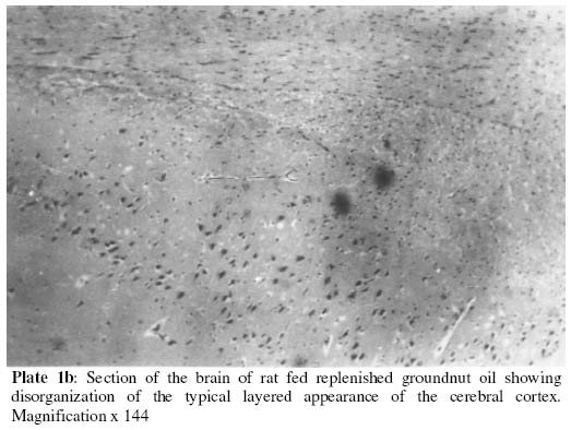
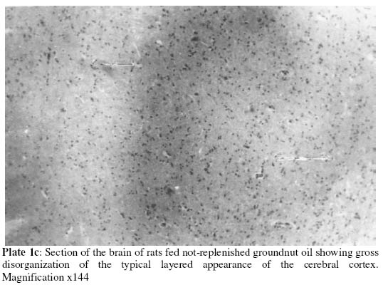
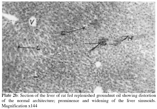
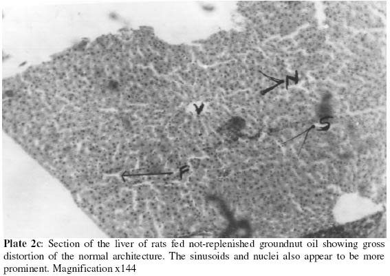
A.Diet of animals fed with fresh groundnut oil; The brain (cerebral cortex), liver and kidney were quickly removed from sacrificed rats after which the kidney was decapsulated. They were cleansed of blood using 0.25M sucrose solution. They were then fixed separately in 10% formalin solution before being taken to the Chemical Pathology Department, University of Ilorin Teaching Hospital, Ilorin, Nigeria, where the histopathology was carried out. Histological proceduresFixed tissues were dehydrated through ascending grades of ethanol to absolute ethanol. They were cleaned in xylene, impregnated and embedded in paraffin wax (melting point 56oC). Sections were cut at 5mm on a rotatory microtome. They were flattened on warm water and mounted onto albumerised slides and dried overnight. The sections were dewaxed in xylene and hydrated through descending grades of ethanol to water. They were initially stained in Harris haematoxylin and differentiated in acid alcohol and thereafter stained with methylene blue. They were then dehydrated in 95% alcohol, stained in 10% alcohol eosin, dehydrated in absolute alcohol, cleaned in xylene and mounted in Canada balsam. The resulting slides are then viewed under the light microscope. The photomicrographs were printed at a total magnification of x144. RESULTSPlates 1 a, b, c show the sections of the brain following the various treatment regimen described earlier. Section of control rat brain showed the typical layered appearance of the cerebral cortex labelled 1-6 as follows: 1 - molecular layer; 2 - external granular layer; 3 - pyramidal cell layer; 4 - internal granular layer; 5 - ganglionic layer; 6 - multiform layer. Section of the brain of rats fed replenished groundnut oil showed disorganization of normal architecture (1b). A section of the brain (1c) of rats fed with not replenished groundnut oil showed gross disorganization of normal architecture. Plates 2 a, b, c show the sections of the liver following the various treatment regimen described earlier. Plate 2a shows a section of control rat liver depicting essentially normal liver architecture, with hepatocytes (H) radiating from the central vein (V). A section of the liver of rats fed replenished groundnut oil (2b) shows distortion of the normal architecture; prominence and widening of the liver sinusoids. The liver section of animals fed with not-replenished groundnut oil (2c) shows gross distortion of the normal architecture, the sinusoids and nuclei also appear to be more prominent. Plates 3 a, b, c show the sections of the kidney following the various treatment regimen described earlier. Plate 3a shows a section of the kidney of control rat depicting normal architecture of tubules (T) and glomeruli (G). Ingestion of replenished groundnut oil led to disorganization of normal architecture (3b) tubules and glomeruli are no longer clearly distinguished. Plate 3c shows gross disorganization of normal architecture, the tubules and glomeruli cannot be distinguished. DISCUSSIONMicroscopic examinations of the different tissues (brain, liver and kidney) showed significant changes. There is a paucity of information on tissue pathology in studies relevant to the present one. In this investigation, the disintegration of the cytoplasmic membrane of the glomeruli and kidney tubules were observed (i.e. the glomeruli and tubules can no longer be distinguished) when rats were placed on diet containing oxidized groundnut oil. Disruption of cellular membranes has been reported as one of the consequences of ingestion oxidized lipids (Braughler and Hall, 1989; Kagan 1988; Odutuga et al, 1997; Ologan, 2002). This is as a result of peroxidation of membrane phospholipid due to free radical attack (Halliwell and Gutteridge, 1989; van Ginkell and Sevanian, 1994). The disintegration of cytoplamsmic membrane may likely lead to disruption in the filteration and concentration of urine, it may also affect the fluid and electrolyte balance of the rat as well as regulation of total body homeostasis. In the liver, mild to severe necrosis of the cells of animals fed oxidized oil were observed in this study. The sinusoids of animals fed oxidized groundnut oil were found to be widened and the nuclei appear to be more prominent. This is as a result of disintegration of the normal cellular structure observed in the control group. Necrosis of the liver has been reported as one of the consequences of oxidized oil ingestion (Alexander, 1978; Sander, 1983). Morphological changes caused by cell injury are known to become apparent only after some critical biochemical systems within the cell have been deranged (Ngaha, 1979; Akanji, 1986). Disruption of the liver cell as reflected by altered morphological structure is therefore suggested as the cause of raised serum level of liver enzymes as previously reported (Jimoh and Odutuga, 2001. There is paucity of information on morphological changes in the brain due to dietary oxidized oil ingestion. We report here the fact that, the typical layered appearance of the cerebral cortex was disrupted in the groups of rat fed thermally oxidized groundnut oil. These changes would probably be the result of oxidized oil ingestion and might imply change in function of this part of the brain. Kieckebu et al (1962) had earlier reported exaggerated responses of the central nervous system to stimuli as well as spontaneous movement as arising from the ingestion of oxidized oil. The result of the different sections of the tissues of animals fed oxidized oil showed that the diets containing oxidized groundnut oil has deleterious effects on the architecture of the tissues. The effect is more pronounced in animals fed with not replenished groundnut oil. REFERENCES
The following images related to this document are available:Photo images[bk04001p2c.jpg] [bk04001p2a.jpg] [bk04001p3a.jpg] [bk04001p1a.jpg] [bk04001p3c.jpg] [bk04001p1b.jpg] [bk04001p1c.jpg] [bk04001p3b.jpg] [bk04001p2b.jpg] |
| |||||||||
