
|
Biokemistri
Nigerian Society for Experimental Biology
ISSN: 0795-8080
Vol. 17, Num. 2, 2005, pp. 57-71
|
Biokemistri, Vol. 17, No. 2, Dec, 2005, pp. 57-71
Review
Article
An overview of toxic
freshwater cyanobacteria in South Africa with special reference to risk, impact and
detection by molecular marker tools
Paul J. OBERHOLSTER1, Anna-Maria
BOTHA*1,2,,3 and T. Eugene CLOETE1
1Department of
Microbiology and Plant Pathology, 2Department of Genetics, University
of Pretoria, Hillcrest, Pretoria, ZA0002, South
Africa;
3Department of Soil and Crop Sciences, ColoradoStateUniversity,
Fort Collins,
CO80521, USA
*Author to whom
all correspondence should be 1addressed. E-mail: ambothao@postino.up.ac.za; Tel:
+27124203945;Fax::+27124203947
Received 13 May 2005
Code Number: bk05010
Abstract
Toxic
cyanobacteria found in eutrophic, municipal and residential water supplies are
an increasing environmental hazard in South Africa. Cyanobacteria produce
lethal toxins, and domestic and wild animal deaths are caused by drinking water
contaminated by these toxins. Among the species causing death of livestock,
blooms of Microcystis aeruginosa are the most common in South Africa.
More than 65 microcystins have been isolated to date and they are the most
abundant cyanobacterial toxins. Hazards to human health may result from chronic
exposure via contaminated water supplies. Microcystins are powerful tumour
promoters and inhibitors of protein phosphatase 1 and 2A and they are suspected
to be involved in the promotion of primary liver cancer in humans. In this
minireview, we discuss the significance of toxic cyanobacteria in South Africa
as well as the detection of potential microcystin-producing cyanobacteria
strains in South African reservoirs with a mcyB molecular marker. It
would be of economic and public health value to be able to detect early stage
blooms of cyanobacteria, especially if it is on a sufficiently timely basis for
municipalities and recreation facilities to implement a response plan.
Key words: water quality, Microcystis
aeruginosa, longterm exposure, purification processes
Introduction
Southern Africa
is generally an arid to semi-arid region, with an average rainfall of a little
under 500 mm per annum. There are practically no freshwater lakes in South Africa;
exploitable water supplies are therefore confined to rivers, artificial lakes
behind dams, and groundwater. The total runoff from South Africa
is estimated at 53 500 million m3 per annum, of which about 33 000
million m3 could practically be exploited. The many demands for
water, and the erratic flow of most South African rivers, have led to the
creation of artificial lakes, i.e. impoundments on all the major rivers, in
order to stabilize flow and therefore guarantee annual water supply. The total
capacity of state impoundments amounts to more than 50 per cent of South Africa’s
total average annual river runoff1.
Urban complexes in Gauteng especially Pretoria and Johannesburg, generate large amounts of sewage, which even if
treated give rise to effluents that are high in salts, phosphates and nitrates.
When effluents containing high levels of nutrients reach artificial lakes, they
stimulate growth of algae including cyanobacteria leading to accelerated
eutrophication, disturbances of relationships among organisms, biodiversity and
levels of oxygen concentrations. Extensive growth of cyanobacteria in compound
reservoirs can create severe problems in the maintenance of water supplies and
in meeting the ever-increasing demand for potable water2,3.
Large cyanobacteria blooms may rapidly clog not only
the fine sand filters but even the primary coarse fast filters of water
treatment plants. Secondly, cyanobacteria may release substances in the water
that are harmful or toxic, which cause unnatural colouration of the raw water
or which add an objectionable odour or taste to drinking water2.
Cyanobacterial
toxins and health effects
Toxins of cyanobacteria are grouped in two main
categories by Carmichael4 namely, biotoxins and cytotoxins based on
the types of bioassays used to screen for their activity. Cytotoxins are
detected by mammalian cell lines and biotoxins are assayed with small animals,
e.g. mice or aquatic invertebrates. Because cytotoxins are not highly lethal
to animals, and no reports in South Africa have been published indicating they
were responsible
for livestock deaths in the field, they will not be discussed further. In the
toxicity standards, biotoxins are considered supertoxic (Table 1). Biotoxins
of
cyanobacteria are water-soluble and heat stable and they are released upon
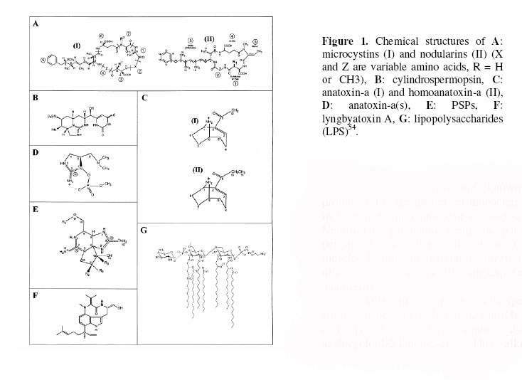
aging or lysis of the cells. The primary types of cyanobacterial biotoxins
include hepatotoxin (microcystins, nodularins, cylindrospermopsins), neurotoxin
(anatoxins, saxitoxins) and dermatotoxins (lyngbyatoxin A, aplysiatoxins,
lipopolysaccharides) (Fig. 1).
Hepatotoxins
Hepatotoxins are low molecular weight cyclic peptide
toxins that affect the liver and have been the predominant toxins involved in
the case of freshwater algae toxicosis.
Microcystins and nodularins
Microcystin are a cyclic heptapeptides with about 65
different isoforms identified, with diverse levels of toxicity5.
Nodularins are pentapeptides with only four forms been described. The
hepatospecificity of these toxins is due to the requirement for uptake by a
bile acid transporter. Microcystin and nodularins have been shown to be
inhibitors of serine/threonine protein phosphatase 1 and 2A. This inhibition
leads to hyperphosphorylation of proteins associated with the cytoskeleton in
hepatocytes6.
Cylindrospermopsins
Cylindrospermopsins is an alkaloid containing a
tricyclic guanidine combined with hydroxymethyl uracyl and is stable to boiling.
Studies on the mechanism of action of cylindrospermopsin have shown that in
mouse hepatocytes in vivo the toxin disrupts protein synthesis7.
The main target of this toxin is the liver, but unlike the microcystins, it can
affect other organs such as the lungs, kidneys, adrenals and intestine8.
Genotoxic activity is caused by the ability of cylindrospermopsins to induce
strand breaks at the DNA level and loss of whole chromosomes 9.
Table 1: Comparison
of toxicities of some biological toxins
|
Toxins
|
Sources
|
Lethal doses (LD50)
|
Reference
|
|
Saxitoxin
|
Aphanizomenon flos-aquae
|
10
|
Oshima, 1995.98
|
|
Anatoxin-a(s)
|
Anabaena flos-aquae
|
20
|
Falconer, 1998.8
|
|
Cobra toxin
|
Naja
naja
|
20
|
Bagchi, 1996.69
|
|
Nodularin
|
Nodularia spumigena
|
30
|
Rhinehart et al., 1994.70
|
|
Microcystin-LR
|
Microcystis aeruginosa
|
50
|
Rhinehart et al., 1994.
70
|
|
Anatoxin-a
|
Anabaena flos-aquae
|
200
|
Carmichael, 1992.10
|
|
Brevetoxin
|
Karenia brevis (dinoflagellate)
|
500
|
Morohashi et al., 1999.71
|
|
Ciguatoxin
|
Gambierdiscus toxicus
(dinoflagellate)
|
0.25
|
Bagnis et al., 1980.72
|
|
Cylindrospermopsins
|
Cylindrospermopsins raciborskii
|
2 100
|
Ohtani et al., 1992.73
|
|
Styrchnine
|
Strychnos nuxvomica
|
2 000
|
Bagchi, 1996.69
|
The neurotoxins are known to be produced
by freshwater cyanobacteria strains include anatoxin-a, anatoxin-a(s) and
saxitoxins. Neurotoxins producing death by paralysis of peripheral skeletal
muscles, then respiratory muscles leading to respiratory arrest in a few
minutes to a few hours following exposure.
Anatoxins
Anatoxin-a are produced by species and strains of the
genera Anabaena and Oscillatoria and is a secondary amine,
2-acetyl-9-azabicyclo(4.2.1)non-2-ene. This alkaloid, a structural analogue of
cocaine, is a potent post-synaptic cholinergic nicotinic agonist, which causes
a depolarizing neuromuscular blockade, followed by fatigue and paralysis10.
Anatoxin-a(s) is unrelated to anatoxin-a. Structurally
it is a unique N-hydroxyguanidine methyl phosphate ester. It can be called a
natural organophosphate because of its ability to irreversibly inhibit
acetylcholinesterase, causing the same clinical end result as anatoxin-a10.
Blood, lung and muscle acetylcholinesterases are inhibited, whereas retina and
brain acetylcholinesterase activities are normal11.
Saxitoxins
Saxitoxins or paralytic shellfish poisons are produced
by species and strains of freshwater cyanobacteria Anabaena and Aphanizomenon,
but are better known as the products of dinoflagellates, the marine algae
responsible for red-tide paralytic shellfish poisoning. The saxitoxins or
paralystic shellfish poisons inhibit nerve conduction by blocking sodium
channels in axons, thereby preventing the release of acetylcholine at
neuromuscular junctions with resultant muscle paralysis. The paralysis of the
respiratory muscles leads to the death of animals within a few minutes12.
Dermatotoxins
Dermatotoxins lyngbyatoxin A and aplysiatoxin are
produced by the cyanobacterium Lyngbya majuscula, a marine benthic
cyanobacterium with different metabolite constituents in deep and shallow water
varieties. While the deep water varieties produce inflammatory substances and
tumor promoters, the shallow water forms produce lipophilic substances,
malyngamides A, B and C. Clinical signs include skin, eye and respiratory
irritation13. .
Historical
perspective in South Africa
The genus of most concern for toxin-producing strains
is the cosmopolitan Microcystis, predominantly Microcystis aeruginosa,
with other genera being Oscillatoria, Anabaena, Aphanizomenon
and Nodularia14,15. In South
Africa almost all cases of animal
poisoning have been associated with Microcystis aeruginosa16,17(Table
2). Early researchers on the algal flora of South
Africa have commented on the ubiquitous
nature of Microcystis throughout the country18,19. It was
around this period (1927) that the first cattle intoxications by Microcystis
in the Transvaal province were recorded. In agricultural practice,
poisoning of farm animals occurs when the animals are prevented from reaching
clean water by the specific layout of fences restricting them to shorelines
contaminated by cyanobacteria. Because access to drinking water may be the
limiting feature of livestock production in arid climates such as those of South Africa or
Australia, poisoning episodes have been reported more often from those countries20,21,22.
Yearly duration of exposure is also shorter (3-5 months) in countries where the
water bloom growth season is shorter, like the United States and Canada compared
to those with milder climates such as Australia and South
Africa (6-10 months)23. As
far as can be ascertained, cases of poisoning in South Africa have only been
described from the former Transvaal (currently Gauteng and Mpumalanga),
former Orange Free State (currently Free
State)16,17 and Western Cape
provinces24. In Gauteng, cyanobacteria poisoning periodically occurs around
the Bon Accord and Hartbeespoort Dams25. Kellerman et al. 25
ranked the plant poisonings and mycotoxicoses occurring in South Africa in
order of importance and regarded Microcystis aeruginosa poisoning as the
fifth most important type of poisoning in the Gauteng Province, and the tenth
most important in Mpumalanga.
Chronic
studies of cyanotoxins; implications for humans
Where climate and other environmental factors permit,
there may be continuous water blooms of toxic cyanobacteria in drinking water
reservoirs and other surface water supplies (Table 3). The dominance of
cyanobacteria may be due to their low need for uptake of nutrients during the
benthic life phase and overwintering26 or additionally, buoyancy
control enables them to outcompete other algal species for light and nutrients27.
While water supply authorities often control these blooms, the conventional
method of algicide treatment lyses the organisms, releasing toxic cell
contents into the water. The chronic administration of Microcystis extract
in the drinking water of mice resulted in increased mortality, particularly in
male mice, together with chronic active liver injury. The deaths were largely
due to endemic bronchopneumonia, indicating an impairment of disease resistance.
Only six tumors were seen in the 430 mice killed at intervals up to 57 weeks
of age; however, four of the six tumors were in females that ingested
the highest Microcystis concentration23.
Table 2:
Some reported animal poisoning incidents related to cyanobacterial blooms in South
Africa
|
Cases attributed to cyanotoxins in raw
drinking water
|
|
Date
|
Description
|
|
1913-1943
|
Location;
Free State and Southeast
Transvaal. Affected animals;
Thousands of livestock (horses, sheep, cattle and rabbits); Symptoms and
findings; liver damage, photosensitivity Organism; Microsystis toxica
(=aeruginosa)21,74.
|
|
1973-1974
|
Location;
Hartbeespoort Dam. Affected animals; cattle deaths, Symptoms and findings;
microcystin poisoning Organism; Microcystis aeruginosa75.
|
|
1979
|
Location;
Klipvoor Dam. Affected animals; Death of 3 White Rhinoceroses (Ceratotherium
simum). Symptoms and findings; Necrosis of the liver. Organism; Microcystis
aeruginosa76.
|
|
1980
|
Location;
Vaal
Dam. Affected animal; Cattle deaths Symptoms and findings; Microcystis
poisoning Organism; Microcystis aeruginosa77.
|
|
1984
|
Location;
Willem Pretorius Game Reserve (Free
State). Affected animals; Death of
several Black Wildebeest (Connochaetes gnou) Symptoms and findings; Microcystis
poisoning, Organism; Microcystis aeruginosa76.
|
|
1987
|
Location;
Eastern Transvaal. Affected animal; Death of 47 cattle. Symptoms and
findings; Microcystin poisoning Organism; Microcystis aeruginosa 17.
|
|
1989
|
Location;
Bloemhof Dam, Sandveld Nature Reserve (Free
State). Affected animals; Seven
Giraffe deaths Symptoms and findings; Microcystin poisoning Organism; Microcystis
aeruginosa78.
|
|
1989
|
Location;
Klipdrif Dam. Affected animals; Livestock Symptoms and findings; Microcystis
poisoning Organism; Microcystis aeruginosa79.
|
|
1994
|
Location;
Zeekoevlei. Affected animal; Bull terrier bitch Symptoms and findings;
Hepatic necrosis, first reported incident of nodularin in South Africa.
Organism; Nodularia spumigena15.
|
|
1994
|
Location;
Paarl, Western Cape. Affected animals; Death of 11 sheep and
induced-photosensitivity in a further 20 animals Symptoms and findings;
Hepatotoxin, microcystin-LR Organism; Microcystis aeruginosa24.
|
|
1996
|
Location;
Tsitsikamma-Kareedouw district, South
Cape. Affected animals; Death of 290
dairy livestock and induced-photosensitivity in a further 70 Symptoms and
findings; Microcystin poisoning Organism; Anabaena spp. and Oscillatoria
spp.80.
|
|
1998
|
Location;
Erfenis Dam, Free State Affected animals; Death of livestock Symptoms and
findings; Neurotoxicosis Organism; Anabaena spp.81.
|
|
2000
|
Location;
Orange River. Affected animals; Fish kills along the rivier
Symptoms and findings; First reported incident of Cylindrospermopsis
raciborskii in South Africa. Organism; Cylindrospermopsis raciborskii, Anabaena
sp., Oscillatoria sp.82.
|
This result led to an investigation of the
tumor-promoting activity of orally administered Microcystis in mice that
had dimethylbenzanthracene applied to their skin. Results of these trials
showed that there were significant increases in the growth of skin papillomas
in mice given Microcystis but not Anabaena to drink28.
The finding that microcystin activated phosphorylase A preceded studies
showing that microcystin-LR, -YR, and -RR, and nodularin are potent inhibitors
of protein phosphatases type 1 and type 2A29. This inhibition leads
to hyperphosphorylation of proteins associated with the cytoskeleton in
hepatocytes. The rapid loss of the sinusoidal architecture and attachment to
one another leads to the accumulation of blood in the liver, and death most
often results from hemorrhagic shock. These experiments clearly indicate that
microcystin are a health threat in drinking water supplies10.
Removal
of Microcystis toxins in water purification processes
Hoffman30 demonstrated that dissolved
substances like microcystin deriving from Microcystis aeruginosa samples
of the Hartbeespoort Dam were not removed to below ‘active levels’ by
conventional water treatment like flocculation, sedimentation, rapid sand
filtration and chlorination. These findings are in accordance with results
presented by James and Fawell31 and Rositano and Nicholson32 that
flocculation was effective in removing cells, but not in eliminating free
microcystins and other extra-cellular secondary metabolites which remained
constant after flocculation with aluminium sulphate33,34 or ferric
chloride35. In another study Pietsch et al.36
reported that flocculation and filtration resulted in an increase of
extracellar toxin after experiments with Microcystis aeruginosa and Planktothrix
rubescens. The researchers suggested turbulences in pipes and pressure
gradients in the filter as reasons for the increase of the toxin level. The
efficacy of chlorine (0.5 mg/l) to eliminate microcystin is also doubtful37.
Water treatment studies conducted at the laboratory and pilot plant-scale have
concluded that granular activated carbon filtration is effective in removing
the cyanobacterial toxins from water38,39. This treatment add
considerably to the expenses of water treatment and only a few purification
water treatment plants in South Africa is equipped with granular activated
carbon systems, the rest make use of conventional water treatment practices
that remove live cyanobacterial cells and debris but not biotoxins in solution.
In rural areas the choice of water supply may be limited, depending on the
stage of development of the country. Similarly, in urban areas if the
reticulated drinking water is of doubtful quality, the only choice may be
bottled water, which is financially out of reach for the poorer majority of the
population. Thus, the potential for injury from cyanobacteria toxins in water
supplies will to some extent depend on the level of development of the country
and to some extent on the socio-economic status of the family40.
Survey analysis of utility waters in the United States and
Canada
were confirmed to contain microcystin during the sampling period of June 1996
to January 1998. Of the 677 samples collected, 539 (80 percent) were positive
for microcystin when tested using ELISA. Of the positive samples, 4.3 percent
were higher than the WHO drinking water guideline levels of 1μg/L. Only two of the plant outlet samples submitted exceeded the 1-μg/L WHO drinking water guideline. This indicates that, although almost
all water treatment plants had adequate procedure to reduce microcystin to safe
levels in the finished water during the test period, the majority of source
waters with cyanobacteria do contain microcystin23. Surveys of
different cyanobacterial blooms for given geographical areas have shown that
the frequencies of toxic cyanobacterial blooms in raw water ranged from 22 to
95%5. For example, the frequency is an average of 74% for some
Mediterranean countries including Portugal, France (Brittany) and Greece41,42,43. The screening of
cyanobacterium strains isolated from rice fields, irrigation and drainage water
canals in the Nile Delta in Egypt showed that 23% of these isolates were found as
active producers of microcystins with an amount of more than 500 ng-144.
A survey of cyanobacterial water blooms carried out from 2000 to 2004 in South
Africa reservoirs confirmed an average frequency of 95% toxicity in field
samples tested by the ELISA method45(Table 3).
Human
health risks of long-term exposure to low levels of microcystin
Little information is available on the effects of
long-term exposure to low levels of microcystin toxins in humans. We know that
in experiments performed on a time-scale of minutes or hours, microcystin has
obvious effects on the functions of plant and animal cells at concentrations as
low as 3-10 nM that is equivalent to 3-10 μg for an adult
female liver. In cells that take up microcystin freely, the maximum effects are
visible at concentrations of around 1μM, the point at
which all of the cellular PP1 and PP2A is saturated with toxin. This means that
approximately 1 mg (equivalent to drinking two liters of water per day at 32 μg/L microcystin over two weeks) would bind all of the PP1 and PP2A in
an adult female human liver, provided that the PP-microcystin complexes were
stable46. However most of the available data about uptake and
turnover of microcystins has been obtained from experiments carried out with
rodents. In this regard, it should be noted that PP1 and PP2A from mice and
humans amino acid sequences are 100 percent identical47. In the case
of mice low doses of microsystin cause progressive changes in liver tissue over
time, including chronic inflammation, focal degeneration of hepatocytes and the
accumulation of metabolites such as bilirubin in the blood, and tend to
increase mortality48. In South
Africa, liver damage and death of
vervet monkeys has occurred following toxic Microcystis administration
with signs of poisoning similar to those observed in live stock and mice49.
These demonstrations of the susceptibility of primates to cyanobacterial poisoning
are consistent with the results of an epidemiological study of a human
population of the city of Armidale, New South
Wales, Australia, which obtains its
drinking water from the Malpas Dam reservoir. A clear pattern of admission of
patients to the local hospital with liver complaints was identified which
coincided with the seasonal production of a hepatotoxic Microcystis
aeruginosa bloom in the reservoir. This correlation was confined to
patients who had taken their drinking water from the Malpas Dam50.
Yu51 in 1995 reported that the incidence of
liver cancer is significantly higher for populations using
cyanobacteria-infested surface water than those drinking groundwater in China. In Shanghai and
its nearby regions where epidemiological studies showed that increased
incidence of primary liver cancer is related to the consumption of microcystin
contaminated water, the concentrations of microcystins in samples of pond-ditch
water were within the range of 0.09-0.46 μg/l52.
However, Zegura et al.53 showed that microcystin-LR induced
oxidative DNA damage in HepG2 human cells at low concentrations (0.01μg/ml) and this might be a mechanism by which chronic exposure to low
concentrations of mycrocistins contribute to increase the risk for liver
cancer development. A recent study in mice has shown that Microcystis
aeruginosa extract provided in drinking water increased the area of
aberrant crypt foci in the colon, suggestive that microcystins promote
preneoplastic colonic lesions55.
Monitoring
toxigenicity of cyanobacterial strains by molecular assay
Monitoring the quality of water destined to public
supply includes identification of potentially toxic cyanobacteria and their
population density. Identification of such microorganisms based on
morphological features only, though widespread, has proven problematic, mainly
for the genus Microcystis, due to its extensive phenotypic plasticity56.
Identification of a cyanobacterial genus by microscopic morphology or molecular
analysis does not indicate the potential for toxin production. Different
strains of one species can be morphologically identical but differ in
toxigenicity. Microcystis aeruginosa for example has both toxic and
nontoxic strains57.
Table 3. Acute
intoxications of humans from cyanobacteria
|
Cases attributed to cyanotoxins in drinking
water
|
|
Year
|
Report
|
|
1931
|
United
States; A massive Microcystis
bloom in the Ohio and Potomac rivers caused illness in 5 000 to 8 000 persons
whose drinking water was taken from these rivers. Low rainfall has caused the
water of a side branch of the river to develop a cyanobacterial bloom, which
was then washed by new rainfall into the main river. Drinking water treatment
by precipitation, filtration, and chlorination was not sufficient to remove
the toxins83, 84.
|
|
1960
to 1965
|
Zimbabwe, Harare; Cases of acute gastroenteritis among European
children admitted to the local hospital in Salisbury, Rhodesia (now
Harare, Zimbabwe). In this instance, several supply reservoirs
provided water to different regions of the city, but only the reservoir
containing blooms of Microcystis supplied water to the affected
population85.
|
|
1968
|
United
States; Numerous cases of gastrointestinal illness after exposure to mass
developments of cyanobacteria were compiled by Schwimmer and Schwimmer (1968)86.
|
|
1975
|
United
States; Hindman et al. (1975)87 reported the results of an
investigation into 49 pyrogenic reactions in patients undergoing
haemodialysis treatment in Washington, DC. They concluded that ‘the cause of these reactions
was traced to an increase in endotoxin contamination of the tap water used to
prepare dialysate, possibly caused by an increase in the algae levels in the
local water source.
|
|
1979
|
Australia;
Combating a bloom of Cylindrospermopsis raciborskii in a drinking
water reservoir on Palm Island with copper sulfate led to liberation of
toxins from the cells into the water, thus causing serious illness with
hospitalization of 141 persons supplied from this reservoir88,89.
|
|
1981
|
Australia; In the city of Armidale, liver enzyme activities were elevated in the blood
of the population that was supplied from surface water polluted by Microcystis
spp.50.
|
|
1992
|
United
States; Carmichael (1992)10 compiled case studies on nausea,
vomiting, diarrhea, fever and eye, ear, and throat infections after exposure
to mass developments of cyanobacteria.
|
|
1993
|
Australia;
Ressom et al. (1994)28 estimated that more than 600,000
person-days are lost annually due to absence of their water source due in
turn to toxic cyanobacterial blooms.
China; The incidence of very high rates of liver cancer
is related to water sources. The incidence is significantly higher for
populations using cyanobacteria-infested surface waters than those drinking
ground water. A cohort study showed that people who drank pond and ditch
water had 121 deaths per 100 000 compared with 0 for those who drank well
water51, 90.
|
|
1994
|
Sweden
Near Malmo; Illegal use of untreated river water in a sugar factory led to an
accidental cross-connection with the drinking water supply for an uncertain
number of hours. The river water was densely populated by Planktothrix
agardhii, and samples taken a few days before and a few days after the
incident showed these cyanobacteria contained mycrocistins. Of 304
inhabitants of the village, 121 became ill with vomiting, diarrhea, muscular
cramps, and nausea91.
|
|
|
Cases
attributed to cyanotoxins in recreational water
|
|
Date
|
Description
|
|
1959
|
Saskatchewan, Canada; In spite of livestock deaths and warnings against
recreational use, people did swim in a lake infested with cyanobacteria.
Thirteen persons became ill (headaches, nausea, muscular pains, painful
diarrhea). In the excreta of one patient – a medical doctor who had
accidentally ingested 300 ml of water-numerous cells of Microcystis spp.
And some trichomes of Anabaena circinalis could be clearly identified92.
|
|
1989
|
England;
In Staffordshire ten out of 20 soldiers became ill after swimming and
canoe-training in water with a heavy bloom of Microcystis spp.; two of
them develop severe pneumonia attributed to the inhalation of a Microcystis
toxin and required hospitalization and intensive care. Sixteen develop sore
throat, headache, abdominal pain, dry cough, diarrhoea, vomiting and
blistered mouths93. Swimming skills and the amount of water
ingested appear to have been related to the degree of illness.
|
|
1995
|
Australia;
Epidemiological evidence of adverse health effects after recreational water
contact from a prospective study involving 852 participants who showed
elevated incidence of diarrhea, vomiting, flu symptoms, skin rashes, mouth
ulcers, fevers, and eye or ear irritations within 2 to 7 days after exposure.
The sensitivity of individuals to allergic-type reactions at low
cyanobacteria cell densities is greater than can be attributed to the toxin
content of cyanobacteria94.
|
|
|
Cases
due to other exposure routes
|
|
Date
|
Description
|
|
1996
|
Caruaru
in Brazil; One hundred and twenty six dialysis patients were exposed to
microcystin through the water used for dialysis, and 60 of them eventually
died, principally of liver failure, 6 had died by 2 weeks after exposure, 30
by 6 weeks, 44 by 10 weeks, and 55 by 27 weeks. At least 44 of these victims
showed the typical common symptoms associated with microcystin, now referred
to as ‘Caruaru Syndrome’ and the liver microcystin content corresponded to
that of laboratory animals that received a lethal dose of microcystin95,96,97.
|
There have been numerous attempts to refine the
identification of strains by using amplified fragment length polymorphism
markers58, and specific gene analysis. Examples include the use of
PCR-based methods for amplification of the phycocyanin intergenic spacer
(PC-IGS) between the α and β subunits of the phycocyanin
operon in environmental samples59, the 16S-23S rRNA internally
transcribed spacer region60 and the DNA-dependent RNA polymerase
(rpoCI) gene61. Although these molecular techniques have improved
the accuracy of strain identification, they have not been able to distinguish
toxigenic from nontoxigenic strains of the same species.
The biosynthetic pathway for production of microcystin
has now been elucidated62 and this has enabled the development of
specific oligonucleotide primers for gene common to production of microcystins62.
To better detect microcystin-producing cyanobacterial strains, Neilan et al.63
and Nishizawa et al.64 have developed genetic probes
directed, respectively, to the mcyB gene and to adenylation domains within
the microcystin synthetase gene cluster. The mcy gene cluster contains
55kb of DNA encoding six large open reading frames, mcyA-E and -G,
together with a further four small open reading frames mcyF and H-J,
placed in the chromosome62. The insertional inactivation of
microcystin peptide synthetase gene mcyB of a Microcystis aeruginosa
strain (PCC 7806) resulted in loss of microcystin production, showing their
involvement in microcystin synthesis. It was also observed by Dittmann et
al.65 that all isoforms of the cyclic heptapeptide were
disrupted by inactivation of the microcystin synthetase gene sequence mcyB.
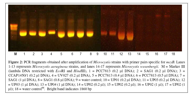
Recently, Oberholster66 reported for the
first time in South Africa, the use of microcystin molecular markers for the
detection of toxic cyanobacteria, both in cultivated strains and environmental
samples in Gauteng and the North
West provinces. Microcystis
aeruginosa and Microcystis wessenbergii strains from Rietvlei,
Hartbeespoort and Roodeplaat Dams in Gauteng and the North
west provinces were analyzed by
polymerase chain reaction (PCR) with oligonucleotide primers for the mcyB
gene of the operon that encodes a microcystin synthetase (Fig.
2). The presence
of the gene mcyB in three of the four environmental strains indicates
that the strains produce microcystin.
By using the mcyB gene in PCR assays, applied
directly to environmental samples provide a useful indicator that the analyzed
strains have the genetic potential to produce microcystin. Although HPLC
provides a direct measure of toxins present, it does require a large capital
investment and considerable sample preparation. The PCR-based assays detect
toxigenic cells rather than toxins and require little sample preparation and
modest capital costs. Detection of toxic Microcystis aeruginosa strains
through molecular markers for microcystin may have great use-potential in
routine analysis of aquatic ecosystems. Thus, it may make water monitoring more
feasible and allow the early application of corrective action before
cyanobacteria blooms start to die or disintegrate. The PCR-based assay is
effective at a level of 10 cells ml-1 and can indicate a possible
toxic bloom well before the cell count reaches the action alert at a cell
density of 2 000 ml-1, as recommended by the Australian Drinking
Water Guideline67, and a high alert level of 20 000 cell ml-1,
where blooms may contain sufficient toxin to be of concern for human health68.
Acknowledgements
The
authors would like to express their gratitude to the Water Research Commission
and the National Research Foundation of South Africa for their financial
assistance.
REFERENCES
- Noble, T. and Hemens, M. (1978) Inland water ecosystems in South Africa-a
review of reseach needs. South African National Scientific Programmes Report
no 34. pp. 21-80.
- Meyer, C. M. (1987) Contamination of water by the toxic blue-green alga Microcystis aeruginosa.
Suid-Afrikaanse Tydskrif vir Wetenskap. 83: 517-518.
- Oberholster,
P. J., Botha, A-M. and Grobbelaar, J. U. (2004) Microcystis aeruginosa:
source of toxic microcystins in drinking water. Afr. J. Biotech. 3:
159-168.
- Carmichael, W. W. (1994) The toxins of cyanobacteria. Scientific American 11: 64-72.
- Sivonen, K. and Jones, G. (1999) Cyanobacterial toxins, In: Toxic
Cyanobacteria in Water, a Guide of Their Public Health Consequences,
Monitoring and Management. I. Chorus and J. Bartram (eds.), pp. 41-111.
E &FN Spon, London.
- Tiovola, D. M., Goldman R. D.,
Garrod, D. R. and Eriksson, J. E. (1997) Protein phosphatases maintain
the organization and structural interactions of hepatic keratin intermediate
filaments. J. Cell Sci. 100: 23-33.
- Tereo, K., Ohmori, S., Igarashi, K., Ohtani, I., Harada, K-I., Ito, E.
and Watanabe, M. (1994) Electron microscopic studies on experimental poisoning
in mice induced by cylindrospermopsin isolated from blue-green alga Umezakia
natans. Toxicon. 32: 833-843.
- Falconer, I. R. (1998). Algal toxins and human
health. In: J.H. Rubec (ed). The handbook of environmental chemistry. 5 Part
C. Quality and treatment of drinking water II. Springer-Verlag, Berlin.
pp. 53-82.
- Humpage, A. R., Fenech, M., Thomas, P. and Falconer, I. R. (2000a)
Micronucleus induction and chromosome loss in transformed human white cells
indicate clastogenic and aneugenic action of the cyanobacterial toxin,
cylindrospermopsin. Mutation Research/DNA Repair 472:
155-161.
- Carmichael, W.W. (1992) Status Report on
Planktonic Cyanobacteria (Blue Green Algae) and their Toxins. EPA/600/R-92/079.
Cincinnati, Ohio; USEPA.
- Beasley, V. R., Haschek-Hock, W. M., Carmichael, W. W., Cook, W. D., Dahlem,
A. M., Hooser, S. B., Lovell, R. A., Schaeffer, D. J., Thorn, P. M. and
Valentine, W. M. (1990) Pathophysiology and toxicokinetic studies of
bleu-green algae intoxication in the swine model. Annual report to the U.S.
Army Medical research and Development Command, FortDetrick, Frederick, Maryland.
21702-5102:R.
- Runnegar, M. T., Jackson, A. R. B.
and Falconer, I. R. (1988) Toxicity to mice and sheep of a bloom of the
Cyanobacterium (blue-green alga) Anabaena circinalis. Toxicon 26:
599-602.
- Osborne, N. J., Webb, P. M.,
Shaw, G. R. (2001) The toxins of Lyngbya majuscula and their
human and ecological health effects. Environ. Int. 27 : 381-392.
- Codd, G. A., Bell,
S. G. and Brooks, W. P. (1989) Cyanobacterial toxins in water. Water Sci.Technol. 21:
1-13.
- Harding, W.R., Rowe, N.,
Wessels, J. C., Beattie, K. A. and Codd, G. A. (1995) Death of a dog
attributed to the cyanobacterial (blue-green algal) hepatotoxicosis nodularin
in South Africa. J.S. Afr. Vet. Assoc. 66: 256-259.
- Kellerman, T. S., Coetzer, J. A.
W. and Naude, T. W. (1988) Plant poisonings and mycotoxicoses of livestock
in Southern Africa. (1st ed.). Cape Town; OxfordUniversity
Press. pp. 49-52.
- Scott, W.E. (1989) Occurrence and significance of toxic cyanobacteria in
South Africa.
Water Sci. Technol. 23:
175-180.
- Huber-Pestalozzi, G. (1929) Das Plankton naturlicher und kunstlicher Seebecken
Sudafrikas, Verh. Int. Ver. theor. angew. Limnol. 4: 343-390.
- Nygaard, G. (1932) Contributions to our knowledge of the freshwater algae
of Africa 9. Freshwater algae and phytoplankton from the Transvaal. Trans. R. Soc. S. Afr. 20: 101-148.
- Francis, G. (1878) Poisonous Australian lake. Nature 18:
11-12.
- Steyn, D.G. (1945) Poisoning
of animals and human beings by algae. S. Afr. J. Sci. 41:
243-244
- McBarron, E. J. and May, V.
(1966) Poisoning of sheep in New
South Wales by the blue-green alga Anacystis
cyanea (Kuetz.) Dr. and Dail, Aust. Vet. J. 42: 449-453.
- Carmichael, W. W. (2001) Assessment of blue-green
algal toxins in raw and finished drinking water. AWWA Research Foundation
and American Water Works Association. ISBN 1-58321-076-8. 1-49
- Van Halderen, A., Harding, W. R.,
Wessels, J.C., Schneider, D. J., Heine, E. W. P., Van der Merwe, J. and
Fourie, J. M. (1995) Cyanobacterial (blue-green algae) poisoning of livestock
in the Western Cape Province of South Africa. J. S.Afr.Vet.Assoc. 56:
49-51.
- Kellerman, T. S., Naude, T. W.
and Fourie, N. (1996) The distribution, diagnosis and estimated economic
impact of plant poisonings and mycotoxicoses in South Africa. Onderstepoort
J. Vet. Res. 63: 65-90.
- Perakis, S.S. (1994) ‘Bentic
Blue-green Algal recruitment in response to alum and environmental factors’.
M.Sc. thesis, Univerity of Washington, Department of Civil Engeneering,
Seattle, WA. interactions. Hydrobiol. 144: 183-192.
- Grobbelaar, J. U., Botes, E., Van den Heever, J. A., Botha, A-M. and
Oberholster, P. J. (2004) Toxin production by cyanobacteria. Scope and
Dynamics of toxin produced by cyanophytes in the freshwaters of South Africa and the human and other
users. WRC Report no. 1029/1/04. ISBN No: 1-77005-191-0. pp. 1-9.
- Ressom, R., Soong, F.
S., Fritzgerald, J., Turczynowicz, L., El Saadi, O., Roder, D., Maynard,
T. and Falconer, L. R. (1994) Health effects of toxic cyanobacteria
(blue-green algae). National Health and Medical Research Council. Looking
Glass Press,
Australian Govt. Pub. Service, Canberra, Australia, ISBN 0-644-32908-4,
pp. 108
- MacKintosh, C., Beattie, K. A.,
Klumpp, S., Cohen, P. and Codd, G. A. (1990) Cyanobacterial microcystin-LR
is a potent and specific inhitor of protein phosphatases 1 and 2 A from
both mammals and higher plants. FEBS Lett. 264: 187-192.
- Hoffmann, J. R. H. (1976) Removal of Microcystis toxins in water purification processes. Water S.A. 2: 58-60.
- James, H. and Fawell, J. (1991) Detection and removal of cyanobacterial
toxins from freshwaters. FR O211, Foundation for Water Research, Marlow.
- Rositano,
J., Nicholson, B. (1994) Water treatment techniques for the removal of
cyanobacterial toxins from water, 2/94, Australian Centre for Water Quality.
- Velzeboer, R., Dirkas, M.,
Donati, C., Burch, M. and Steffensen, D. (1995) Release of geosmin by Anabaena
circinalis following treatment with aluminium sulphate. Water Sci.
Technol. 31: 187-194.
- Chow, C. W. K., Drikas, M.,
House, J., Burch, M. D. and Velzeboer, R. M. A. (1999) The impact of
conventional water treatment processes on cells of the cyanobacterium Microcystis
aeruginosa. Water Research 33: 3253-3262.
- Chow, C. W. K., House, J., Velzeboer, R. M. A., Dirkas, M., Burch, M. D.
and Steffensen, D. A. (1998) The effect of ferric chloride flocculation on
cyanobacterial cells. Water Res. 32: 808-814.
- Pietsch, J., Bornmann, K.,
Schmidt, W. (2002) Relevance of intra and extracellular cyanotoxins for
drinking water treatment. Acta Hydrochimica et. Hydrobiologica 30:
7-15.
- Hitzfeld, B. C., Hoeger, S. J. and Dietrich, D. R. (2000) Cyanobacterial
toxins, removal during drinking water treatment, and human risk assessment. Environmental
Health Perspectives 108: 113-122.
- Newcombe, G., Cook, D., Morrison, J. and Brooke, S. (2001) Water
treatment options for saxitoxins: oszonation or activated carbon adsorption. Fifth
International Conference on Toxic Cyanobacteria, Noosa, Australia. p. 12
- Falconer, I. R.
(1989) Effects on human health of
some toxic cyanobacteria (blue-green algae) in reservoirs, lakes and rivers. Toxicity
Assessment 4: 175-184.
- Falconer, I. R.
(1999) An overview of problems caused
by toxic blue-green algae (Cyanobacteria) in drinking and recreational
water. Environ.
Toxicol. 14: 5-12.
- Lanaras, T., Tsittsamis, S., Chlichlia, C. and Cook, C. M. (1989) Toxic
cyanobacteria in Greek freshwaters. J. Appl. Phycol. 1: 67-73.
- Vasconcelos, V. M. (1994) Toxic cyanobacteria (blue-green algae) in
Portuguese fresh waters. Arch. Hydrobiol. 130: 439-451.
- Vezie, C., Brient, L., Sivonen, K., Betru, G., Lefeuvre, J. C. and
Salkinoja-Salonen, M. (1997) Occurrence of microcystin containing
cyanobacterial blooms in freshwaters of Brittany (France). Arch. Hydrobiol. 139: 401-413.
- Yanni, Y. G. and Carmichael, W. W. (1998) Screening of cyanobacteria
isolated from soil, rice fields and water resources of Nile river delta for
production of cyanotoxins. In: Harmful Algae B. Reguera, J. Blonco,
M.L. Fernandez and T. Wyatt (eds.), pp. 493-494. Xunta de Galicia & Intergovernmental
Oceanographic Commission (IOC) of UNESCO, Santiago de Compostela, Spain.
- Botha,, A-M. and
Oberholster, P. J. (2004) Using molecular markers for the detection of
toxic cyanobacterial strains in Gauteng and North West provinces, ZA. WRC
Report 1502/4, Department of Water Affairs and Forestry. pp. 14.
- MacKintosh, C. (1993) ‘Protein Phosphorylation’ A Practical Approach.
D.G.Hardie (ed.) pp. 1-197. Chapter 9. IRL Oxford .
- Barker, H.M., Craig, S. P., Spurr, N. K. and Cohen, P. T.
W. (1993) Sequence of
human protein serine/threonine phosphatase 1-gamma and localization of
the gene (PPP1CC) encoding it to chromosome bands 12q24.1-q24.2. Biochim. Biophys.
Acta 1178: 228-233.
- Hermansky, S. J., Stohs, S. J., Markin, R.S., Murray, W.J. (1990) Hepatic
lipid peroxidation sulfhydryl status, and toxicity of the blue-green algal
toxin
microcystin-LR in mice. J. Toxicol. Environ. Hlth. 31: 71-91.
- Tustin, R. C., Van Rensburg,
S. J. and Eloff, J. N. (1973) Hepatic damage in the primate following
ingestion with toxic algae. In: Proceedings of an International Liver
Conference with Special Reference to Africa. S. Saunders and J. Terblanche
(eds.), pp. 383-385. University
of Cape Town,
South
Africa.
- Falconer, I. R., Beresford, A. M. and Runnegar, M. T. C. (1983) Evidence
of liver damage by toxin from a bloom of the blue-green alga, Microcystis
aeruginosa Med. J. Aust. 1 : 511-514
- Yu S-Z. (1995) Primary Prevention of Hepatocellular Carcinoma. J. Gastroenterol.
Hepatol. 10: 674-682.
- Ueno, Y., Nagata, S., Tsutsumi,
T., Hasegawa, A., Watanabe, M. F., Park, H. D., Chen, G. C., Chen, G.
and Yu, S. Z. (1996) Detection of microcystins, in blue-green alga, hepatotoxin
in drinking water sampled in Haimen and Fusui, endemic areas of primary
liver
cancer in China, by highly sensitive immunoassay. Carcinogenesis 17: 1317-1321.
- Zegura, B., Sedmak, B. and Filipic, M. (2002) Microcystin-LR induces
oxidative DNA damage in human hepatoma cell line HepG2. Toxicon. 41:
41-48.
- Briand, J-F.,
Jacquet, S., Bernard, C., Humbert, J. F. (2003) Health hazards for
terrestrial vertebrates from toxic cyanobacteria in surface water ecosystems.
Vet. Res. 34: 361-377.
- Humpage, A.R., Hardy, S.J., Moore,
E.J., Froscio, S.M. and Falconer, I.R. (2000b) Microcystins (cyanobacterial
toxins) in drinking water enhance the growth of aberrant crypt foci in
the colon. J. Toxicol.
Environ. Hlth. 61: 101-111.
- Kondo, R., Yoshida, T., Yuki, Y.
and Hiroishi, S. (2000) DNA-DNA reassociation of a bloom-forming
cyanobacterial genus Microcystis. Int. J. Syst. Evol. Microbiol. 50: 767-770.
- Meiβner, K., Dittmann, E. and Bormer, T. (1996) Toxic
and non-toxic strains of the cyanobacterium Microcystis
aeruginosa contain sequences homologous to peptide synthetase genes. FEMS
Microbiol. Lett. 135: 295-303.
- Oberholster, P. J., Botha, A-M., Muller, K. and
Cloete, T. E. (2005) Assessment of
the genetic diversity of geographically unrelated Microcystis aeruginosa strains
using amplified fragment length polymorphisms (AFLPs). Afr. J. Biotech. 4: 389-399.
- Baker, J.A., Neilan, B. A.,
Entsch, B. and McKay, D. B. (2001) Identification of cyanobacteria and
their toxigenicity in environmental samples by rapid molecular analysis.
Environ. Toxicol. 16: 472-482.
- Otsuka, S., Suda, S.,
Li, R., Watanabe, M., Oyaizu, H., Matsumoto, S. and Watanabe, M. M. (1999) Phylogenetic
relationships between toxic and non-toxic strains of the genus Microcystis based
on 16S and 23S internal transcribed spacer sequence. FEMS Micr Biol.
Lett. 172: 15-21.
- Fergusson, K. M. and Saint, C. P.
(2000) Molecular phulogeny of Anabaena circinalis and its
identification in environmental samples by PCR. Appl. Environ. Microbiol. 66: 4145-4148.
- Tillett, D., Parker, D. L. and
B.A. Neilan (2001) Detection of toxigenicity by a probe for the microcystin
synthetase A gene (mcy A) of the cyanobacterial genus Microcystis;
comparison of toxicities with 16S rRNA and phycocyanin operon (phycocyanin
intergenic spacer) phylogenies. Appl. Environ. Microbiol. 67 :
2810-2818.
- Neilan, B.A., Dittmann, E.,
Rouhiainen, L., Bass, R.A., Schaub, V., Sivonen, K. and Borner, T. (1999) Non-ribosomal
peptide synthesis and toxigenicity of cyanobacteria. J.
Bacteriol. 181: 4089-4097.
- Nishizawa, T., Asayama, K.,
Fujll, K., Harada, K. and Shirai, M. (1999) Genetic analysis of the peptide
synthetase gene for a cyclic heptapeptide microcystin in Microcystis spp.
J. Biochem. 126: 520-529.
- Dittman, E., Neiland,
B. A., Erhard, M., von Döhren, H. and Börner, T. (1997) Insertional
mutagenesis of a peptide synthetase gene that is responsible for hepatotoxin
production in the cyanobacterium Microcystis aeruginosa PCC 7806. Mol.
Microbiol. 26: 779-787.
- Oberholster,
P. J. (2004) Assessing genetic diversity and identification of Microcystis
aeruginosa strains through AFLP and PCR-RFLP analysis. M.Sc. Thesis, pp.
1-114 University of the Free State, ZA.
- National
Health and Medical Research Council/Agriculture and Resource Management Council
of Australia and New
Zealand. (1996) Australian drinking water guidelines. National Health
and Medical Research Council, and Agriculture and Resource Management Council
of Australia and New Zealand, Canberra, Australia.
- Fitzgrald, D.J., Cunliffe, D.A.
and Burch, M.D. (1999) Development of health alerts for cyanobacteria and
related toxins in drinking water in South
Australia. Environ. Toxicol. 14:
203-209.
- Bagchi, S.N. (1996) Cyanobacterial toxins. J. Sci. Ind. Res. 55: 715-727.
- Rhinehart, K.L.,
Namikoshi, M. and Choi, B.W. (1994) Structure and biosynthesis of
toxins from blue-green algae (cyanobacteria). J. Am. Phycol.6:
159-176.
- Morohashi, A.,
Satake, M., Noaki, H., Kaspar, H.F., Oshima, Y. and Yasumoto, T. (1999) Brevetoxin
B4 isolated from greenshell mussels Perna canaliculus, the
major toxin involved in neurotoxic shellfish poisoning in New Zealand. Nat. Toxins 7:
45-48.
- Bagnis, R., Chanteau,
S., Chungue, E., Hurtel, J., Yasumoto, T. and Inoue, A. (1980) Origins
of ciguatera fish poisoning: a new dinoflagellate, Gambierdiscus toxicus Adachi
and Fukuyo, definitively involved as a causal agent. Toxicon 18:
199-208.
- Ohtani, I., Moore, R. E. and Runnegar, M. T. C. (1992) Cylindrospermopsin:
A potent hepatotoxin from the blue-green alga Cylindrospermopsis raciborskii. J.
Am. Chem. Soc. 114: 7941-7942
- Steyn, D. G. (1943)
Piosoning of animals by algae on dams and pans. Farming S. Afr. 18:
489
- Toerien, D. F., Scott, W. E. and
Pitout, M. J. (1976) Microcystis toxins, isolation, identification,
implications. Water S. Afr. 2: 160-162.
- Soll, M. D. and Williams, M. C.
(1985) Mortality of a white rhinoceros (Ceratotherium simum)
suspected to be associated with the blue-green algae Microcystis aeruginosa.
J. S.Afr.Vet.Assoc. 56: 49-51.
- Scott, W. E., Barlow, D. J. and
Hauman, J. H. (1981) Studies on the ecology, growth and physiology of toxic Microcystis aeruginosa in South
Africa. In The water environment.
Algal toxins and health. W.W. Carmichael (ed.), pp. 49-69. Plenum Press,
New York.
- Theron, C.P. (1990a) Kameelperdvrektes en die teenwoordigheid van Microcystis in Bloemhof
Dam. DWAF-HRI Report N4/84/1.
- Theron, C.P. (1990b) ‘n
Ondersoek na die voorkoms van Microcystis in Klipdrif Dam. Institute for
Water Quality Studies Report N4/84/1. Department of Water
- Harding, W.R. (1997) New
regional emergence of cyanobacterial toxicosis. Harmful Algae News 16:
12-13.
- Van Ginkel, C.E. and Hohls, B.C.
(1999) Toxic Algae in Erfenis and Allemanskraal Dams. Occasional report
by the Institute for Water Quality Studies, Department of Water Affairs
and
Forestry. N/C400/03/DEQ/1999.
- Harding, W. R. and Paxton, B. R. (2001) Cyanobacteria in South Africa:
A review. WRC Report No, TT 153/O1.
- Tisdale, E. S. (1931) Epidemic of intestinal disorders in Charleston, W.Va.
occurring simultaneously with unprecedented water supply conditions. Am. J. Pub. Hlth. 21:
198-200.
- Veldee, M. V. (1931) An
epidemiological study of suscepted water-born gastroenteritis. Am. J. Publ.
Health. 21: 1227-1235.
- Zilberg, B. (1966) Gastroenteritis in Salisbury European children - a
five-year study. Cent. Afr. J. Med. 12: 164-168.
- Schwimmer, M. and Schwimmer, D. (1968) Medical Aspects of Phycology. In Algae,
Man and the Environment. D.F. Jackson (ed.), pp. 368-412 Syracuse, SyracuseUniversity
Press. New York.
- Hindman, S. H.,
Favero, M. S., Carson, L. A., Petersen, N. J., Schonberger, L. B.
and Solano, J. T. (1975) Pyrogenic Reactions during Haemodialysis caused
By Extramural Endotoxins. Lancet
2: 732-734.
- FalconerI. R. (1993a) Algal toxins in seafood
and drinking water. pp. 1-224. Academic Press, London.
- Falconer, I.R. (1993b) Health problems from exposure to cyanobacteria
and proposed safety guidelines for drinking and recreational water. In: Proceeding
of the first international symposium on detection methods for cyanobacterial
(blue-green algal) toxins, 27-29 September, University of Bath, UK. G.A.
Codd, T.M.
Jefferies, C.W. Keevil and E. Potter (eds.), pp. 3-10. Detection Methods
for Cyanobacterial Toxins. Special Publication No. 149. pp. 3-10. The Royal
Society
of Chemistry, Cambridge, U.K.
- Yu, S-Z. (1994) In toxic cyanobacteria, current status of research and
management, proceedings of an internasional workshop, Adelaide, Australia,
1994, D.A. Steffensen, B.C.Nichols (eds.), Australian Centre for Water Quality
Research, Private Mail Bag, Salisbury, Australia 5108.
- Cronberg, G., Annadotter, H., Lawton, L. A., Hansson, H-B., Gothe,
U. and Skulberg, O. M. (1995) A large outbreak of gastroenteritis associated
with the toxic cyanobacteria Planktothrix (Oscillatoria) agardhii.
Presented at 1st International Symposium on Toxic Cyanobacteria, Bornholm, Denmark.
- Dillenberg, H. O. and Dehnel, M. K. (1960) Toxic waterbloom in Saskatchewan,
1959. Can. Med. Assoc. J. 83: 1151-1154.
- Turner, P. C., Gammie, A. J.,
Hollinrake, K. and Codd, G. A. (1990) Pneumonia associated with
cyanobacteria. Brit. Med.J., 300: 1440-1441.
- Pilotto, L.G., Douglas, R.M.,
Burch, M.D., Cameron, S., Beers, M., Rouch, G.R., Robinson, P., Kirk, M.,
Cowie, C.T., Hardiman, S., Moore, C. and Attwell, R.G. (1997) Health
effects of exposure to cyanobacteria ( Blue-green algae) due to recreational
water-related activities. Aust. N.
Zealand J. Pub. Hlth. 21: 562-566.
- Carmichael, W.W., An,
J.S., Azevedo, S.M.F.O., Lau, S., Rinehart, K.L., Jochimsen, E.M., Holmes,
C.E.M. and Da Silva, Jr. J.B. (1996) Analysis for microcystins involved
in an outbreak of liver failure and death of humans at a hemodialysis center
in Caruaru, Pernambuco Brazil. In: Proc.
Brazilian Society Toxinology. Recife. Univ. Fed. Pernambuco (UFP).
- Jochimsen, E. M., Carmichael, W.
W., An, J. S., Cardo, D. M., Cookson, S. T., Holmes, C. E., Antunes, M.
B., De Melo Filho, D. A., Lyra, T. M., Barreto, V., Azevedo, S. M. and
Jarvis, W. R.
(1998) Liver failure and death following exposure to microcystin toxin
at a hemodialysis center in Brazil. New Engl. J. Med. 338: 873-878.
- Pouria, S., Deandrade, A.,
Barbosa, J., Cavalcanti, R.L., Barreto, V.T.S., Ward, C.J., Preiser, W.,
Poon, G.K., Neild, G.H., Codd, G.A. (1998) Fatal microcystin intoxication
in haemodialysis unit in Caruaru, Brazil. Lancet 352: 21-26.
- Oshima, Y. (1995) Post column derivatization liquid chromatography method
for paralytic shell fish toxins. J. AOAC. Int. 78: 528-532.
© 2005 Nigerian Society for
Experimental Biology.
The following images related to this document are available:
Photo images
[bk05010f2.jpg]
[bk05010f1.jpg]
|
