 |
| About Bioline | All Journals | Testimonials | Membership | News |
|
||||||
|
||||||
Biokemistri, Vol. 18, No. 1, June, 2006, pp. 45-51 Pathogenicity and cell wall-degrading enzyme activities of some fungal isolates from cowpea (Vigna unguiculata [L] Walp) Oluyemisi B. FAWOLE1, Oladimeji AHMED1 and Olusegun S. BALOGUN2* Departments
of 1Agronomy and 2Crop Protection, University
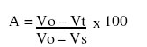
of Ilorin, Ilorin Nigeria Received December 17, 2005 Code Number: bk06008 Abstract Nine fungal species isolated from cowpea seeds were used as inocula on four cowpea varieties commonly distributed to farmers in Ilorin, Kwara state, Nigeria by the National Seed Service, a subsidiary of the Federal Ministry of Agriculture and Natural Resources. The effects of fungi on germinability and seedling health were determined using seedling symptom test. Two of the virulent species were screened for the production of cell wall degrading enzymes using viscometric method. All the fungi reduced germination rate in all the cowpea varieties and different types of seedling symptoms were noted for the fungi. The symptoms included seed rot, chlorotic leaf development, stunted growth etc. Production of pectinases and cellulases by Aspergillus flavus and Penicillum sp. was observed and the virulence of the two organisms could be attributed to the activities of these cell wall degrading enzymes. Keywords:Cowpea, Seed mycoflora, Aspergillus sp, Penicillium sp, Pectinases and Cellulases INTRODUCTION Cowpea, Vigna unguiculata (L) Walp (Fabaceae), has always been an important grain legume in tropical countries especially Nigeria and a veritable source of dietary protein for the teeming population of human and livestock1,2. The dry seed consists of about 25% protein and 67% carbohydrate3. In the 1970’s, about 94% of the total world crop was produced in Africa4. The situation remains much the same today but the optimization of production, which is still grossly at the subsistence level in this region continues to be hampered by pests5,6,7 and diseases8,9. Some of the diseases of cowpea are known to be caused by seed-borne pathogens most of which are fungi6,8,10,11. Many phytopathogenic fungi and bacteria have long been known to produce enzymes capable of hydrolyzing the polymeric carbohydrate constituent of higher plants cell wall. This factor might be responsible for the penetration of the fungi into the cowpea seeds12. In Nigeria, there is still a dearth of information on cowpea seed-borne fungi and their significance in disease development. Thus, the present study aimed at determining the pathogenicity of some cowpea seed-borne fungi and screening some of the virulent ones for their cell wall degrading enzyme activities. MATERIALS AND METHODS Source of organisms Nine fungal species namely; Alternaria sp., Aspergillus flavus, Aspergillus niger, Cladosporium sp., Fusarium oxysporum, Fusarium semitectum, Fusarium solani, Fusarium sp. and Penicillum sp. isolated on Potato Dextrose Agar (PDA) by the standard procedure of culturing and subculturing, from cowpea seeds were used for the study. The cowpea seeds were obtained from the National Seed Service Ilorin, Kwara state, Nigeria and were stored under refrigeration (4 oC) until needed for use. Preparation of inocula from the organisms Spore suspension was prepared from highly sporulating organisms such as Aspergillus flavus, and Aspergillus niger by washing the spores from agar slants with sterile water. The number of spores was adjusted to 5x10⁵ spores/ml by dilution. Mycelia suspensions of other isolates were prepared by punching several small pieces from 10 day old cultures using 5mm cork borer. The mycelia discs (5mm) were comminuted in 150ml sterile water13 for 10 seconds in a blender. Inoculation of seeds To study the effect of seed borne fungi on germination and seedling growth, the method of Baggett and Fraizer14 in which seeds were coated with fungal cultures was adopted. Surface-sterilized seeds were mixed with the individual inocula in corked conical flasks for approximately one hour using mechanical shaker. After shaking, the seeds were left to stand in the inoculum for 8-10 hours. The seeds were then dried overnight on sterile paper towel. Effect of organisms on seed germination and seedling health Inoculated seeds were sown in small plastic pots filled with steam- sterilized soil. Sterile water was used for wetting the soil while being kept at room temperature for 6-10 days. The pots were later examined for germination and disease symptoms on emerging seedlings. Cell wall degrading enzymes assay Two demonstrably virulent fungal isolates of the nine isolates were selected for the assay of cell wall degrading enzyme enzyme activities. The selected organisms were prepared on PDA slants for 3 days at 28 ± 2 oC. The cultures were then separately washed with sterile water to dislodge the spores and the resulting spore suspension was used for preparing enzyme filtrate. Preparation of enzyme Seeds of cowpea cultivar (ITA 90-22-2K) susceptible to attack by the virulent fungi were ground in a blender. The ground seeds were sterilized in an autoclave at 15 lb pressure for 15 minutes to get rid of all seed-borne fungi and other contaminants. Two separate 50g of the sterile ground seeds was moistened with 50ml of each inoculum separately and the mixture incubated for 3-5 days at about 28±2oC. Sterile water (150ml) was later added in each case and the mixture agitated for 30 minutes with mechanical shaker. The resulting mixture was filtered using sterile Whatman No. 1 filter paper. The filtrates were kept under refrigeration until needed. Enzyme assay Viscometric method was used in measuring the activity of the enzymes. Viscosity was determined using the method of Endo15. The reaction mixture contained 15ml of carboxymethyl cellulose (CMC) (1% w/v) in citrate phosphate buffer (pH 5.5) and 15ml of enzyme filtrate for cellulose assay. For pectinase assay, the mixture contained 15ml pectin (2% w/v) in citrate phosphate buffer (pH 5.5) and 5ml of the enzyme filtrate. Measurements were made in glass viscometer after the mixtures have been incubated for 2 hours at 30oC. The enzymatic activities were expressed in terms of percentage viscosity change. The reducing rate of viscosity was calculated from the following equation:
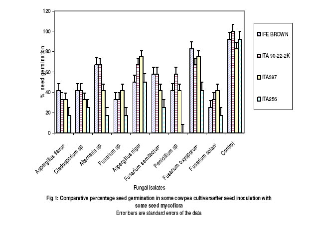
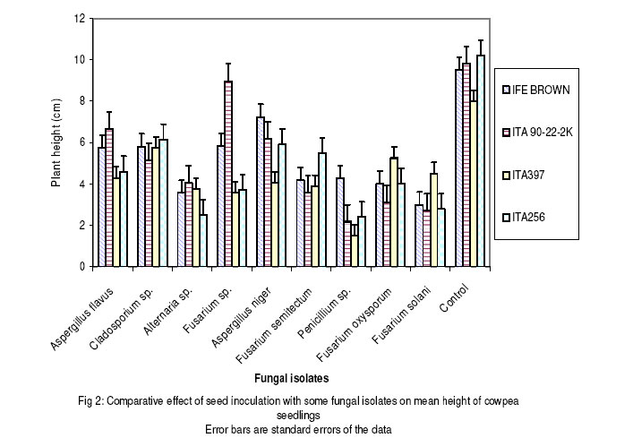
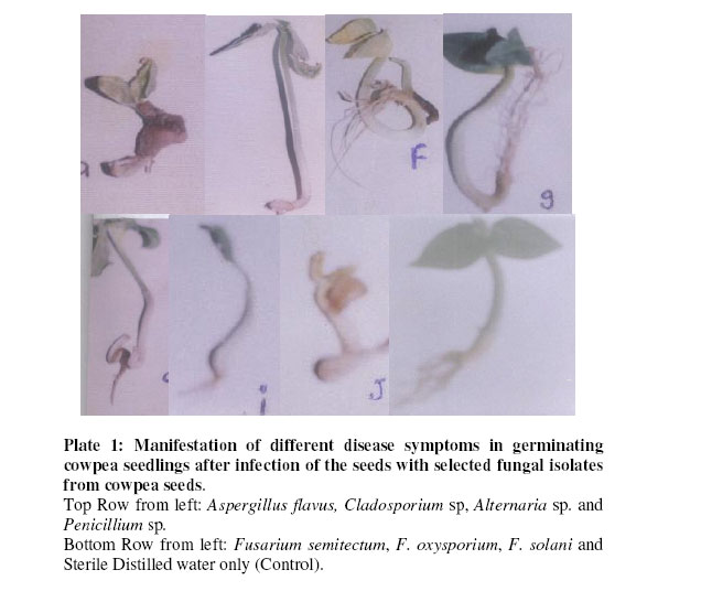
Where, Vo = flow time in seconds of pectin/cellulose + inactivated enzyme. Vt = flow time in seconds of pectin/cellulose + active enzyme. Vs = flow time in seconds of solvent (water) + inactive enzyme. One unit of viscosity reducing activity (VRU) is defined as the quantity of enzyme necessary for 50% viscosity change of 20ml of the reaction mixture at 30oC. Data collected from repeated experiments were pooled and analyzed for variance at 5% level of significance. RESULTS AND DISCUSSION Pathogenic effects of seed mycoflora All the fungi tested reduced percentage seed germination and average height of seedling of the four cowpea varieties compared with the control as shown in Figures 1 and 2. Among seed borne organisms, fungi cause maximum seed damage, which include reduced germination and vigour16. Reduced germinability of seeds may be attributed to damaged embryos from deep seated infection of seeds. All the fungi used in this study induced disease symptoms on germinating seedlings. The symptoms observed ranged from chlorosis to necrotic spots development on the leaves stems and roots while the control remained healthy ( Plate 1). The impact of fungal inocula on germinating seeds and seedling health which includes seed rot, stunted seedlings and yellowing of leaves is outlined on Table 1. Many seed borne fungi on cowpea in India have been reported to reduce seed germination and produce symptoms on infected seedlings17. It was further observed17 that fungi such as Aspergillus flavus and Fusarium solani were associated with damage to plumule, radicle and hypocotyl of germinating seedlings. This kind of observation explains the reducing effects of such fungi, which were also used in this study on seedling development. Cell wall degrading enzyme activities of some test fungi The results of the assay for pectolytic and cellulolytic enzyme production by two of the virulent fungi (Aspergillus flavus and Penicillium sp) are presented in Table 2. A significantly (P≤ 0.05) higher volume of enzyme preparation from A. flavus than that of Penicillium sp was required to bring about 50% viscosity changes in carboxymethyl cellulose (CMC) indicating a higher cellulase activity in the preparation from Penicillium sp. Conversely, pectinase activitiy of enzyme preparation from A. flavus was significantly higher than that of Penicillium sp. Nowithstanding the difference in quantities, the ability of these moulds to produce cellulases and pectinases explains their virulence. The cell wall-degrading enzymes must have facilitated the penetration of the fungi and hence their ability to cause deep seated infection of the seeds and consequent symptom manifestation in the seedlings. Table 1: Pathogenic effect of fungal isolates on cowpea seedlings
Table 2: Pectolytic and cellulolytic activities of enzyme preparations from Aspergillus flavus and Penicillium sp
V.R.U (Viscosity Reducing Unit) is defined as quantity of enzyme necessary for 50% viscosity change of 20 ml of the reaction mixture at 30oC. Means ± std. dev. followed by different letter in a column are significantly different at P< 0.05. In general, the seeds of all the four varieties of cowpea used for this study were to varying extent susceptible to fungal infections and they exhibited the ability to carry over such infections to the seedling stage. The observations made in this study have further underscored the need for regular seed testing and seed treatments with effective protective and systemic fungicidal preparations, by the seed services, before disbursement to growers. REFERENCES
© 2006 Nigerian Society for Experimental Biology The following images related to this document are available:Photo images[bk06008f1.jpg] [bk06008f3.jpg] [bk06008f2.jpg] | |||||||||||||||||||||||||||||||||||||||||||||||||||||||||||||||||
| |||||||||
