 |
| About Bioline | All Journals | Testimonials | Membership | News |
|
||||||
|
||||||
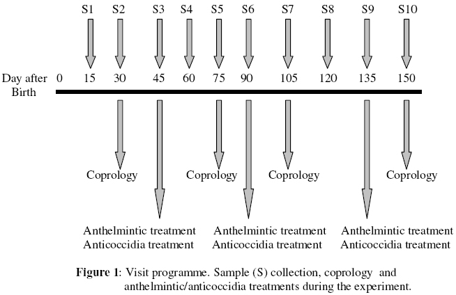
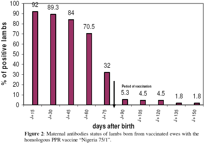
Biokemistri, Vol. 18, No. 2, December, 2006, pp. 99-103 Assessment of the duration of maternal antibodies specific to the homologous peste des petits ruminant vaccine “Nigeria 75/1" in Djallonké lambs Sanne C. BODJO, Emmanuel COUACY-HYMANN*, Mathurin Y. KOFFI and Thérèse DANHO LANADA / Laboratoire Central de Pathologie Animale de Bingerville, BP 206 Bingerville – Côte-d’Ivoire *Author to whom correspondence should be addressed. Received 23 November 2006 Code Number: bk06015 Abstract The duration of maternal immunity was determined from 112 lambs born from vaccinated ewes with the homologous PPR vaccine “Nigeria 75/1" at day 90 and day 120 of pregnancy. Serum samples were collected from lambs starting from day 15 to day 150 after birth and analyzed using the PPR specific competitive ELISA. At day 75 and day 90 after birth, 70% and 95% of these lambs respectively became negative. So it is recommended to vaccinate lambs against PPR in the interval from 75 to 90 days after birth. Keywords:Small ruminants, Vaccine, PPR, Maternal Immunity, Morbillivirus INTRODUCTION The development of small ruminant production in Côte-d’Ivoire started in 1970 initially focusing on sheep. However, this was regularly jeopardized by various diseases. In particular, peste des petits ruminants (PPR), with a national prevalence rate of 19%, remains a major threat to small ruminants in Côte-d’Ivoire1. Veterinary field services in the central region have recorded a high mortality rate (about 40%) in lamb flocks, up to 4 months-old. Observed signs were respiratory symptoms. PPR is also regarded as the main cause of respiratory diseases in small ruminants2,3. PPR is an acute infectious disease of small ruminants with goat being the most sensitive species. It is characterised by high morbidity and mortality rates. It continues to cause serious economic losses in these species in Africa and the Middle-East where it is endemic4,5. It is caused by the peste des petits ruminant virus (PPRV), a member of the morbillivirus genus belonging to the paramyxoviridae family. In the past, the heterologous attenuated rinderpest vaccine6 was used for the protection of small ruminants against PPR4. A homologous PPR vaccine was developped7 and is presently used to protect small ruminants against the disease8,9,10,11. The objective of the study was to assess the duration of maternal antibodies and to determine the appropriate optimum age to vaccinate lambs against PPR with the homologous PPR vaccine Nigeria 75/1. MATERIALS AND METHODS Animals Two flocks of 300 Djallonke sheep were selected in the central region of Côte-d’Ivoire from a list established by the National Programme for Ovine Selection. In each flock, 56 ewes, 3 years old, and negative for rinderpest and PPR specific antibodies were randomly selected. The serology status of selected animals was determined by performing the comparative viral neutralising test12. Vaccination of ewes At day 90 of pregnancy, selected ewes were vaccinated subcutaneously with 1 ml of homologous PPR vaccine7 at a concentration of 103TCID50/ml (Tissue Culture Infectious Doses 50%). A booster vaccination was administratered to target animals at day120 using the same concentration of vaccine. Vaccinated and pregnant ewes were followed clinically during pregnancy period. The ingestion of colostrum by young lambs was verified by four attendants (2 for each lot). Each lamb received 300 mg of AlbendazoleTM and 50 ml of AmprolTM diluted in 50ml of distilled water, against internal parasites at day 45, day 90 and day 135. Sample collection Serum samples were taken from all 112 vaccinated ewes at day 140 of pregnancy to verify the seroconversion after vaccination. Serum samples from lambs flocks were taken 10 times from day 15 to day 150 after birth (Figure 1). Faeces were randomly collected from 30% of lamb population (17 lambs from each flock) at day 30, day 75, day 105 and day 150. The faeces were stored in a saturated solution of NaCl and investigated using sedimentation and quantitative MacMaster counting. Serological analysis The competitive PPR ELISA (cELISA)13 was used for to detect of PPR specific antibodies. Briefly this technique was performed as described hereafter following the recommended protocols: a commercial (BDSL, UK) competitive ELISA (cELISA) kit was used to detect specific antibodies against PPR. Fifty µl volumes (50µl) were used throughout. Maxisorp 96-wells plates were coated with antigen diluted 1/100 in Phosphate Buffer Saline (PBS) (0.01M, pH 7.2-7.4) and incubated at 37°C for 1 hour on an orbital shaker. After a cycle of three washes in PBS, test serum (10µl), was added to 40µl of blocking buffer (PBS 0.01M. pH 7.2-7.4; 0.1% Tween 20 [v/v]; 0.3% negative bovine/sheep serum [v/v]) followed immediately by the addition of 50µl of the specific monoclonal antibody (MAb) at a dilution of 1/100 in blocking buffer. Control sera included were, strong positive, weak positive, negative and a MAb control (0% competition). The plates were incubated and washed as above. Anti-mouse horse radish peroxidase enzyme conjugate diluted 1/1000 in blocking buffer was added and plates incubated as before. The plates were washed. Substrate/chromogen (H2O2/OPD) was added and the colour allowed to develop for 10 min after which any reaction was stopped by the addition of sulphuric acid (1M). Plates were read on an ELISA reader (Multiskan MK II Titerkek, Finland) at an absorbance of 492 nm. Optical density (OD) readings were converted to percentage inhibition (PI) values using the following formula: PI%=100 – (OD in test well/OD in 0% control well)x100 PI% values greater than or equal to 50% were considered as positive. RESULTS
Clinical observations No specific signs of any diseases were recorded from lambs and ewes during the observation period. The coproscopic analysis revealed the presence of oocystes of Coccidia at a detection limit of 10,000 oocysts g-1 showing that lambs were infested by Coccidia. Strongyloides were also found (detection limit: 1,000 eggs g-1). The treatment with the anthelmintic, AlbendazoleTM and with the anticoccidia, AmprolTM, cleared these young animals from infestation. The last control at day150 showed that lambs became negative regarding coccidia and strongyloides. Serological analysis Sera collected from 112 ewes after vaccination and tested using PPR cELISA technique were highly positive indicating high levels of PPR specific antibodies. All vaccinated ewes seroconverted. Serum samples collected from lambs from day 15 to day 150 and analysed with PPR cELISA showed that 103 lambs out of 112 (92%) seroconverted at day15; 89% (100/103) at day 30; 70.5% (79/103) at day 60 while the number of positive young animals decreased to 32% (36/103) and 5.3% (6/103) at day 75 and day 90 respectively (Figure 2). Only two lambs remained positive (1.8%) at the end (day 150) of the experiment. DISCUSSION Vaccination of small ruminants with the homologous PPR vaccine “Nigeria 75/1" produces high titers of PPR specific antibodies as desmontrated in previous studies7,8,9,10,11. The vaccine is also safe for pregnant females1,7. The protection of offspring against many infectious agents during the first months of life depends on the maternally acquired immunity through the ingestion of colostrum. The duration of maternal antibodies influences the optimal age for vaccination. In the present study, 103 lambs out of 112 (92%) had detectable specific antibody to PPRV at day15 after birth. The remaining, 9 (negative) lambs, did not probably ingest any colostrum despite the surveillance of attendants. At day75, 36 lambs were still positive while at day90, only 6 target animals remained positive (5.3%). This study highlights clearly that 3 months after birth, 95% of lambs have lost their maternal antibodies. This result could explain why field technicians recorded high cases of mortality in lamb flocks aged up to 4 months. As maternal antibodies titer declines quickly and young animals before vaccination remain naive, these cases of mortality are more probably due to PPR which is endemic in Côte-d’Ivoire. A similar study conducted on 11 lambs born from ewes vaccinated with the heterologous rinderpest vaccine6 has shown that the passive immunity protects lambs for more than 3 months. Nevertheless the authors recommended vaccination of lambs at 3 months of age against PPR14. Bidjeh et al.9 suggested the vaccination of lambs and kids at 4 to 5 months of age. A recent investigation on the duration of maternal antibodies specific to PPR in lamb and kid flocks demonstrated that they failed to be protected after 3.5 and 4.5 months in lambs and kids, respectively. Therefore, it was recommended to vaccinate them at 4 and 5 months of age, respectively11. Different serological techniques, viral neutralising test for the previous studies and competitive ELISA used in this present one, to investigate the duration PPR maternal antibodies in offspring, showed their decay mainly after 3 months duration leaving afterwards these young animals without any protection. The data generated from the present study suggest that the optimal age for the vaccination of lambs and also goat kids against PPR is from day 75 to day 90 after birth. Acknowledgements We thank the farmers Mr. N’DA Kramo and OUATTARA Seydou for their cooperation allowing us to work with their animals. This work is funded by ANADER (Agence Nationale pour le Développement Rural). REFERENCES
© 2006 Nigerian Society for Experimental Biology. The following images related to this document are available:Photo images[bk06015f2.jpg] [bk06015f1.jpg] |
| |||||||||
