 |
| About Bioline | All Journals | Testimonials | Membership | News |
|
||||||
|
||||||
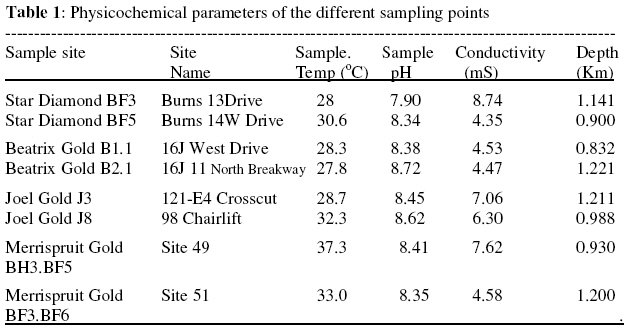
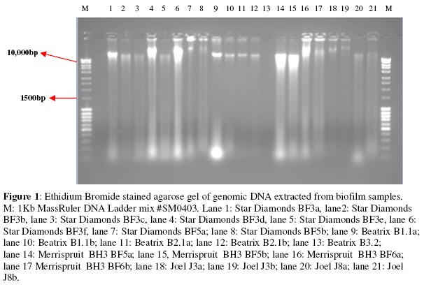
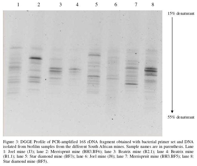
Biokemistri, Vol. 20, No. 2, Dec, 2008, pp. 53-62 Bacterial diversity of biofilm samples from deep mines in South Africa Abdulmajeed I. Raji*, Christelle Möller, Derek Litthauer, Esta van Heerden and Lizelle A. Piater Department of
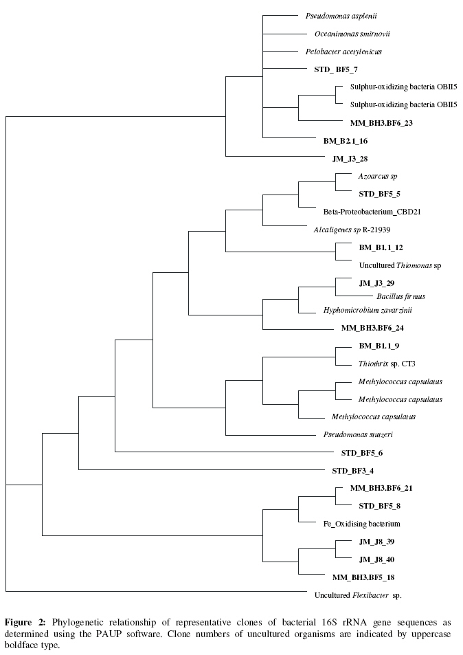
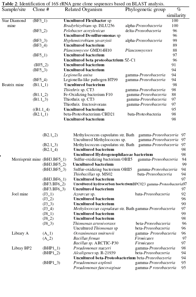
Biotechnology, University of The Free State, Box 339, Bloemfontein 9300. South Africa Received 26 October 2008 Code Number: bk08009 Abstract The Au, Pt and diamond mines of South Africa provide access to microorganism bearing fluids emanating from fractures at depths ranging from 0.7 to 3.2 km. Due to the unique characteristic of mine environment as demonstrated by extreme pH, pressure, temperature and/or salinity, it is anticipated that it could hold the promise for novel gene sequences and hence gene products of industrial and pharmaceutical importance. To provide insight into the microbial diversity of mines in South Africa, biofilm samples were collected from Goldfield and diamond mines and their bacterial diversity determined using molecular approaches. 16S rRNA genes were amplified from DNA extracted from these samples using polymerase chain reaction with universal bacterial primers 27F (5’- AGA GTT TGA TCM TGG CTC AG-3’) and 1492R (5’- GGT TAC CTT GTT ACG ACT T-3’). Metagenomic clone libraries were constructed and restriction fragment length polymorphism (RFLP) analysis of >100 derived clones resulted in four major restriction patterns from which 40 clones were chosen for sequencing. More than half (53%) of the sequences were affiliated with the bacterial phylum Proteobacteria, forty-one percent (41%) of the sequences with yet uncultured bacteria and the phyla Firmicutes and Planctomycetes were accounted for by 4% and 2% of the sequences respectively. DGGE analysis of PCR-amplified 16S rRNA genes showed characteristic fingerprints for each sample. The differences in community structure observed account for the uniqueness of each of the mines with respect to its microbial diversity. Keywords:PCR; DGGE; Bacterial diversity; RFLP; Metagenome INTRODUCTION Gold, platinum and diamond mines have been known to provide unique environment with high salinity, extreme temperature, pH and high pressure that may select for exceptional microorganisms housing proteins with unique sequences1. However, there have been major obstacles to understanding the subsurface biosphere. Prominent among such obstacles are, our limited abilities to access the environment, acquiring pristine samples from such environment and placing our knowledge of microorganisms (functional genes and proteins) into an environmental context2,3. Microorganisms that are able to develop under such extreme conditions have recently attracted considerable attention because of their peculiar physiology and ecology4, and important biotechnological applications5-8. The peculiarity of these microorganisms could be due to the unique sequences they posses which they can translate to proteins with superior biocatalytic properties9,10. The discovery of novel microorganisms in deep accessible subsurface habitats has provided the opportunity to discover new pharmaceuticals, understand microbial biosynthetic processes and enhance remediation of contaminated environments3. The microbial diversity in these extreme environments has been the key to the successful isolation of novel microorganisms with unique characteristics. In the light of this, South African deep mines which encompasses yet unexplored extreme niches of novel populations of diverse microorganisms are suitable environment for investigation11. However, the conventional approach of studying microbial diversity through their growth on culture media have been shown to have much limitations as less than 1% of the total microbial species are culturable under particular conditions8,12-16. The pioneering works of Woese and Fox17 and Woese18 on comparative analysis of small-subunit ribosomal RNAs have pioneered the determination of evolutionary relationships between organisms using a molecular approach and thereby establishing diversity as sequence divergence on a phylogenetic tree. This has proved important in the determination of microbial diversity in many environments19. This study was therefore, aimed at investigating the bacterial diversity of biofilm samples with pH ranging from 7.9 to 8.62, temperature, 28°C to 32.3°C and conductivity of between 4.35 to 8.74mS. Samples were collected from South African gold and diamond mine environments with depth ranging from 0.9km to 1.2km. The phylogenetic diversity was studied using culture-independent techniques, namely: extracting DNA directly from the samples, construction of metagenomic 16S rRNA gene libraries, sequencing and analysis of PCR-amplified 16S rRNA genes using denaturing gradient gel electrophoresis (DGGE) technique. MATERIALS AND METHODS Sample collection Duplicate biofilm samples were collected from one diamond and three gold mines in South Africa namely: Star Diamond, Beatrix, Merrispruit and Joel Gold mines respectively over a period of three months. Samples were collected within these mines from sites rife with slime deposits at different depth ranging from 0.900Km to 1.204Km below sea level. The physicochemical properties of the various biofilm samples from each mine environment were determined (Table 1). Samples were collected aseptically into sterile 50ml Falcon tubes and transported immediately to the laboratory in ice boxes. DNA was extracted immediately from the samples while in the laboratory, and where that was not feasible the samples were stored at -80°C prior to DNA extraction. The Falcon tubes were filled to the brim with samples to avoid air entrapment, and surface contamination of samples was avoided as much as possible. DNA Extraction Genomic DNA was extracted from the biofilm samples using the method of Towner20, with some modifications. All stock and working solutions, the Milli-Q water used for reagent preparation and all plasticwares were autoclaved. Biofilm samples were suspended in TE-buffer in a ratio of 0.5g sample matter: 40ml TE-buffer. After centrifugation at 3000 x g for 5min at 4°C, the pellet was resuspended in 3.2ml of resuspension buffer [50mM Tris-HCl buffer (pH 8.0) and 0.7mM sucrose] and 0.6ml lysozyme (20mg/ml). This mixture was allowed to stand on ice for 5min, after which 0.6ml of 0.5M EDTA (pH8.0) and 0.5ml SDS(10%w/v) were added and mixed gently. The mixture was further placed on ice for 5min.Ten milliliters of digestion buffer [50mM Tris-HCl, pH 9.0; 1% (w/v) SDS; 0.1M EDTA;0.2M NaCl; 0.5mg/ml Proteinase K] was added and incubated for 16h at 55°C with gentle end-over-end inversion intermittently. One volume of chloroform was added and gently mixed for 3h at 25°C followed by centrifugation at 4000 x g for 10 min. One volume of chloroform-isoamylalcohol (24:1) was added to the resultant aqueous phase followed by centrifugation at 3000 X g for 10min at room temperature, and subsequent incubation on ice for 10min. Nucleic acid was precipitated overnight at -20°C by adding 0.1volume of 3M sodium acetate (pH 5.6) and equal volume of ice-cold isopropanol. Nucleic acid was pelleted by centrifugation, washed with 70% ethanol, air-dried and resuspended in sterile Milli-Q water. 16S rRNA Gene Library Construction The genomic DNA extracted from the different biofilm samples were PCR amplified for 16SrRNA genes using the universal bacterial primers 27F (5’- AGA GTT TGA TCM TGG CTC AG- 3’) and 1492R (5’- GGT TAC CTT GTT ACG ACT T-3’). This primer combination amplifies a 1500bp 16SrDNA fragment. Fifty to One hundred nanogram of DNA extract was added to a final volume of 50uL of PCR reaction mixture containing 1.5mM MgCl2, 1X Reaction buffer (without MgCl2) (promega), 200uM of each dNTP, 0.20uM of each primer and 1.0U Taq polymerase (Promega). PCR was performed in an automated thermal cycler with an initial denaturation at 95°C for 5min. followed by 30 cycles of 95°C for 30sec (denaturation), 52°C for 45sec (annealing), 72°C for 1.5min (extension) and 72°C for 10min (final extension). PCR products were run on 1% agarose in TAE buffer [40mM Tris, 20mM Acetic acid, 1mM EDTA (pH8.0)] to confirm that the right product (1500bp) was formed. The PCR product was purified using the QIAGEN PCR purification kit and ligated into pGEM-T Easy vector (Promega) following the manufacturer’s protocol. The ligation mixture was transformed into competent E.coli JM 109 cells, plated on LB/Ampicillin/IPTG/X-Gal plates and incubated for 16hr at 37°C. Two hundred successfully transformed clones were picked and stored at -80°C as glycerol stocks (20% glycerol in LB-ampicillin). Plasmid DNA was isolated from 120 randomly chosen clones (distributed among all the sampling points) using FastPlasmid miniprep kit (Eppendorf, Germany). RFLP Analysis, Sequencing and Phylogenetic Analysis Cloned 16S rRNA inserts were characterized by single digestion with the restriction endonuclease EcoR1. The diversity of the restriction patterns were observed on 1% agarose gel and similar patterns were grouped together. Randomly chosen clones from each RFLP group were selected for sequencing (Inqababiotech SA). Sequences were edited and their similarities determined against known sequences in the NCBI Genbank database using the Basic Local Alignment Search Tool (BLAST) algorithm21. For phylogenetic analysis, the sequences were first aligned using ClustalW, the resulting alignment checked and the sequences used for phylogenetic tree construction using the PAUP software. Parsimony, neighbor- joining and maximum likelihood analysis with different positional conservation filtering was performed to evaluate tree topologies. PCR – DGGE This method utilizes the principle that the melting behaviour of the double stranded DNA is dependent on its base-pair composition22. A highly variable region (V3) of the 16SrRNA gene was amplified using GC-clamped PCR primers. One primer (forward), which complemented a region conserved among members of the domain bacteria (E.coli) positions 341 to 358, had incorporated at the 5’end a 40bp GC-clamp (5'- CGCCCGCCGCGCGCGGCGGGCGGGGCG GGGGCACGGGGGGCCTACGGGAGGCAGCAG -3'). The specificity of this primer is imparted by the underlined region. The other primer (reverse) is based on a universally conserved region (E.coli positions 517-534: 5'-ATTACCGCGGCTGCTGG - 3')23. This primer combination amplified a 233bp fragment suitable for DGGE analysis. Fifty to one hundred nanogram of biofilm DNA extract was added directly to a PCR reaction mixture containing 0.2uM of each primer, 200uM of each dNTP, 1.5mM MgCl2 (promega), 1X reaction buffer (without MgCl2) (promega) and 1.0U Taq polymerase (promega). PCR was performed in an automated thermal cycler with an initial denaturation at 95°C for 4min. followed by 30cycles of 1min. at 95°C, 2min. at 55°C and 2min. at 72°C before the final extension at 72°C for 10min. DGGE was performed on the PCR product using a Hoefer SE 600 DCode system (Hoefer, Inc.USA) following the procedure first described by Muyzer et al.23. The 8% (w/v) Polyacrylamide gels(acrylamide : bisacrylamide ratio 37.5:1) were made with a denaturing gradient ranging from 15% - 55% (100% denaturant corresponds to 7M urea and 40%(w/v) formamide). Electrophoresis was performed in 1X TAE buffer at a constant voltage of 70V and a temperature of 60°C for 12hr. After electrophoresis, gels were stained with Ethidium bromide (10mg/ml) for 45min and viewed with a UV transilluminator (Biorad, Italy). RESULTS AND DISCUSSION In this study, we have undertaken to determine the bacterial diversity in 3 South African gold and 1 diamond mine using culture-independent approaches. The successful extraction of microbial DNA from the biofilm samples collected at different locations from the mines is a positive indication of the presence of microbial flora in the environment (Fig.1). Even though there are no major differences in the physicochemical properties of the different biofilm samples despite the difference in sampling depth (Table 1), there are major phylogenetic differences in their bacterial population. The analysed clone sequences were affiliated to three phyla namely: Proteobacteria (n=27; 53%), Firmicutes (n=2; 4%) and Planctomycetes (n=1; 2%) and more than a third of the entire clone sequences were affiliated to yet uncultured bacteria (n=21; 41%). The extent of the bacterial diversity in the studied South African mines is also demonstrated by the level of divergence of the pylogenetic tree constructed using parsimony, neighbor-joining and maximum likelihood analysis with PAUP software (Fig.2). The community structure within the Proteobacteria phylum changed with the mines. Clone sequences belonging to the gamma-Proteobacteria lineage dominated, and were represented in all the mine samples except in Joel mine. Clone sequences belonging to the beta-Proteobacteria were isolated in all the samples except with samples collected from Star Diamond mine and from Library A. Clone sequences that are not close matches to database sequences of known bacteria (uncultured bacteria) were identified across the mine samples analysed (Table 2). This confirms that a variety of organisms that have not been successfully identified exist in the mine environment. The result of the short-gun sequencing of some of the clones (Table 3), revealed between 86% and 100% closeness to some proteins of industrial importance. This is a possible indication that the group of bacteria in these mine samples may represent novel species, genera or even families of bacteria or archaea that may possess properties and characteristics that are unique. This agree with other workers8,9,15,24, that have reported that groups of yet uncultured bacteria may be housing unique genes responsible for the expression of novel enzymes in the field of pharmaceuticals, bioremediation and in the studying of biosynthetic processes. However, the sequences have not been submitted to any gene bank as further work on the identification is still on The use of DGGE method to determine community structure of microorganisms in particular environments has been reported22,25,26. In this respect, DGGE analysis of the PCR-amplified 16S rRNA fragment was used to further provide information on the genetic diversity of microbial communities of the mines (Fig. 3). In general, the number of amplified bands varied with biofilm samples rather than with mines. This resulted in different samples from the same mine giving diverse banding pattern, with no specific finger printing for each specific mine. Samples from Beatrix gold mine had fewer numbers of bands, which in this case was interpreted to be fewer operational taxonomic units (OTU) even though the physicochemical properties and the depth of sampling did not vary much from the other sites. The operational taxonomic units (OTU) as depicted by the banding pattern thus observed for all the samples shows a diverse community structure with a few organisms showing dominance with respect to their band intensity. However, a few bands had the same electrophoretic mobility irrespective of the sample. This could indicate the presence of these organisms in all the mine samples. Table 3: Shortgun sequencing of selected clones for gene identification
Since none of the DGGE bands were excised and sequenced, estimation of the microbial diversity in this respect was limited to the finger prints on the DGGE gel. It can therefore be concluded that our use of a culture-independent approach has revealed a diverse community structure in the sampled gold and diamond mines of which many of the organisms are yet unidentified and could therefore be the carrier of novel genes for future biotechnological breakthroughs. REFERENCES
© 2008 Nigerian Society for Experimental Biology The following images related to this document are available:Photo images[bk08009f3.jpg] [bk08009f2.jpg] [bk08009t1.jpg] [bk08009f1.jpg] [bk08009t2.jpg] |
| |||||||||
