 |
| About Bioline | All Journals | Testimonials | Membership | News |
|
||||||
|
||||||
Biokemistri, Vol. 20, No. 2, Dec, 2008, pp. 63-70 Antihyperglycaemic effect of aqueous extract of Daniella oliveri and Sarcocephalus latifolius roots on key carbohydrate metabolic enzymes and glycogen in experimental diabetes Adaku V. Iwueke* and Okwesili Fred C. Nwodo Department of
Biochemistry, Faculty of Biological Sciences, University of Nigeria, Nsukka, Enugu, Nigeria Received 10 December 2008 Code Number: bk08010 Abstract The blood sugar lowering effect of the aqueous extract of Sarcocephalus latifolius S. M. (Rubiaceae) and Daniella oliveri Rolfe (Caesalpiniaceae) roots (250mg/kg) was investigated on normoglycaemic and alloxan induced diabetic rats. In addition, hexokinase, glucokinase and phosphofructokinase activities as well ashepatic glycogen content were assessed. The extract’s potency was compared with a standard drug, glibenclamide. The test dose of the extract caused a significant (p<0.05) lowering of blood sugar level in alloxan diabetic rats within six hours; from 261.00±3.02 to 65.00±5.40 and from 302.75±79.62 to 119.00±20.30 after three weeks. The same dose did not show blood sugar lowering effect in normoglycaemic rats. Diabetic rats showed significant (p<0.05) decrease in the activities of hepatic hexokinase, glucokinase, phosphofructokinase and hepatic glycogen content. The extract significantly (p<0.05) restored both hepatic glycogen content and the activities of hexokinase, glucokinase and phosphofructokinase. The oral LD50 of the extract in mice was greater than 5000 mg/kg. Phytochemical analysis of the extract revealed the presence of high levels of flavonoids, alkaloids, saponins, glycosides, steroids and terpenoids and moderate levels of tannins. The results indicate that the decoction of the roots of S. latifolius and D. oliveri has antihyperglycaemic effect. The probable mechanism of action of the extract is discussed and we suggest that the combination of the root extracts of S. latifolius and D. oliveri is a good option for alternative medicine in the management of diabetes mellitus. Keywords:Daniella oliveri, Sarcocephalus latifolius, diabetes, carbohydrate metabolism INTRODUCTION In Nigeria, herbal therapies occupy a very special position in health delivery, especially among the rural populace. Easy accessibility and low cost of treatment enhance patronage. Diabetes mellitus is the commonest non-communicable endocrine disease and is considered one of the leading causes of death all over the world; affecting over 100 million people worldwide1. Many traditional plant treatments for diabetes mellitus are used throughout the world, and some of these plants have been scrutinized while a good number of them are yet to receive scientific scrutiny.2 A decoction of the roots of Sarcocephalus latifolius (Rubiaceae) and Daniella oliveri (Caesalpiniaceae) is used in parts of the south eastern Nigerian folk medicine as herbal remedy for hyperglycaemia. Although, the antihyperglycaemic potential of the leaves3 of S. latifolius has been demonstrated, there is no scientific report on the blood sugar lowering activity of the roots of these plants in combination. A preliminary study in our laboratory showed the reduction of blood glucose in diabetic rats treated with root extract of S. latifolius4. This study was initiated with the primary aim of investigating the effect of aqueous extract of a combination of S. latifolius and D. Oliveri roots on the fasting blood glucose level. In addition, the effect of the extract on the activities of key glucose metabolic enzymes: hexokinase, glucokinase, phosphofructokinase and glycogen concentration in the livers of non-diabetic and alloxan-induced diabetic rats were also evaluated in the bid to understand the probable mechanism of antihyperglycaemic action of the extract. MATERIALS AND METHODS Materials Fresh roots of S. latifolius and D. oliveri were collected in November, 2006 from the plants in Nsukka, Enugu State, Nigeria. The plants were identified and authenticated by Mr. A. O. Ozioko of the International Centre for Ethnomedicine and Drug Development, Nsukka where the Voucher Specimens (InterCEDD 76AO and 158AO) respectively were deposited. Preparation of the Plant Extract The fresh roots of S. latifolius and D. oliveri were separately cleaned, chopped into pieces and shade-dried to a constant weight. 250 g each of the S. latifolius and D. oliveri root chips were collectively subjected to exhaustive extraction in 4 volumes (w/v) of distilled water by decoction. The extract was filtered with cheese cloth. The filtrate was dried at 40°C yielding about 38.49 g of a dark brown residue of Sarcocephalus/Daniella Aqueous (SDA). The extract was refrigerated and dissolved in distilled water before use. The extract was administered at a dose of 250 mg/kg body weight. This dosage was determined from a preliminary dose response study which showed that the 250 mg/kg body weight had the greatest hypoglycaemic effect. Animals Adult Sprague-Dawley (SD) rats weighing 300 g and albino mice (15 – 35 g) of either sex were obtained from the Laboratory Animal Facilities of the Faculties of Veterinary Medicine and Biological Sciences, University of Nigeria, Nsukka. The animals were administered for about 7 days under standard environmental conditions with a 12 hour light/12 hour dark cycle and maintained on a regular feed (vital feed) and water ad libitum.The Institution’s Animal Ethics Committee of the Faculty of Veterinary Medicine approved the study. Preliminary Phytochemical Screening Preliminary phytochemical tests were carried out on the extract using standard reagents and procedures5,6. In general, tests for the presence of phytochemical compounds involve the addition of appropriate chemical reagent(s) to the extract in separate test tubes. The mixture was then shaken and/or heated as the case may be. The presence or absence of saponins, flavonoids, tannins, alkaloids, etc was observed. Acute Toxicity Test The oral median lethal dose (LD50) of the extract was determined in albino mice using the method of Lorke7. This study was conducted in two stages; an initial dose-range determination stage involving 9 animals (3 groups of 3 animals each) treated p.o. with 10, 100 and 1000 mg/kg of the SDA extract. The animals were observed for number of deaths and behavioural changes for seventy two hours. Based on the percentage survival rates, the second stage was carried out in which three groups of mice were treated with 1500, 2900 and 5000 mg/kg of the extract and observed as in the first stage. The LD50 was then calculated as the geometric mean of the highest non-lethal and the lowest lethal doses. Induction of Experimental Diabetes Mellitus Rats fasted for 16 hours, but with free access to water were made diabetic by an intravenous injection of alloxan monohydrate (Sigma-Aldrich, U.S.A.) (65 mg/kg) given in the tail vein. The alloxan was dissolved in sterile normal saline just prior to injection; the injection volume was 1 ml/kg. Since alloxan is capable of producing fatal hypoglycaemia as a result of massive pancreatic insulin release, the rats were treated with 20% glucose solution after 3 hours. The animals were further kept for the next 12 hours with 5% glucose solution bottles in their cages to prevent hypoglycaemia8. After 12 hours of alloxan injection, surviving rats with fasting blood glucose levels ≥ 200 mg/dl were included in the study. Animals had free access to food and water after alloxan injection. Repeated Administration of SDA to Alloxan-Diabetic Rats Before treating the animals as shown in Table 1, the animals were fasted for 16 hours and their fasting blood glucose levels were determined using One Touch Ultra Blood glucose monitor (Lifescan, a Johnson and Johnson Company, U.K.). The treatment was administered orally by intubation twice daily to the various groups for 21 consecutive days. At the end of the treatment days, the animals fasting blood glucose levels were determined again. The animals were then euthanized using and their livers were surgically removed immediately, washed with ice cold KCl (1.15%) and refrigerated. The liver tissues were used for the biochemical assay of hexikinase, glucokinase, phosphofructokinase and hepatic glycogen within one week of collection. Hepatic Hexokinase (EC. 2.7.1.2) Activity Liver tissues (0.5 g) were homogenized with ten times their weight in volume of the homogenizing buffer (50 nM Tris/HCl, pH 7.5, 2 mM mgCl2, 1mM EDTA and 30 mM DTT). The homogenates were centrifuged at 20,000 g for 15 mins at 4°C and the supernatants used for the enzyme assay. The enzyme activity, using a coupled assay with gluco-6-phosphate dehydrogenase, was monitored at 37°C by following the appearance of NADPH at 340 nm for 3 minutes at 30 seconds interval according to the method of Lapeir and Rodnick9 as reported by Ugochukwu and Babady10. Hepatic Glucokinase (EC 2.7.1.3) Activity Liver tissues (0.5g) were homogenized in nine volumes of the homogenizing buffer (100 mM KCl, 10 mM DTT, 50 mM Tris/HCl and 1 mM EDTA at pH 7.4). The hepatic glucokinase activity was determined according to the method of Newgard et al.11, which was modified and reported by Ugochukwu and Babady10. The production of glucose-6-phosphate by glucokinase in the presence of ATP was linked to the reduction of NAD to NADH by glucose-6-phosphate dehydrogenase from Leuconostoc mesenteroides. Hepatic Phosphofructokinase (EC 2.7.1.11) Activity Liver tissues (0.5 g) were homogenized with ten times their weight in volume of (50 mM Tris/HCl, pH 8.5, 5 mM MgSO4.7H2O, 1 mM EDTA and 10 mM DTT). The homogenates were centrifuged at 20,000 g for 15 minutes at 4OC. Supernatants (50 μl) were used for the enzyme assay. The supernatants and the other components of the reaction mixture12 in a final volume of 1 ml were incubated at 37°C and the change in absorbance (appearance of NADH) was followed at 340 nm for 3 minutes at 30 seconds interval. Table 1: Experimental Design
Hepatic Glycogen Content Glycogen content was measured as described by Ong and Khoo13. Liver tissues (0.5 g) were homogenized in 10 ml volumes (w:v) of ice-cold 30% KOH and boiled at 1000C for 30 minutes. Glycogen was precipitate with ethanol, pellet, washed and resolubilised in distilled water. Glycogen content was determined by treatment with anthrone reagent and measured at 625 nm. Protein Determination Protein content of the liver tissues was determined by the Biuret method as described by Gornal et al.14 using Bovine Serum Albumin (BSA) as standard. Statistical Analysis The results are presented as mean ± SEM for 4 – 6 rats in each group. The results were analysed for statistical significance by One-Way ANOVA test and was further subject to Fischer LSD post Hoc test using the SPSS Genstat Release (Windows 98) Software Package Version 11. Differences between means were considered significant at p<0.05. RESULTS Phytochemical Test Phytochemical investigation revealed the presence of biological active constituents in the extract. The extract gave positive reactions for flavonoids, alkaloids, saponins, tannins, carbohydrates, reducing sugars, glycosides, steroids and terpenoids as shown in Table 2. Acute Toxicity Test The safety of the extract is evidenced by the high LD50 value of the extract (>5g/kg). In addition, there were no significant modification in the general behaviour of the animals nor were there death after 72 hours at the highest administered dose (5g/kg) of the extract. Fasting Blood Glucose Level Prior to treatment, the fasting blood glucose levels of the alloxan-induced diabetic rats were significantly (p<0.05) higher than the fasting blood glucose level (FBGL) of the non-diabetic control rats. Within seven days of treatment there was a significant increase (p<0.05) in the FBGL of the group of rats treated with the extract (Table 3). Continued daily administration of the extract for 21 days caused a significant reduction (p<0.05) in the blood glucose level when compared with the diabetic control group of rats. Similarly, repeated administration of glibenclamide (5mg/kg) for 21 days produced a significant reduction in the FBGL in alloxan-induced rats relative to the diabetic control group and the initial day values. Table 2: Phytochemical constituents of the extract
++++ Abundantly Present Table 3: Fasting blood glucose level in alloxan diabetic rats
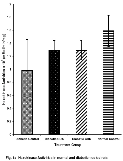
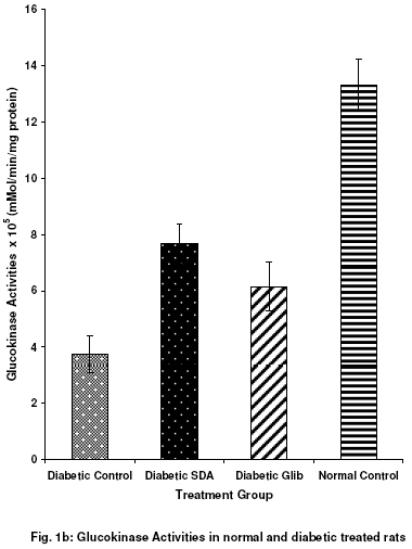
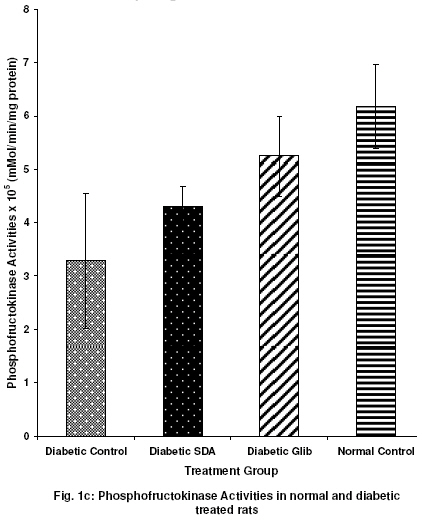
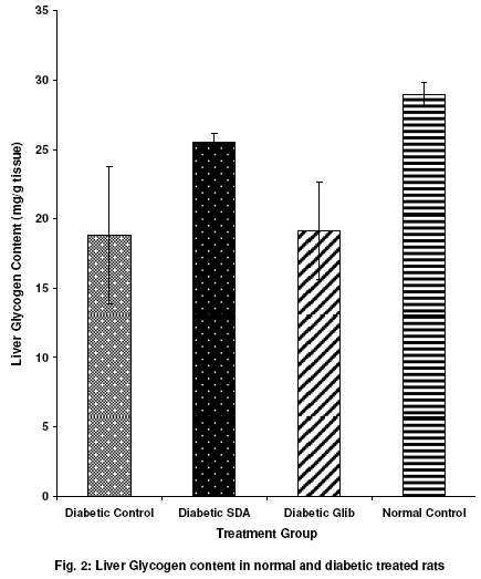
Diabetic-SDA (Diabetic treated with extract), Diabetic-Glib (Diabetic-treated with glibenclamide water), Diabetic control (Diabetic treated with distilled water). Values are expressed as mean ± SEM (n = 4). *P<0.05 indicates a significant difference compared with the initial value. Hepatic Enzymes The activities of hepatic hexokinase (HK), glucokinase (GK) and Phosphofructokinase (PFK) in normal and diabetic rats treated repeatedly for 21 days with the extract, glibenclamide and distilled water were determined and the results shown in Figures 1a, b and c. As compared to the normal control values, mean levels of the enzymes (HK, GK and PFK) activities decreased in the diabetic control. Treatment with the extract and glibenclamide led to a rise in the activities of these enzymes when compared with the diabetic control group of rats. Hepatic Glycogen Content Glycogen content of the liver tissues was estimated after 21 days of repeated treatment with the extract (SDA), glibenclamide and distilled water in normal and diabetic rats as shown in Fig. 2. In diabetic control animals, hepatic glycogen content decreased significantly when compared with the non-diabetic controls. Treatment with the extract (SDA) and glibenclamide increased the hepatic glycogen content with SDA being significant at p<0.05 in comparison to the diabetic control. DISCUSSION Available drug regimens for the management of diabetes mellitus have certain disadvantages 15,16, and so there is need to develop new therapies that can improve hyperglycaemic management, but with reduced side effects. Our results indicate that the extract of the two plants S. latifolius and D. oliveri in combination has rich phytochemical constituents and one or more of these may be responsible for the hypoglycaemic activity. Studies carried out to access the safety of this extract using mice revealed a high margin of safety LD50 > 5 g/kg. Alloxan produces hyperglycaemia by selective cytotoxic effect on pancreatic β-cells17,18, causing permanent destruction of β-cells. The 65 mg/kg dose of alloxan used in this study, caused moderate diabetes19. It has been reported that glibenclamide was not very effective when complete destruction of ß-cells has occurred and hence more effective in moderate than in severe diabetes20. Daily administration of SDA (250 mg/kg) and glibenclamide (5 mg/kg) to the diabetic rats twice daily for 21 days resulted in a significant reduction (61% and 53% respectively) in fasting blood glucose levels. Repeated administration of this extract twice daily elicited an initial increase in the fasting blood glucose levels of the diabetic rats which subsequently decreased. This finding explains the assertion of tradomedical practitioners; that the combination therapy demonstrates initial rise in FBGL which goes down after sometimes and then stabilizes. In view of this, unlike the sulphonylureas or insulin, the onset of action of the extract is slow and so cannot be used for the management of emergency situations. The anti-hyperglycaemic activity of SDA exceeded that of glibenclamide and hence the inference that this extract may be more efficacious as more than one component may be responsible for the activity. Diabetes mellitus is known to be associated with a reduced capacity of the ß-cells of the pancreas to release sufficient insulin which induces the activity of glucose metabolizing enzymes21. Our results indicate that the activities of hexokinase, glucokinase and phosphofructokinase were significantly depressed in the untreated diabetic rats. These results are consistent with the reports of other researchers for the activities of hexokinase and glucokinase 22,19,21,23,24,25 and phosphofructokinase26. Treatment of the diabetic rats with the extract (SDA) showed an increased hexokinase, glucokinase and phosphofructokinase activities. The mechanism(s) of action of SDA is not yet known exactly, but from its effect on these glycolytic enzymes, it seems to increase flux of glucose into the glycolytic pathway in an attempt to reduce high blood glucose concentration. Reduction in these enzyme activities in diabetic animals has been reported to give rise to a depletion of liver glycogen 27,28. Some studies have demonstrated that hepatic glycogen content in untreated diabetic rats was higher than in treated and untreated non-diabetic rats29,10. In this study, the hepatic glycogen content was reduced significantly in diabetic controls as compared to the normal control animals. This is in agreement with earlier findings and was also supported by the findings of 30,19,31 who demonstrated that glycogen deposition from glucose is impaired in diabetic animals proportional to the severity of insulin deficiency32,33. Some workers have found no difference though34. Treatment of the diabetic rats with SDA inhibited the said depletion in glycogen content and almost normalized it. This is possibly due to either the stimulation of insulin release from ß-cell35 or insulinomimetic activities of the extract giving rise to direct peripheral glucose uptake or a combination of the two. In conclusion, we suggest that the combination of the roots extracts of S. latifolius and D. oliveri is a good option for alternative medicine in the management of diabetes mellitus. ACKNOWLEDGEMENTS The authors wish to thank Mr. A. O. Ozioko for identifying the plants, Dr. (Mrs.) C. Ezekwesili for supplying the plant material and Mr. Joshua Parker for secretarial assistance. REFERENCES
© 2008 Nigerian Society for Experimental Biology. The following images related to this document are available:Photo images[bk08010f1b.jpg] [bk08010f1c.jpg] [bk08010f2.jpg] [bk08010f1a.jpg] | |||||||||||||||||||||||||||||||||||||||||||||||||||||||||||||||||||
| |||||||||
