 |
| About Bioline | All Journals | Testimonials | Membership | News |
|
||||||
|
||||||
Biokemistri, Vol. 21, No. 1, June, 2009, pp. 25-31 R-α–Lipoic acid and acetyl-L-carnitine optimal combinations in MPP+- induced cellular model of Parkinson’s disease Afolabi A. Akindahunsi Department of Biochemistry,
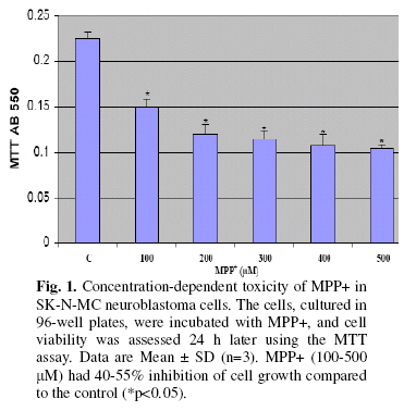
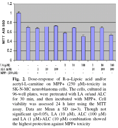
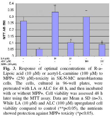
Federal University of Technology, P.M.B. 704 Akure, Nigeria Received 20 September 2008 Code Number: bk09004 Abstract Mitochondrial insufficiency and oxidative damage contribute to the etiopathology of Parkinson’s disease (PD). However, there is a dearth of information on the protective activities against PD of mitochondrial nutrients, safe for coenzyme Q10. In the present study, the PD protective effects of two mitochondrial nutrients, R-α–lipoic acid (LA) and acetyl-L-carnitine (ALC), as well as their combinations using 1-methyl-4-phenylpyridinium ion (MPP+)-treated SKN-MC human neuroblastoma cells as a model of PD was examined. Pretreatment of cells with LA (1-100 μM), ALC (1-100 μM) or LA-ALC (1:10; 10:100; 100:100 μM) combinations showed protective effects against MPP+-induced toxicity of cells. The best concentrations were LA-ALC (1:10 μM) combination, LA (10 μM) and ALC (100 μM) in that order, thus indicating a synergy by the mitochondrial nutrients. This could be a promising strategy in combating PD and other neurodegenerative disorders. Keywords:Parkinson’s disease; R-α–lipoic acid; Acetyl-L-carnitine; 1-methyl-4-phenylpyridinium ion; SKN-MC human neuroblastoma cells INTRODUCTION Neurodegenerative disorders (Parkinson’s disease, Alzheimer’s disease, Down syndrome, stroke, multiple sclerosis, amyotrophic lateral sclerosis, Huntington’s disease, Friedreich’s ataxia, and aging) involve impairments of the mitochondrial citric acid cycle and oxidative phosphorylation proteins and enzymes1-6 However, the major mitochondrial defect in Parkinson’s disease appears to be associated with inhibition of respiratory chain complex I activity7,8 Parkinson’s disease is characterized by a progressive degeneration of dopaminergic neurons in the substantia nigra pars compacta (SNc) leading to a dopamine (DA) depletion in the striatum9-11.Up to now, both the cause and the mechanisms of PD remain largely unknown. In order to gain insight into the mechanisms responsible for the demise of dopaminergic neurons in PD, a number of compounds are used in animal models - reserpine, 6-hydroxydopamine, methamphetamine, and 1-methyl-4-phenyl-1,2,3,6-tetrahydropyridine (MPTP) 12. Of the lot, MPTP is the most popular tool for inducing a model of PD in a number of animal species. MPTP, a proneurotoxin, is converted by Monoamine oxidase-B (MAO-B) to its ultimate neurotoxic species 1-methyl-4-phenylpyridinium (MPP+)13,14. MPP+ acts via inhibition of mitochondria complex I8,15 Since mitochondria are the source and also the target of oxidants, the use of orthomolecular nutrients involved in mitochondrial metabolism could be a way of combating neurodegenerative diseases1,8,16,17. Earlier, it has been shown that feeding the mitochondrial metabolites, acetyl-L-carnitine (ALC) and/or R-α–Lipoic acid (LA), inhibits oxidative damage and restores mitochondrial structure and function in aged rats18,19. It was also shown that ALC can elevate the cardiolipin level in aged animals20. Protection of peroxidation–sensitive cardiolipin may be particularly important in preserving mitochondrial function, as this lipid facilitates the interaction between cytochrome c and cytochrome oxidase.. Bharath et al21 have found that LA effectively prevents mitochondrial complex I deficiency induced by GSH loss in PC12 cells; this suggests that LA may be useful in preventing/treating Parkinson’s Disease. It was then hypothesized that a combination of ALC and LA would potentially maximize the protective action of the two compounds against mitochondrial damage, and thus may prove effective in preventing and/or treating PD. In the present study, the effect of LA and ALC in MPP+ (250 μM)-treated SK-N-MC human neuroblastoma cells PD model was investigated. MATERIALS AND METHODS Materials Penicillin and streptomycin from Invitrogen, Carlsbad, CA., acetyl-L-carnitine (hydrochloride salt) was from Sigma Tau (Pomezia, Italy), R-alpha-lipoic acid (tris salt) was a gift from Dr. K. Wessel, Viatris, Bad Homburg, Germany Cell culture and treatments SK-N-MC neuroblastoma cells were cultured in Gibco Cat. No 41500 MEM with Earle’s salts containing 5 mM glucose, 2 mM L-Glutamine, 1 mM sodium pyruvate, nonessential amino acid, 10% fetal bovine serum, 50 U/ml penicillin and streptomycin. Cellular models Cells were exposed or unexposed to LA, ALCAR, or their combinations. Cells were treated with LA (10, 20, 50, 100, 200 μΜ), ALCAR (50, 100, 200, 500, 1000 μΜ), or combinations of LA and ALCAR (0.05, 0.1, 0.2, 0.5, 1, 10 μΜ) 30 min prior to the addition of MPP+ (250 or 500 μM). LA and ALC were dissolved in sterilized Phosphate Buffered Saline (PBS), and MPP+ in PBS. For control experiments, PBS or DMSO was used. For routine culture, cells were grown in 6- or 96-well plates. There were both acute (24 h) and chronic (3 weeks) models. Cell viability was assessed by measuring the response to the mitochondrial dye 3-[4,5-dimethylthiazol]-2,5-diphenyltetrazolium bromide (MTT), MTT Assay The mitochondrial dye 3-[4,5-dimethylthiazol]-2,5-diphenyltetrazolium bromide is converted into a blue formazan product by metabolically active cells22. Briefly, the medium was aspirated before addition of 50 μl of MTT (5 mg/mL) (MTT stock was filtered through a 0.2 μm filter and store at 2-8 oC), and then incubated at 37 °C for 3 h. Thereafter, the supernatant was aspirated and 200 μl DMSO was added, incubated at 37 °C for 5 min, mixed very well and read at 550 nm using a plate reader spectrophotometer. RESULTS MPP+ inhibits cell growth Concentration-dependent toxicity of MPP+ in SK-N-MC neuroblastoma cells was determined. The cells, cultured in 96-well plates, were incubated with MPP+, and cell viability was assessed 24 h later using the MTT assay. MPP+ (100-500 μM) had 40-55% inhibition of cell growth compared to the control (Fig 1). Dose-response of R-a–Lipoic acid and/or acetyl-L-carnitine on MPP+ (250 μM)-toxicity The dose-response of R-a–Lipoic acid and/or acetyl-L-carnitine on MPP+ (250 μM)-toxicity in SK-N-MC neuroblastoma cells was determined. The cells, cultured in 96-well plates, were pretreated with LA or/and ALC for 30 min, and then incubated with MPP+. Cell viability was assessed 24 h later using the MTT assay. Though not significant (P>0.05), LA (10 μM), ALC (100 μM) and LA (1 μM)-ALC (10 μM) combination showed the highest protection against MPP+ toxicity (Fig. 2). Response of optimal concentrations of R-a–Lipoic acid or acetyl-L-carnitine to MPP+ toxicity Response of optimal concentrations of R-a–Lipoic acid/or acetyl-L-carnitine to MPP+ toxicity in SK-N-MC neuroblastoma cells was equally investigated. The cells, cultured in 96-well plates, were pretreated with LA or ALC for 48 h, and then incubated with or without MPP+. Cell viability was assessed 48 h later using the MTT assay. While LA (10 μM) and ALC (100 μM) upregulated cell viability compared to control, the nutrients showed protection against MPP+ toxicity (Fig. 3). DISCUSSION Parkinson’s disease (PD) is a progressive neurological disease, which is marked by the extensive loss of dopaminergic neurons in the substantia nigra pars compacta (SNc). A leading model to study idiopathic PD in animals involves the use of 1-methyl-4-phenyl-1,2,3,6-tetrahydropyridine (MPTP), a precursor to the mitochondrial toxin 1-methyl-4-phenylpyridinium (MPP+), which targets nigrostriatal dopaminergic neurons in the substantia nigra in a pattern precipitating pathogenesis analogous to human PD 23, 24 On the other hand, exposure to MPP+ is used as an in vitro cellular model of Parkinson’s disease25-27. Concentration-dependent toxicity of MPP+ in SK-N-MC neuroblastoma cells was determined. MPP+ addition to the cells resulted in a reduction in cell viability. MPP+ (100-500 μM) had 40-55% inhibition of cell growth compared to the control. A 250 μM MPP+ was therefore employed for subsequent assays. The toxicity of MPP+ has been used in numerous cellular models. Soliman et al.23 found a drop in mitochondrial oxygen consumption and ATP during MPP+ toxicity with no restoration of mitochondrial function concurrent to a heightened concentration of somatic ATP during piroxicam rescue. MPP+ was found to induce nuclear damage, changes in the mitochondrial membrane permeability, formation of reactive oxygen species and depletion of GSH, which lead to cell death in differentiated PC12 cells24. Sublethal dose of MPP(+) was found to enhance glutamate toxicity against dopaminergic28 neurons, probably by the facilitation of suppressed NO conversion to ONOO(-) in dopaminergic neurons29 Domingues et al.29 found that MPP+ minimizes the ability of both rho+ and rho0 cells to reduce MTT. Qian et al.30 found that overexpression of mutant alpha-synuclein enhanced the toxicity of MPP+ to PC12 cells and elevated intracellular reactive oxygen species (ROS) levels. On their part, De Simoni et al.31 were able to show that mitochondrial PRDX-depleted cells are more prone to oxidative damages and apoptosis induced by MPP(+), A group of mitochondrial targeting nutrients have been identified and named ‘‘mitochondrial nutrients’’, which can (1) prevent the generation of oxidants; (2) scavenge oxidants or inhibit oxidant reactivity; (3) repair oxidative damage to lipids, proteins/enzymes, and RNA/DNA by enhancing antioxidant defense systems and (4) cofactor function: elevate cofactors of defective enzymes (increased Km) in mitochondria to stimulate enzyme activity, and also protect enzymes from further oxidation2,32. Two of such nutrients namely R-α–lipoic acid (LA) and acetyl-L-carnitine (ALC) were employed in the present investigation. LA (10 μM), ALC (100 μM) and LA (1 μM)-ALC (10 μM) combination showed the highest protection against MPP(+) toxicity, thus indicating a synergy by the nutrients. LA is a coenzyme involved in mitochondrial metabolism. The reduced form of LA, dihydrolipoic acid, is a powerful mitochondrial antioxidant33,34. LA has been hypothesized to play its memory improving effect by enhancing mitochondrial function, scavenging free radicals to decrease oxidative damage, or increase the levels of the antioxidants GSH and ascorbate to enhance the antioxidant defense32. ALC contains carnitine and acetyl moieties, both of which have neurobiological properties. Carnitine is important in the beta-oxidation of fatty acids and the acetyl moiety can be used to maintain acetyl-CoA levels. Other reported neurobiological effects of ALC include modulation of: (1) brain energy and phospholipid metabolism; (2) cellular macromolecules, including neurotrophic factors and neurohormones; (3) synaptic morphology; and (4) synaptic transmission of multiple neurotransmitters35 Aliev et al.36 concluded from their work that feeding ALC with LA may ameliorate age-associated mitochondrial ultrastructural decay, which are consistent with previous studies showing improved brain function. Specifically, Zhang et al.37 found that a 4-week pretreatment with LA and/or ALC effectively protected SK-N-MC human neuroblastoma cells against rotenone-induced mitochondrial dysfunction, oxidative damage, and accumulation of alpha-synuclein and ubiquitin. The treatment of a combination of R-alpha-lipoic acid, acetyl-L-carnitine, nicotinamide, and biotin effectively improved glucose tolerance, decreased the basal insulin secretion and the level of circulating free fatty acid (FFA), and prevented the reduction of mitochondrial biogenesis in skeletal muscle38 L-carnitine and alpha-lipoic acid may offer neurotherapeutic effects (e.g., neurocognitive enhancement) via disparate mechanisms including antioxidant, anti-inflammatory, and metabolic regulation39 Conclusively, MPP(+) at a concentration range of 100-500 μM was found to have 40-55% inhibition of cell growth compared to the control. Though not significant (P>0.05), LA (10 μM), ALC (100 μM) and LA (1 μM)-ALC (10 μM) combination showed the highest protection against MPP(+) toxicity, thus indicating a synergy by the nutrients. ACKNOWLEDGEMENTS The author acknowledges the support offered by the 2004 award of CAS-TWAS Visiting Scholar Fellowship, Prof. Jiankang Liu (Institute for Nutritional Sciences, Shanghai Institutes for Biological Sciences, Chinese Academy of Sciences, China) in whose laboratory the work was carried out and the technical assistance of Dr Haiqun Jia. REFERENCES
© 2009 Nigerian Society for Experimental Biology The following images related to this document are available:Photo images[bk09004f2.jpg] [bk09004f3.jpg] [bk09004f1.jpg] |
| |||||||||
