 |
| About Bioline | All Journals | Testimonials | Membership | News |
|
||||||
|
||||||
Biokemistri, Vol. 21, No. 1, June, 2009, pp. 33-39 Evaluation of toxicological implications of ingestion exposure to gasoline in mammals Chibuike Samuel Ubani, Parker Elijah Joshua* And Valentine C. Umenwanne Department of
Biochemistry, Faculty of Biological Sciences, University of Nigeria, Nsukka, Enugu
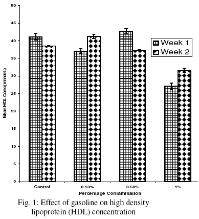
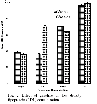
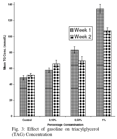
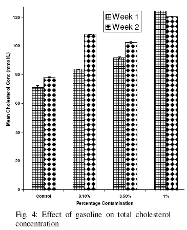
State, Nigeria Received 2 February 2009 Code Number: bk09005 Abstract Epidemiologic and experimental data suggest that exposure to petroleum hydrocarbon exert toxicities on variety of organs of living system such as the lungs, liver and kidney. Because increased LDL cholesterol, decreased HDL cholesterol and alteration in the level of total serum cholesterol have all been implicated as risk factors for atherosclerosis, this present study was designed to determine whether very low percentages of gasoline per kilogramme body weight could cause toxicity in rats. Female albino Wistar rats were divided into four groups. The control group was fed with normal rat diet. Groups 2, 3 and 4 were fed with diet contaminated with 0.10%, 0.50% and 1.00% gasoline respectively for a total of 14 days. Total serum cholesterol, HDL-cholesterol, LDL-cholesterol and triglyceride levels were used as diagnostic markers to assess liver dysfunction. The mean levels of total cholesterol, LDL-cholesterol and triglyceride were significantly (p<0.05) higher in the experimental groups when compared to the control and the mean level of HDL- cholesterol was significantly (p<0.05) lower when higher percentage of gasoline was administered. These results showed that frequent exposure to gasoline fuel may be highly deleterious to the liver cells. Keywords:Gasoline, Low density lipoprotein, High density lipoprotein, Triacyglycerol INTRODUCTION Crude oil can bioaccumulate in food chains where they disrupt biochemical or physiological activities of many organisms, thus causing carcinogenesis of some organs, mutagenesis in the genetic material, and impairment in reproductive capacity and/or causing hemorrhage in exposed population1. The cause/effects of oil pollutant are usually quantified by using biological and point parameters referred to as biomarkers. Contamination of soil arising from spills in one of the most limiting factors to soil fertility and hence crop productivity1. These deleterious effects make it mandatory to have a counter measure for the petroleum hydrocarbon pollutant in the environment. Crude oil is a complex mixture of several polycyclic aromatic compounds and other hydrocarbons2. The major hydrocarbon classes found in diesel fuel are the normal alkanes (rapidly degraded), branched alkanes and cycloalkanes (difficult to identify), the isoprenoids (very resistant to biodegradation), the aromatic (fairly identified and much more soluble than other hydrocarbons), and finally the polar ones containing mainly sulphur, oxygen and/or nitrogen compounds2. Gasoline is a mixture of hydrocarbons, although some may contain significant quantities of ethanol and some may contain small quantities of additives such as methyl tert-butyl ether as anti-knock agents to increase the octane rating3. The hydrocarbons consist of a mixture of n-paraffins, naphthenes, olefins and aromatics. Naphthenes, olefins and aromatics increase the octane rating of the gasoline whereas the n-paraffins have the opposite effect. Gasoline4 originally referred to any liquid used as the fuel for a gasoline-powered engine, other than diesel fuel or liquefied gas; methanol racing fuel would have been classified as a type of gasoline. Hydrocarbons (HCs) are any molecules that just contain hydrogen and carbon, both of which are fuel molecules that can be burnt (oxidized) to form water (H2O) or carbon dioxide (CO2). If the combustion is not complete, carbon monoxide (CO) may be formed. As CO can be burnt to produce CO2, it is also a fuel. Gasoline contains over 500 hydrocarbons that may have between 3 and 12 carbons, and with boiling point ranging from 30C to 220C at atmospheric pressure5. Gasoline is produced in oil refineries. Material that is separated from crude oil via distillation, called virgin or straight-run gasoline, does not meet the required specifications for modern engines, but will form part of the blend. The bulk of a typical gasoline consists of hydrocarbons with between 5 and 12 carbon atoms per molecule6. Millions of people from all around the world, especially the oil producing nations like Nigeria, are on daily basis being exposed to petroleum and its by-products either by inhalation, skin contact or ingestion through food and water. From all indications, petroleum and its by-products are not just harmful to the environment but also to man and animals alike. In this work, we evaluate the toxicological impact of petroleum by-products (gasoline) on mammals when ingested knowingly or unknowingly. MATERIALS AND METHODS The experimental animals used in this study were female Wistar albino rats of between 7 and 9 weeks old with average weight of 114 to 156g. The rats were purchased from Department of Veterinary Pathology and Microbiology, Faculty of Veterinary Medicine, University of Nigeria, Nsukka, Enugu State, Nigeria. The gasoline (one liter) used in the course of this research work was obtained from a local filling station in Nsukka, Enugu state, Nigeria. All reagents used for the assays were commercial kits and products of Quimica Clinica Applicanda (QCA), S. A. Spain. Experimental Design Twenty four (24) female Wistar albino rats were housed in separate cages, acclimatized for seven days and then divided into four groups of six rats each. The route of administration (exposure) was vice per-os.Group 1 was the control group and was fed with the normal rat diet (Vital Feed which was obtained from Bendel Feed and Flour Mill Limited, Nsukka, Enugu State) and water ad libitum. Group II was fed with the rat diet contaminated with 0.1% gasoline (w/w) in conjunction with drinking water. Group III was fed with the rat diet contaminated with 0.5% gasoline (w/w) in conjunction with drinking water while Group IV was fed with the rat diet contaminated with 1% gasoline (w/w) in conjunction with drinking water. The rats were weighed at the beginning of the experiment in order to predetermine the percentage exposure and fed with the contaminated diet for twenty one days. The first analysis was carried out after the first fourteen days while the second analysis was after the twenty first day of exposure. Lipid profile parameters were assayed using the serum of the experimental animals (rats) from the various groups. The parameters analyzed were total cholesterol concentration (T. Chol), triacylglycerol concentration (TAG), high density lipoprotein concentration (HDL) and low density lipoprotein concentration (LDL). Blood sample collection for clinical chemistry determination Blood sample for clinical chemistry determination was collected from the retro-bulbar plexus of the medial canthus of the eye of the rats. A nucrocapillary tube was inserted into the medial canthus of the eye of the rat to puncture the retro-bulbar plexus and thus enable out flow of about 2ml of blood into a clean glass test tube. The blood sample was kept at room temperature for 30 minutes to clot. Afterwards, the test tube containing the clotted blood sample was centrifuged at 3,000 rpm for 10 minutes, using a table centrifuge to enable a complete separation of the serum from the clotted blood. The clear serum supernatant was then carefully aspirated with syringe and a needle and stored in a sample bottle in the refrigerator before carrying out the clinical chemistry determinations. Laboratory analyses For the determination of Serum High Density Lipoprotein (HDL), we used the dextrin sulphate-Mg (II) method for the in-vitro determination of HDL-cholesterol in serum, using Quimica clinica Applcada (QCA) HDL test kit (QCA, S.A. Spain). The serum sample (0.3ml) and one drop of the precipitant solution containing dextrain sulphate and magnesium acetate were added into a set of clean labelled 1ml test tubes. This was appropriately mixed and allowed to stand for 15 minutes at room temperature. The mixture was centrifuged at 3000 rpm for 10 minutes. The cholesterol working reagent (1ml) and 0.01 ml of the supernatant derived from centrifugation of the precipitant- serum sample mixture was added to the appropriately labeled test tubes. These were thoroughly mixed and allowed to stand for 10 minutes at room temperature. The absorbance for the samples and standards was read against the reagent blank at 505nm. The result is expressed in milligrame HDL-Cholesterol per deciliter Following the instructions on the other test kits, the serum LDL, TAG and T-Chol were similarly determined Statistical Analysis The results were expressed as mean ± SD and test of statistical significance were carried out using one-way ANOVA and correlation. The statistical package used was statistical package for social sciences (SPSS) version 15.0 RESULTS Effect of Gasoline on High Density Lipoprotein (HDL) Concentration Fig. 1 shows that there was a significant decrease (p<0.05) in the level of HDL of the test group administered (per os) the high percentage (1%/kg body weight) of gasoline when compared to the control group in Weeks 1 and 2. However, there was no significant difference (p>0.05) between the test groups administered (per os) low and moderate percentages (0.1 and 0.5%/kg body weight) of gasoline when compared to the control group in Weeks 1 and 2. Also there was significant difference (p<0.05) between the test group administered (per os) the high percentage (1%/kg body weight) and the groups administered (0.10% and 0.5%/kg body weight) in Weeks 1 and 2 respectively (Fig. 1). Effect of Gasoline on Low Density Lipoprotein (LDL) Concentration There was a significant increase (p<0.05) in the LDL level of the test groups administered (per os) the percentages (0.5% and 1%/kg body weight (Week 1) and the test groups administered the various percentages (0.1, 0.5 and 1%/kg body weight (Week 2) when compared to the control group. Fig. 2 shows that there was no significant difference (p>0.05) between the test group administered (per os) the low percentage (0.10%/kg body weight) and the control group in Week 1. Also there was a significant (p<0.05) difference between the test group (0.10%/kg body weight) and the group administered (0.5%/kg body weight) in Week 1 while in week 2 there was no significant difference (p>0.05) between the two. However, there was a significant difference (p<0.05) between the group administered (per os) the high percentage (1%/kg body weight) and the groups administered (0.1 and 0.5%/kg body weight) of gasoline as can be seen in Fig. 2. Effect of Gasoline on Triacylglycerol (TAG) Concentration It is shown in Fig. 3 that there was a significant increase (p<0.05) in the TAG level of the test group administered (per os) in various percentages (0.10, 0.50 and 1%/kg body weight respectively) of gasoline when compared to the control group in Weeks 1 and 2. Meanwhile in Week 1, there was a significant (p<0.05) difference between the test group administered (per os) the low percentage (0.10%/kg body weight) of gasoline and the group administered (0.50%/kg body weight) as well as between the test group administered (per os) the high percentage (1%/kg body weight) and the group administered (0.50%/kg body weight) of gasoline (Fig. 3). Also, in Week 2 there was no significant (p>0.05) difference in the TG level between the test group administered (per os) the low percentage (0.10%/kg body weight) and that administered (0.50%/kg body weight) of gasoline. Effect of Gasoline on Total Cholesterol Concentration Fig. 4 shows that there was a significant increase (p<0.05) in the total cholesterol level of the test group administered (per os) in varying percentages (0.1, 0.5 and 1%/kg body weight respectively) when compared to the control group in Week 1 and Week 2. Also, in Week 1, there was a significant difference (P<0.05) between the test group administered (per os) the low percentage (0.10%/kg body weight) of gasoline and that administered (0.50%/kg body weight). The test group administered (per os) the high percentage (1%/kg body weight) of gasoline was also significantly (p<0.05) different from that administered (0.50%/kg body weight) of gasoline. However, in Week 2, there was no significant difference (p>0.05) between the test group administered (per os) the low percentage (0.10%/kg body weight), and the group administered (0.50%/kg body weight) of gasoline (Fig. 4). DISCUSSION The liver is necessary for survival7. It plays a major role in metabolism and has a number of functions in the body, including glycogen storage, decomposition of red blood cells, plasma protein synthesis, and detoxification8. Gasoline is one of the distilled fractions of crude petroleum which contains aliphatic, aromatic and a variety of other branched saturated and unsaturated hydrocarbons8. The toxicological effect of gasoline may be explained as an interference with the cellular or subcellular process, which leads to a disruption of the normal metabolism of a living organism upon exposure to it10. Petroleum hydrocarbon magnified their toxic effects by competing with some endogenous metabolites or blocking some pathways. This interference may or may not be lethal11. Metabolism of aliphatic and aromatic hydrocarbons which are the major constituents of petroleum and petrochemical products, like other xenobiotics are metabolized in the liver to a greater extent3. The serum levels of the controls and the animals exposed to gasoline are shown in Figures 1, 2, 3, 4. The percentage-dependent significant increase in the level of serum total cholesterol and triacylglycerol is an indication that oral exposure to gasoline affect lipid metabolism. On one hand, lipid metabolism is affected once there is liver damage since the disturbance of cell membrane integrity is likely to cause some membrane lipids to be released into circulation; while on the other hand, it causes the tissue to compromise its effectiveness in regulating lipid metabolism. There is therefore a likelihood that exposure to gasoline predisposes the subject to atherosclerotic condition. Deaths caused by petroleum product poisoning have been ascribed more to pulmonary hypoxia than to induced damage in other organs like the liver12. This is likely so because the liver has the ability to regenerate8. Jeffries13 defined hepaloxias as any agent that cause liver injury after a relatively short period, and which many cause liver cell necrosis alone or with altered enzyme activity. However, our work is consistent with the research findings of Dede and Kagbo11 which established a dose dependent hepatocellular necrosis in the liver of rats when alcohol was administered. Most high molecular weight compounds pass through the digestive tract unchanged14. Many of the petroleum hydrocarbons are highly lipophilic and will be stored for varying times in tissues with high lipid content including fat, nervous tissue, and the liver. Some of the absorbed compounds are metabolized into more toxic byproducts for example benzene, toluene and n-hexane5. However the hypercholesteromic effect that we observed was due liver damage because we found alterations due to gasoline in the activities of alanine aminotransferase, plalsma proteins and alkaline phosphatase in the experimental animals. LDL-Cholesterol increased significantly while HDL-cholesterol decreased especially in higher percentage of gasoline. This suggests that the ingestion of gasoline causes a redistribution of cholesterol among the various lipoprotein fractions, with an increase in LDL-cholesterol and a reduction in HDL-cholesterol. This could be as a result of gasoline inducing cellular injury and functional abnormalities in hepatocytes by the process of lipid peroxidation. Because the liver has a central role in the maintenance of lipid homeostasis15, the presence of gasoline may alter the concentration of serum lipids which could increase the risk of atherosclerosis16, given that increased LDL cholesterol and decreased HDL- cholesterol are implicating risk factors for atherosclerosis and related cardiovascular diseases17. Based on our research findings and previously reported data we propose that the presence of gasoline increase the intracellular oxidative stress, perhaps exceeding the protective capacity of the antioxidant systems. Oxidative stress has been related to lipid peroxidation and membrane damage18, oxidation of glutathione and consequently, ATP and NADPH depletion that would cause marked disruption in lipid synthesis and transport19. Membrane peroxidation may alter the activity of liver enzymes involved in cholesterol metabolism and lipoprotein formation resulting in a higher total serum cholesterol concentration20. Our results show that the ingestion of petroleum fractions like gasoline increases the plasma total cholesterol and triglyceride. Also the presence of gasoline in the system causes a redistribution of cholesterol among the various lipoprotein fractions, with an increase in LDL cholesterol and a reduction in HDL-cholesterol in rats fed with gasoline contaminated diet. Thus, when risk factors such as cholesterol, lipoprotein and blood pressure are considered, our results support the hypothesis that exposure to some xenobiotics like petroleum hydrocarbon increases the risk of atherosclerosis and related cardiovascular diseases. REFERENCES
© 2009 Nigerian Society for Experimental Biology. The following images related to this document are available:Photo images[bk09005f3.jpg] [bk09005f2.jpg] [bk09005f1.jpg] [bk09005f4.jpg] |
| |||||||||
