 |
| About Bioline | All Journals | Testimonials | Membership | News |
|
||||||
|
||||||
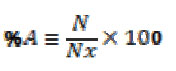
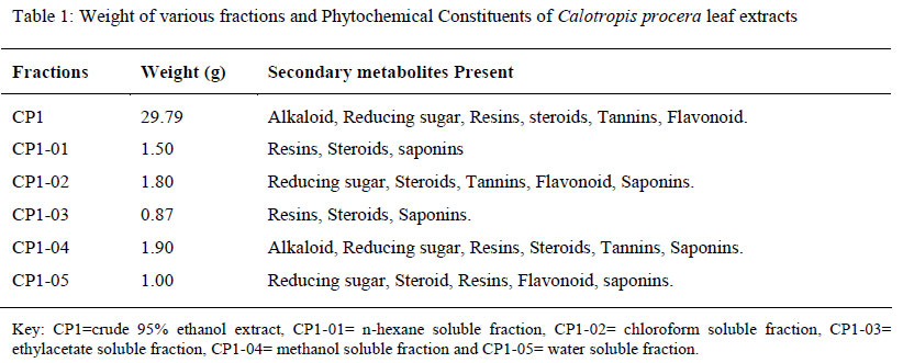
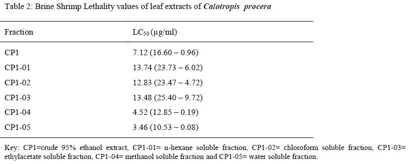
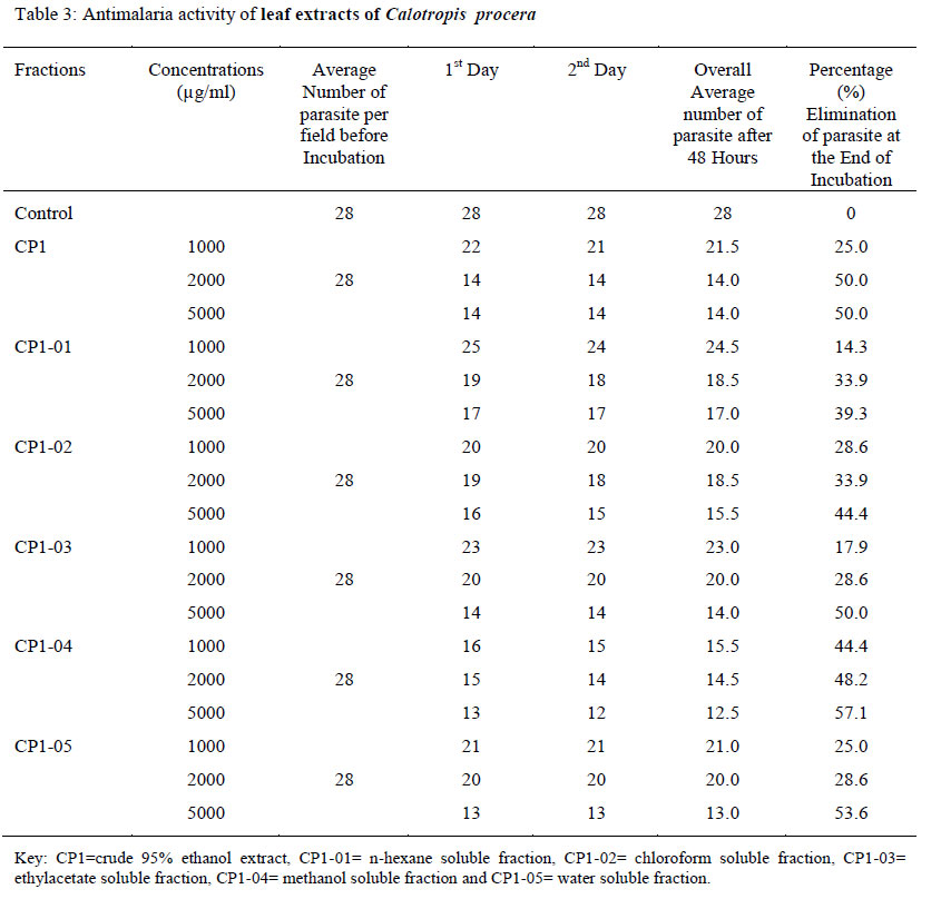
Biokemistri, Vol. 23, No. 1, March, 2011, pp. 29-34 Anti-plasmodia activity of leaf extracts of Calotropis procera Linn S. Y.*1 Mudi and A. Bukar2 1Department of Pure and Industrial Chemistry, Bayero University, Kano, P M B 3011, Kano. (Received January 9, 2011; Accepted March 1, 2011) Code Number: bk11005 ABSTRACT The leaves of Calotropis procera were air dried, grounded and soaked with ethanol. The extracts obtained (29.79g, CP1) was fractionated sequentially using aqueous methanol with petroleum ether, chloroform and ethyl acetate respectively. The residue of ethanol extract (marc) was extracted with 5M HCl, basified and extracted with chloroform. These were labeled as CP1-01 to CP1-05 for the plant. Each of these fractions was phytochemically screened to detect the class of secondary metabolite present. The fractions obtained from the plant were found to be selectively active against brine shrimp larvae. These fractions were also subjected to antimalaria parasites bioassay. Fractions CP1, CP1-04 and CP1-05 were found to be active against tested organisms, withCP1-04 being the most active. CP1-04 was further subjected to activity guided column chromatography that led to the isolation of two pure compounds CP1-04-1 and CP1-04-61. Compound CP1-04-61 was found to be active against the malaria parasite. This was further purified and subjected to qualitative and quantitative analysis. Key Words: Secondary metabolites, Antimalaria, Brine shrimp, Column chromatography, Calotropis procera. Introduction Malaria is a vector-borne infectious disease caused by protozoan parasites. It is wide spread in tropical and subtropical regions including part of the American, Asia and Africa (Snow et al., 2005). Each year, it causes diseases in approximately 515 million people and kills between one and three million people, majority of whom young children in sub-Saharan Africa (Snow et al., 2005). Adults are less likely to die from malaria, but still suffer from the sickness (Adams, 2008). Infection often peaks during the rainy season, and higher rates of bedridden workers affect the agricultural productivity of families, communities, and nations (Newman, et al., 2008). This made the disease to be associated with poverty and regarded as a major hindrance to economic development (Joy et al., 2003). Experts estimate that the annual costs of malaria treatment and lost in productivity due to malaria total between $2 billion and $3 billion in Africa (Newman, et al., 2008). Malaria parasites are transmitted by female anopheles mosquitoes bite. The parasites multiply within red blood cells (RBC) causing symptoms that include anaemia as well as other general symptoms such as fever, chills, nausea, and flu-like illness and in severe cases coma and death (Joy et al., 2003). Malaria transmission can be reduced by preventing mosquito’s bites with mosquito nets and insect repellents and by mosquito control measures such as spraying insecticides inside houses and draining standing water where mosquitoes lay their eggs. Taking preventive drugs (both traditional and orthodox drugs) can reduce risk of infection (Mary et al, 2000). Resistance acquired by the mosquito to insecticides, and the parasite to drugs, has led to new therapeutic challenges, particularly in the treatment of P. falciparum. The plants have shown significant killing capacity against plasmodium parasites, these includes; Cinchona tree, Artemisia tree, Artabotrys uncinatus (China: arteflene), Azadirachta indica (Asia: nimbolide) and Brucea javanica (China: terpenoids). Many more plants might have antimalarial potential (Ying and Yu-Lin, 1998). The research study reports on the active compounds present in Calotropis procera L responsible for its activity against malaria parasite. Materials and Methods Sample Collection Leaves of Calotropis procera were collected at Panshekara town, Kumbotso Local Government Area on September 22, 2007. The leaves were identified at the Department of Biological Science, Bayero University, Kano and with the help of keys (Aliyu, 2004). Extraction and Fractionation The air-dried sample (200g) was percolated using Ethanol (1.3L) for a period of 10 days. The extracts were drained and concentrated under reduced pressure using Rotavapor (R110 at 40°C). The ethanol extract was allowed to dry and its weight was recorded. This was labeled as CP1 as Ethanol extracts C procera. Ethanol extract (10g) was dissolved in 60% aqueous methanol (150ml, Aq. MeOH) and partitioned with petroleum ether (150ml), chloroform (150ml) Ethyl acetate (150ml) sequentially. All fractions obtained were collected in weighted beakers and were labeled as CP1-1 to CP1-5 (Sofowora, 1984). The Residue obtained (marc) after extraction with ethanol was repercolated with 5M Hydrochloric acid (500ml) for the period of 3 days. Aqueous acid fraction obtained was basified with solution of sodium hydroxide and partitioned with chloroform. The chloroform fraction was collected in a weighted beaker (After evaporating excess chloroform using Rotavapor) (Djilani et al, 2006). Column Chromatography of CP1-04 Five grams of the extract (CP1-04) was thoroughly mixed with silica gel until it changed to a non-sticky powder. It was then loaded to a silica gel column (108g; 62 x 2.5 cm). The column was run by eluting solvents of increasing polarity as follows: petroleum ether (60-80), petroleum ether-chloroform (9:1), (8:2), (1:1), (2:8), chloroform, chloroform-ethyl acetate (1:1), (2:8), ethyl acetate, methanol. A varied quantity of the solvents was eluted and the eluant was collected in fraction of 100ml. They were allowed for complete evaporation at room temperature. Weight and Thin Layer Chromatographic analysis on pooled fraction were determined. Malaria parasite assaying was re-conducted on selected samples. Chemical Analysis Plants extracts were phytochemically screened using standard techniques for the qualitative detection of Alkaloid, Flavanoids, Resins, Steroids, Sugars, Tannins and Saponins (El-olemy et al., 1994; Sofowora, 1984; Evans, 1995). Brine Shrimp Lethality Test (BST) Meyer, et al., (1982). The fractions obtained from the two plants were subjected to the test for their activity against Brine Shrimp Larvae (Artemia Salina). Artemia salina (leach) eggs (50mg) were added in a hatching chamber containing ocean sea salt water. The hatching chamber was kept under on inflorescent bulb for 48 hours for the eggs to hatch into shrimp larvae. The fractions CP1-1 to CP1-5 (20mg) were separately dissolved in methanol (2ml) from which 500, 50, and 5ml of each solution was transferred into vials corresponding to 1000, 100 and 10 mg/ml respectively. Each dosage was tested in triplicate. The vials (9 per test fraction) and one control contain 500ml of solvent were allowed to evaporate to dryness in about 48 hours at room temperature 4.5ml of ocean sea salt water was added to each vial. The final volume of solution in each vial was adjusted to 5ml with sea salt water immediately after adding the shrimps 24 hours later, the number of surviving shrimps at each dosage was counted and recorded. The lethality test against Brine shrimp larvae (Artemia salina) were carried out. The Brine shrimp Test (BST) results are expressed in LC50 µg/ml values at 95% confidence interval. Malaria Parasite Assay Sourcing of Malaria Parasites for the Assay Haematology Department, Muhammad Abdullahi Wase Specialist Hospital, Kano provide clinical blood samples containing heavy parasitaemia of Plasmodium falciparum. Venous blood from patients recommended for malaria parasites test (MP test) was carried out using 5ml disposable plastic syringes and needles (BD and 20 SWG). The samples were immediately transferred into K3-EDTA disposable plastic sample bottles with tightly fitted plastic corks and mixed thoroughly and then transported to the Microbiology laboratory at Bayero University in a thermoflask containing water maintained at 4°C as demonstrated by Dacie (1968). Preparation of Plasmodium falciparum Culture Medium Venous blood (2ml) from the main vein of white healthy rabbits pinnae was withdrawn using a disposable 5ml syringe (BD 205 WG). This was defibrinated by allowing it to settle for at least one hour (Dacie and Lewis, 1968). The defribranated blood was centrifuged at 1500rpm using spectre merlin centrifuge for 10minutes and the supernatant layer was collected in a sterilized tube. The sediment was further centrifuged at 1500rpm for five minutes, and the supernatant layer was added to the first test tube. The sediments were discarded and the serum collected was supplemented with the salt of RPMI 1600 medium (KCl 5.37mM, NaCl 10.27mM, MgS04 4.00 mM, NaHPO4 17.73mM, Ca(NO3)2 0.42mM, NaHCO3 2.5mM, and glucose 11.0 mM. (BDH ltd, UK) as demonstrate by Devo et al (1985). The medium was sterilized by 40µg/ml gentamicin sulphate (Trager, 1982). In Vitro Assay of the Activity of the Extracts on Plasmodium falciparum Culture A 0.1ml of tested solution as 0.2ml of the culture medium were added into a tube containing 0.1ml of .5% parasitaemia erythrocytes and mixed thoroughly. The sensitivity of the parasites to the tested fractions was determined microscopically after incubation for 24 and 48 hours at 37°C.The incubation was undertaken in glass bell jar containing a lighted candle to ensure the supply of required quantity of CO2 (about 5% 02 gas, 2% and about 93% nitrogen gas as demonstrated by Mukhtar et al, (2006). Determination of the Activity At the end of the incubation periods 24 and 48 hours, a drop of a thoroughly mixed aliquot of the culture medium was smeared on microscopic slides and stained by Giemsa’s staining techniques. The mean number of erythrocytes appearing as blue discoid cells containing life rings of the parasite (that appeared red pink) was estimated and the average percentage elimination by the samples was determined. The activity of the tested samples was calculated as the percentage elimination of the parasites after incubation period – 24 and 48 hours, using the formula below:
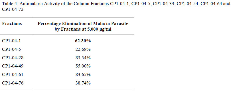
Where, %A = Percentage activity of the extracts Results and Discussion The various extracts/fractions and the phytochemical of Calotropis procera and their weights were shown in Table 1. The phytochemical screening result showed the distribution of the presence of secondary metabolites in the extracts. The ethanol extract (CP1) have shown the presence of alkaloid, flavonoids, resins, saponins, reducing sugars, steroids and tannins. They were reflected in the other fractions, such that resins and saponins appeared in all the fractions, steroids were presence in CP1-01, CP1-02, CP1-03, CP1-04 and CP1-05, while sugars and tannins reflected in CP1-02 and CP1-04. CP2 and CP1-04 showed alkaloid present only. Table 2 showed that all extracts obtained from the plant exhibit activity for all the fractions tested with CP1-05 (LC50 µg/ml value = 3.46 (10.53 – 0.08)) being most active among the fractions followed by CP1-04 (LC50 value = 4.52 (12.85 – 0.19). It is also observed that, the activity for Calotropis procera against Brine shrimp larvae increases with increasing polarity of solvent with the exception of ethyl acetate fraction. The results of antimalaria activity of the extracts are shown in Table 4. Fraction CP1 has demonstrated a remarkable activity at all concentrations. The most interesting anti-plasmodia activity was obtained with CP1-04, in which the microscopic examination of Giemsa’s stained slides for the fraction at 5000ug/ml showed a serious absence of the parasite after 48 hours. These observations suggest that the activity of the extract may be cytotoxic for P. falciparum, thereby inhibiting their development. MP test result on CP1 has shown activity of almost 50% elimination of the parasite, whereas the fractions CP1-04 and CP1-05 have about 57.1% and 53.6% elimination of the parasite respectively (after 48 hours at 5000ug/ml). In Table 4, column fractions CP1-04-28 and CP1-04-61 have shown the highest activity; the later was obtained in an appreciable quantity for spectral analysis (CP1-04-61: a white crystalline compound; 0.14g; Rf =0.67; 1HNMR = 5.4, 5.2 ppm; 13CNMR = 72.05, 140.99, 121.97 ppm; IR = 3246, 1667, 2919 cm-1) was found to be most active against malaria parasite, (83.65% after 48 hours at 5000 µg/ml) and it melts between 139°C-141°C. The qualitative and quantitative analysis of this fraction revealed the presence of carbon (36.25%), hydrogen (62.50%) and oxygen (1.25%). The percentage composition of oxygen shows that it is a mono oxygenated compound. While CP1-04-5 and CP1-04-76 exhibited weak activity against the parasite; 22.69% and 38.74% after 48 hours, for the respective fractions. Conclusion Conclusively, this work provides a scientific basis for using Calotropis procera as antimalaria plant. The fraction CP1-02 led to isolation of active white crystalline compound. ACKNOWLEDGEMENT Our gratitude to Dr. M. D. Mukhtar of Biological Sciences Department, Bayero University Kano for providing the antimalaria protocol as well as putting us through and Malam Ado Dakata of Haematology Department, Muhammad Abdullahi Wasai Hospital, Kano for helping with the infected blood sample.. References
Copyright © 2011 Klobex Academic Publishers The following images related to this document are available:Photo images[bk11005t1.jpg] [bk11005t4.jpg] [bk11005t2.jpg] [bk11005t3.jpg] |
| |||||||||
