 |
| About Bioline | All Journals | Testimonials | Membership | News |
|
||||||
|
||||||
Journal of Culture Collections, Volume
3, 2000-2002, pp. 15-24
ISOLATION AND TAXONOMIC INVESTIGATION OF ACTINOMYCES FROM SPECIFIC BIOTOPES IN BULGARIA Mariana Naidenova and Denitsa Vladimirova* National Bank for Industrial Microorganisms and Cell Cultures, 1113 Sofia, P. O. Box 239, Bulgaria Code Number: cc02002 Summary Investigations were performed for isolation of Actinomyces strains from different extreme biotopes in Bulgaria. According to the morphological, physiological and chemotaxonomic data three of the strains were taxonomically identified to genus, one as a member of genus Frankia and two - from genus Actinomadura. The other ten Actinomyces isolates belonged to genus Streptomyces and were also identified to species. Introduction The Actinomyces are commonly known as producers of bioactive compounds in medicine, agriculture, ecology and industry. In the most of the cases the ability to produce one or another bioproduct is taxonomically determined, which makes taxonomic identification of the isolates so important. Recently, the scientists have turned to investigate some more specific biotopes for new microorganisms, perspective both for industry and for science. Nowadays less explored areas as caves, old mine galleries, salt-cellar and salt lakes, hot-springs, contaminated waters and sediments have attracted scientific attention. This research line led to discovering of many new species and genera Actinomyces, which produced bioactive compounds or were found to be resistant to heavy metals and other pollutants. For example, two new Actinomyces species were isolated from old gold mine in Korea - Catellatospora koreensis and Pseudonocardia kongjuensis and also a new genus - Hongia koreensis [19, 20, 23]. Also there were found two new species in Chinese cave - Knoella and Beutenbergia [8, 10]. More trivial biotope like soil also can give new Actinomyces - Microbispora gen. nov., Bogoriella gen. nov., Gordonia, Cryptosporangium gen. nov., Ornythincoccus gen. nov., Acrocarpospora gen. nov, Terracoccus, Streptomyces thermogriseus [7, 9, 11, 12, 21, 24, 27, 28, 29, 31]. It should be mentioned that some of the isolates came from very specific inhabitats such as deteriorated automobile tyres - Gordonia; salt-cellars - Nocardiopsis; compost – Thermocrismum [4, 15, 18, 23]. Along with these new Actinomyces it was found that many members of genus Streptomyces possessed interesting biological characteristics - ability to accumulate chromium, mercury resistance, anti-bacterial activity and production of different secondary metabolites [1, 6, 14, 25]. In order for isolation of new, scientifically important and with practical application Actinomyces strains, water, soil and plant samples from unexplored and polluted regions in Bulgaria have been investigated. Materials and MethodsMaterial. Water, soil and plant samples have been taken from different regions in Bulgaria (Table 1). Sampling was carried out from 1988 to 1991. Different diagnostic media for Actinomyces isolation were inoculated with the samples after some initial treatment. Table 1. Sample characteristics and isolation media.
Microorganisms. Thirteen Actinomyces strains were isolated and were deposited in the National Bank for Industrial Microorganisms and Cell Cultures (NBIMCC) with the following numbers: strain D-1 (3747), D-9 (3727), M-1 (3748), M-9 (3749), M-13 (3761), M-14 (3760), M-15 (3762), M-16 (3781), M-18 (3782), A-4 (3744), A-43 (3746), Elsha-7 (3743), Siva I (3745). These strains were obtained as pure cultures by using standard microbiological method by growing suspensions of ten times successive dilutions of the samples. The cultivation was carried out at 14 and 28°C for 7 to 21 days. Suitable colonies were re-cultivated several times for purity. Three test-microorganisms were used to test the antibiotic activity of the isolates - Bacillus subtilis, Escherichia coli and Candida albicans. They were maintained on specific media at 25°C for yeasts and 37°C for the bacterial strains. Culture media. The Actinomyces strains were isolated by growing on different diagnostic media: yeast-starch agar, Chapek medium with peptone, Benet medium, soil agar I and II, Nocardia medium, Mineral agar I, L-agar containing kanamycin, starch-casein agar, glycerol-asparagine agar, Actinomyces isolation agar, F-medium. For determination the morphological and cultural characteristics of the strains, they were cultivated on following media - Mineral agar I and II, CP I and II, glycerol-nitrate agar, ISP-2, ISP-3, ISP-4, ISP-5 media [29]. The assimilation of carbohydrates was studied by using the medium ISP-9, containing 14 different carbohydrates at a concentration of 1% as only carbon source. Physiological and biochemical characteristics of the isolates were examined by cultivation on ISP-6 and ISP-7 media for melanin synthesis; skimmed milk for milk coagulation and peptonisation and gelatine for gelatine liquation [20, 33]. Media used for cultivation of the test microorganisms were nutrient agar and beer agar. Taxonomic identification. For the purposes of morphological, cultural, physiological and biochemical identification of the Actinomyces strains, the manuals of Gause, Krasilnikov, Waksman, Bergey, Shirling and Gottlieb were used [2, 8, 20, 29, 33]. The characteristics of the colonies were described after cultivation on different culture media; the colour of the aerial and substrate mycelium and those of the soluble pigment were determined according to the Bondatsev colour scale [3]. The type of the spore chains was studied by compound microscope with 20x and 40x objectives. The observations of the spore surface were performed by transmission electron microscopy. Antibiotic activity was tested by using method of agar plate diffusion and was measured as sterile zone in millimetres [5]. Chemotaxonomy. For the purposes of diaminopimelic acid isomers and sugars determination, Actinomyces strains have been cultivated in 500 ml shake flasks with 100 ml complex medium at 220 rpm for 24 h at 28°C. The biomass was freeze-dried after being washed several times in distilled water and harvested by centrifugation. The lyophilized cultures were then used in determination of isomers of diaminopimelic acid and sugars in whole cell hydrolysate by the method of Komagata and Suzuki [7]. Results and discussionThe place from where the samples have been taken, the sample type, date and isolation medium for the 13 Actinomyces strains are shown in Table 1. Most of the strains grew very well on different culture media, except strain 3760, which showed poor growth (Table 2). The temperature optimum for cultivation was 28°C. All thirteen strains formed aerial mycelium on Mineral agar I and ISP-2 media, which colour varied from white through creamy-pinkish, grey-violet, purple to dark grey. The colour of substrate mycelium was in creamy-yellow-brown series but in some strains it could be dark brown or even black. Strains 3745, .3746, 3747, 3727, 3748 and 3782 did not produce any soluble pigment, while its colour among the other isolates was in yellow-greenish-brown series and only in strain 3743 a pinkish-red pigment on Mineral agar I was found [3]. The diameter of the colonies ranged from 1 to 5-6 cm and the surface and shape of the colonies depended on the culture medium and the strain (Table 2). Table 2. Morphological
characteristics of Actinomyces strains.
*Growth: weak (+), good (++), very good (+++), excellent (++++); **Spore
chains: retinaculiaperti (RA), rectiflexible (RF), spiral (S). Table 2. (continued).
*Growth: weak (+), good (++), very good (+++), excellent (++++);
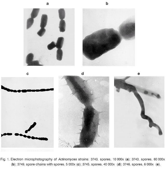
**Spore chains: retinaculiaperti (RA), rectiflexible (RF), spiral (S); The spore chains, observed with 20x and 40x objectives, were spiral with different turn’s number in strains 3743, 3745, 3746, 3748, 3782. Rectiflexible and retinaculiaperti chains had the strains 3727, 3760, 3761, 3781; rectiflexible chains were those of strains 3749 è 3762 and three type spore chains could be found in strain 3744. The spore surfaces in nine of the isolates were smooth (Table2); warty was in strain 3743, with small spines in 3782 è 3745. The spore shape was oval and prolonged in strains 3727, 3761 and cylindrical and prolonged in 3762 (Fig.1). The abilities of all thirteen strains to assimilate 14 different carbon sources are represented in Table 3. The best growth was shown on media with maltose, fructose, rhamnose and xylose. The strains assimilated well glucose, mannitol, sucrose and lactose except strain 3760, which assimilated them poorly. Cellulose, inositol, sorbitol and dulcitol showed weak Actinomyces growth on them and only 6 strains were tested on raffinose. All in all the strains assimilated well most of the tested carbon sources and strain 3743 was the best, assimilating 10 of 14 hydrocarbonates. On the last position was strain 3760, which showed poor growth on most of the sources. Table 3. Assimilation* of different carbon sources by Actinomyces strains.
*Assimilation: weak (+), good (+), very good (++), not determined (ND). The physiological and biochemical characteristics of the strains are summarised in Table 4 [20, 33]. Melanoid pigments were formed only by strain 3761. Amylolytic activity was performed by 5 strains - 3727, 3748, 3749, 3760, 3762 and gelatine was poorly liquefied only by strain 3746. Milk peptonisation and coagulation was performed by strains 3743, 3744, 3745, 3747, 3727, 3761 and 3760; only coagulation - 3748 è 3761; and only strains 3746, 3762, 3781, 3782 performed peptonisation of skimmed milk. The antibiotic activity of the thirteen strains against 3 test-microorganisms was studied by agar plate diffusion (Table 5). Only four strains performed any activity: against B. subtilis - 3761, 3782, 3743 and 3746; against E. coli - 3743 and 3746, and against C. albicans - 3727 and 3749. However, the diameter of sterile zones was small - the biggest one is 22 mm by strain 3743. Table 5. Antibiotic activity of Actinomyces strains.
According to the type of diaminopimelic acid isomers in whole cell hydrolysate, it’s LL-form was that found in strains 3747, 3748, 3749, 3745, 3727, 3760, 3761, 3762, 3781, 3782. The DL-isomer was detected in strains 3743, 3746, 3744. Strains 3746 è 3744 had glucose, galactose and madurose in their cell wall, which supposed their belonging to genus Actinomadura. Data from the chemotaxonomic analysis, along with the morphological, cultural and physiological characteristics have allowed us to refer two of the studied strains to the genus Actinomadura (3744 and 3746) and one - 3743 to genus Frankia. All of them were isolated from soil samples, taken under trees. These Actinomyces genera are part of the natural rhizospheric microflora. The other ten strains were identified as members of genus Streptomyces and more precisely to the following species: Streptomyces aureorectus (3760), Streptomyces noboritoensis (3761), Streptomyces scabies (3762), Streptomyces fulvoviridis (3781), Streptomyces griseoruber (3782), Streptomyces bluensis (3745), Streptomyces flavogriseus (3747), Streptomyces libani (3748), Streptomyces viridogenes (3749), Streptomyces spiroverticillatus (3727). Comparing these results with those of other authors, it could be said that members of Streptomyces family are the most common among the isolates from polluted regions and from plants, inhabitating polluted waters [1, 6, 14, 25]. This is, probably, due to their remarkable resistance to bad environmental conditions and to different pollutants. References
Copyright 2002 - National Bank for Industrial Microorganisms and Cell Cultures - Bulgaria The following images related to this document are available:Photo images[cc02002f1.jpg] | ||||||||||||||||||||||||||||||||||||||||||||||||||||||||||||||||||||||||||||||||||||||||||||||||||||||||||||||||||||||||||||||||||||||||||||||||||||||||||||||||||||||||||||||||||||||||||||||||||||||||||||||||||||||||||||||||||||||||||||||||||||||||||||||||||||||||||||||||||||||||||||||||||||||||||||||||||||||||||||||||||||||||||||||||||||||||||||||||||||||||||||||||||||||||||||||||||||||||||||||||||||||||||||||||||||||||||||||||||||||||||||||||||||||||||||||||||||||||||||||||||||||||||||||||||||||||||||||||||||||||||||||||||||||||||||||||||||||||||||||||||||||||||||||||||||||||||||||||||||||||||||||||||||||||||||||||||||||||||||||||||||||||||||||||||||||||||
| |||||||||
