 |
| About Bioline | All Journals | Testimonials | Membership | News |
|
||||||
|
||||||
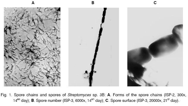
Journal of Culture Collections, Volume 4, No. 1, 2004-2005, pp. 36-42 TAXONOMY OF STREPTOMYCES SP. STRAIN 3B Stefka Antonova-Nikolova*, Nikoleta Tzekova and Ljubomira YochevaSofia University, Biological Faculty, Department of General and Industrial Microbiology, 8 Dragan Tzankov st., 1164 Sofia, Bulgaria Code Number: cc05004 Summary Strain Streptomyces sp. 3B produces high activity extracellular proteolytic complex, in which serine- and metalloproteases are identified. The general and some other significant properties for the taxonomy of the strain were studied by the methods of the International Streptomycetes Project. The results of the investigation of Streptomyces sp. 3B and the comparative references about Streptomyces species with similar taxonomic characteristics identified it as Streptomyces albovinaceus. The antibiotic activity and sensitivity was also tested. Strain 3B was resistant to the group of the penicillin antibiotics. IntroductionProteases constitute one of the most important groups of industrial enzymes and find applications in the production of deter-gents, foods, leather, silk, chemical and agricultural preparations [3, 12]. In the last years they are used in medicine successfully. This enzyme accounts for 60 % of the world market. The streptomycetes produce numerous compounds (secondary metabolites), within which antibiotics are of commercial relevance. As corresponds to their habitat, these bacteria are nutritionally quite versatile and the most produce extracellular hydrolytic enzymes that permit the utilization of high molecular weight biopolymers such as proteins, polysaccharides, fats and other substrates. Streptomycetes are great producers of proteolytic enzymes. The composition of these protein complexes is determined by the taxonomic belonging of the producer. The present paper as a part of a complex investigation of strain Streptomyces sp. 3B and the produced proteolytic enzymes describes its taxonomic determination and biological characterization [4]. Materials and methods The object of the investigation, Streptomyces strain 3B from the collection of the Department of General and Industrial Microbiology, was isolated from protein rich samples, collected around Sevlievo. Its proteolytic activity was clearly expressed. Strain Streptomyces sp. 3B produces extra-cellular enzymes from which serine- and metalloproteases were established [1, 7]. Taxonomic properties of the strain were determined according to the methods and media of the International Streptomycetes Project (ISP) [9]. The morphology of the strain was studied in yeast-malt extract agar (ISP-2), oatmeal agar (ISP-3), starch agar with inorganic salts (ISP-4) and glycerol-asparagine agar (ISP-5). The production of the melanin was tested on peptone-yeast extract iron agar (ISP-6) and tyrosine agar (ISP-7). The utilization of carbon sources was carried out according to the modified method of Pridham and Gottlieb (isp-9) with addition of the following sugars: D-glucose (positive control), L-arabinose, sucrose, D-xylose, I-inositol, D-mannitol, D-fructose, rhamnose, raffinose and cellulose, and in absence of carbon source (negative control). The form of the sporophores was studied with light microscope by directly observation and by replica’s method. The morphology of the spores and the spore chains were investigated by electron microscope. The color of the aerial and vegetative mycelia was determined according to Bondartsev color scale [2]. The morphological determination of the strain was made on mature culture. The production of the melanin was observed on 2d and 4th day. The results from utilization of the carbon sources were measured on 10th and 16en day. Species identification of strain 3B was based on Nonomura’s key [8] and the species description was according to ISP [10, 11]. In addition the following biological characteristics were studied: decomposition of gelatin, hydrolysis of starch [5]; growth on milk agar, effect to the milk, antibiotic activity against test-microorganisms Bacillus subtilis and Escherichia coli according to the method of the agar plates, sensitivity to antibiotics by paper disks method [6]. The following antibiotics (“BulBio”) were tested: erythromycin (15 μg/disk), ampicillin (10 μg/disk), streptomycin (10 μg/ disk), gentamycin (10 μg/disk), penicillin (10E/ disk) and chloramphenicol (30 μg/disk). Results and discussionMicromorphological characteristics. Vegetative mycelium of the studied strain Streptomyces sp. 3B did not fragment. Aerial mycelium formed monopodially situated sporophores with right or lightly curved shape. The sporophores contained more than 10 spores. The spores were elongated with smooth surface (Fig. 1). Macromorphological characteristics. Streptomyces sp. 3B was clearly polymorphic.The strain formed three types colonies on the investigated media: round flat colonies with unfledged aerial mycelium and white concentric lines, smooth surface and edge; completely covered by the aerial mycelium formless protruding colonies with rugged surface, undulating edge and slightly convex central part; big good formed round colonies with concentric folds around the center of the colony and a fosse or umbilicus in the center, the margin of the colony was entire or fibrillated depending on its age. Cultural properties. On more media Streptomyces sp. 3B showed a good development. The color of the aerial mycelium was white. It changed from white to gray-white opal pink depending on the medium. The coloring of the vegetative mycelium varied from pale-yellow to yellow-brown according to the cultural age (Table 1). The strain did not form melanoid pigment on any of the investigated for melanin media. Other soluble pigments were not observed. Physiological and biochemical properties. The utilization of carbon sources is shown in Table 2. The strain 3B grew very well on mineral-salt media, containing D-glucose, L-arabinose, D-xylose, D-fructose, rhamnose and raffinose. On medium with sucrose the growth was weak or absent. The strain did not assimilate I-inositol, D-mannitol and cellulose. It did not hydrolyze starch but decomposed gelatin. The strain 3B caused peptonization of milk but did not its coagulation. It did not attack tyrosine (Table 3). Table 1. Cultural properties of strain Streptomyces sp. 3B.
Table 2. Utilization of carbon sources by strain Streptomyces sp. 3B.
Table 3. Physiological characteristics of strain Streptomyces sp. 3B.
Antagonistic properties and sensitivity to antibiotics.Streptomyces sp. 3B did not possess antibiotic activity against B. subtilis and E. coli. It was sensitive to the more antibiotics used in this investigation. The strain was resistant to the action of β-lactame antibiotics (Table 4). Taxonomic comments. Using references given by ISP [10, 11], strain Streptomyces sp. 3B was compared to Streptomyces cultures similar by morphological, cultural, physiological and biochemical properties (Table 5). More Streptomyces strains assigned to the white series show differences as on the degree of the utilization of the sugars, as well as on the morphological and cultural properties from Streptomyces sp. 3B strain. Table 4. Sensitivity of strain Streptomyces sp. 3B to different antibiotics.
Table 5. Comparison of general taxonomyc properties of Streptomyces sp. 3B strain with relative streptomycetes spesies.
The Streptomyces strain 3B is much closed to S. albovinaceus [10]. Similarities: both Streptomyces sp. 3B and S. albovinaceus belong to section Recti-Flexibilis (RF); the mature spore chain contains more than 10 spores with smooth surface; the color of the aerial mycelium is white; the vegetative mycelium is yellowish; the strains do not produce melanin and other soluble pigments; they grow very well on medium containing L-arabinose, D-xylose, D-fructose and rhamnose; they do not grow on medium with I-inositol and grow weakly on sucrose; Differences: Streptomyces strain 3B does not show growth on medium with D-mannitol but on medium containing raffinose it grows very well. Streptomyces sp. 3B is similar to S. orientalis [10] by morphological properties and by the utilization of some sugars. In contrast to strain 3B, S. orientalis grows on media containing I-inositol and D-mannose but does not utilize raffinose. S. orientalis can form blue aerial mycelium on ISP-3 and its spore chain contains more than 50 spores. The Streptomyces strain 3B also resembles to S. candidus [11]. The differences are that S. candidus has spore chain with more than 50 spores; it grows very well on media with D-mannitol and does not utilize raffinose. Morphologically, Streptomyces sp. strain 3B is similar to S. sindenesis [11] from which differs by the length of the spore chain and by the coloring of the aerial mycelium. The mature spore chain of S. sindenesis contains 3 to 10 spores. It grows well on media containing D-mannitol but does not utilize rhamnose, sucrose and raffinose. The results of the investigation of Streptomyces sp. 3B and the comparative references about Streptomyces species with similar taxonomic characteristics according to ISP [10, 11] gave us the reason to identify it as Streptomyces albovinaceus with the name S. albovinaceus 3B. Short characteristic of Streptomyces albovinaceus 3B.The strain 3B produces monopodially situated right or lightly curved sporophores, fragmenting in spore chains with more than 10 spores. The spore surface is smooth. The color of the aerial mycelium is white with different nuances depending on the medium. The color of the vegetative mycelium is pale - yellow to yellow - brown, depending from the age of the culture. The growth temperature range is 18-45 °C. Melanoid and other soluble pigments do not produce. The strain liquefies gelatin and peptonizes milk, has not tyrozinase activity and does not hydrolyze starch. It utilizes D-glucose, L-arabinose, D-xylose, D-fructose, rhamnose and raffinose but does not utilize I-inositol and D-mannitol. Strain 3B has not antibiotic activity against B. subtilis and E. coli. It is sensitive to the antibiotics - gentamycin, erhytromycin, streptomycin and chloramphenicol. The strain 3B is resistant to the group of the penicillin antibiotics. Acknowledgments. We express our thanks to Vassilis Kapurdov from the National Center of Hygiene for the generous supply of electron microscope. References
Copyright 2005 - National Bank for Industrial Microorganisms and Cell Cultures - Bulgaria The following images related to this document are available:Photo images[cc05004f1.jpg] | |||||||||||||||||||||||||||||||||||||||||||||||||||||||||||||||||||||||||||||||||||||||||||||||||||||||||||||||||||||||||||||||||||||||||||||||||||||||||||||||||||||||||||||||||||||||
| |||||||||
