 |
| About Bioline | All Journals | Testimonials | Membership | News |
|
||||||
|
||||||
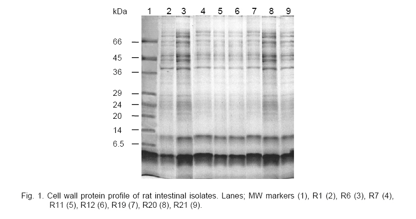
ISOLATION OF BACTERIOCINOGENIC LACTIC ACID BACTERIA FROM RAT INTESTINE Mahantesh Patil*, Ajay Pal, Vijai Pal and Rajini Kumari Yaddula Food Biotechnology Discipline, Defence Food Research Laboratory, Siddartha Nagar, Mysore-570011, *Corresponding author, e-mail: dfrlmysore@sancharnet.in Code Number:cc06008 Summary Lactic acid bacteria were isolated from rat intestinal contents and screened for bacteriocin production. Out of twenty-three isolates, eight were found to produce bacteriocins. The morphological and biochemical properties of these isolates indicated that they belong to the genus Enterococcus. The bacteriocins were extracted and partially purified by Gel Permeation Chromatography (GPC) using G-25, and the antimicrobial spectrum of these bacteriocins was studied against indicator organisms, including Lactococcus diacetylactis, Staphylococcus aureus, Escherichia coli and Pseudomonas sp. Key words: lactic acid bacteria, bacteriocins, Enterococcus, intestine. Introduction Lactic acid bacteria (LAB) are industrially important organisms because of their fermentative ability as well as health and nutritional benefits. Moreover, they are generally regarded as safe for incorporation into food products [2]. The ability of lactic acid bacteria to inhibit the growth of various Gram positive and/ or Gram negative bacteria is well known and attributed to the production of organic acids such as lactic acid and acetic acid [4], hydrogen peroxide, bacteriocins, bacteriocin like substances and possibly biosurfactants [14]. Bacteriocins are ribosomally synthesized, extracellular peptides/proteins produced by bacteria, exhibiting bacteriostatic or bacteriocidal activity against closely related bacteria. These molecules are produced by bacteria belonging to several genera, however, only some of them have been extensively studied. Bacteriocins produced by lactic acid bacteria have received considerable attention in recent years, because of their possible use as biopreservatives in food processing industry with resultant reduction in the use of chemical preservatives [15]. Another advantage of bacteriocins for use as a biopreservatives is that they can be easily digested in the human gastrointestinal tract [11]. Earlier, several researchers have isolated LAB from dairy and meat products, fermented vegetables and mucosal surfaces of animals [5, 8, 16]. Bacteriocin producing species have now been identified among all the genera that comprise the LAB including Lactobacillus, Pediococcus and Carnobacterium [6, 12] as well as several Enterococcus species [1, 9]. In the present study LAB have been isolated and characterized from the intestinal contents of Wistar albino rat and screened for bacteriocin production. Materials and Methods Chemicals. General chemicals and solvents of analytical grade were procured from SRL, Bacterial strains. The indicator organisms viz, Staphylococcus aureus and Lactococcus diacetylactis were procured from Microbial Type Culture Collection (MTCC), Isolation of lactic acid bacteria. LAB were isolated from intestinal contents of 10 months old Wistar rat. The rat was fed with the Nutrilab-rodent feed procured from Tetragon Chemie Pvt Ltd, Screening of LAB for antimicrobial activity. All the isolates maintained in frozen stocks were propagated twice in MRS broth and used for further study. For screening the isolates for bacteriocin production all isolates were grown in MRS broth and incubated at 37 oC for 60 h. The cell free supernatants were adjusted to pH 5.0 with 2N NaOH and concentrated to one tenth of the original volume by rotary flash evaporator. These samples were filter sterilized by passing through 0.22 mm membrane filter (Millipore, Characterization of LAB isolates. Fresh (12-18 h) cultures of bacteriocin producing isolates were Gram stained and examined microscopically. Catalase activity was tested by adding few drops of 3 % hydrogen peroxide to a test tube containing 24 h old culture of each isolate. Growth was assessed in MRS broth at 15, 37 and 45 oC and at pH 4.4, 7.0, 8.6 and 9.6 with incubation at 37 oC. Salt tolerance was tested with 6.5, 10 and 15 % (w/v) NaCl in MRS broth. Production of acid and CO2 was tested in MRS broth containing inverted Cell-wall protein extraction and analysis. LAB isolates were grown in MRS broth. The cell mass was harvested and resuspended in 3 ml of distilled water (A600 ~ 2) and centrifuged. Cell wall proteins were extracted from the final cell pellet with 0.5 ml of 0.01 M Tris-HCl, 0.01M EDTA, 0.01M NaCl, 2 % SDS, pH 8.0 at 100 oC for 5 min [3]. After treatment the supernatants were centrifuged at 11,600 g for 10 min and analyzed by Tris-Glycine SDS-PAGE [9] with 5 % stacking and 12 % separating gel. Extraction and partial purification of bacteriocins. Cell adsorption-desorption method [7] was employed for bacteriocin extraction from 72 h old cultures. Bacteriocins were adsorbed on to the cells at pH 6.5 and desorption was carried out at pH 2.0-2.5, in the presence of 0.1 N NaCl. The contents were further concentrated by rotary flash evaporator followed by adjusting pH to 4.5. The concentrated samples were further purified by passing through a Sephadex G-25 column and eluted with ammonium acetate buffer (0.05M, pH 4.5). Various fractions around shoulder, peak and valley were pooled separately, further concentrated and checked for the inhibittory activity against S. aureus. Subsequently, these active fractions were used to verify the antimicrobial activity against various indicator organisms. Results and Discussion Lactic acid bacteria are known to produce a large variety of bacteriocins and some of them are extensively used to extend the shelf-life of food products by inhibiting the growth of food born pathogens and spoilage microorganisms. In this study we have attempted to isolate LAB from intestinal contents of Wistar albino rat for production of potential bacteriocins. Twenty-three bacterial isolates were screened for bacteriocin production, out of which only eight produced bacteriocins active against S. aureus, E. coli and L. diacetylactis. Further, these isolates were characterized for their biochemical and physiological properties. The morphological and biochemical properties of these isolates have been shown in Tables 1 and 2. All the isolates were Gram-positive cocci with slight variation in the cell arrangement. Isolates R1, R6, R7, R11 and R19 were found to exist in pairs and small chains while isolates R20 and R21 were short chains and isolate R12 existed in pairs only. These isolates were catalase negative and did not produce CO2 from glucose and were found to be homofermentative. All the isolates were able to produce ammonia from arginine and acid from glucose. The isolates R1, R7, R11, R19 and R20 were able to grow even at 15 oC and 45 oC with maximum growth at 37 oC. In case of R6, R12 and R21 no growth was noticed at 15 oC. The growth of all isolates was found to be weak at pH 4.4 as well as at 6.5 % salt concentration. All the isolates grew luxuriously at pH values 8.6 as well as 9.6 and there was no growth at 4 oC and 10 % salt concentration in MRS medium. All the isolates produced acid from cellobiose, fructose, galactose, glucose, glycerol, lactose, mannose, maltose, mannitol, ribose, salicin and hydrolyzed the esculin. Isolates R1, R6, R11, R19, R20 and R 21 were able to ferment melizitose. In contrast, isolates R7 and R12 were able to ferment mellibiose. Table 1. Morphological, physiological and biochemical characteristics of bacteriocinogenic rat isolates.
Legend: growth (+), no growth (-), luxurious growth (++), weak growth (w), arginine (Arg), glucose (Glc), homofermentative (Ho). Table 2. Carbohydrate utilization pattern of bacteriocinogenic rat isolates.
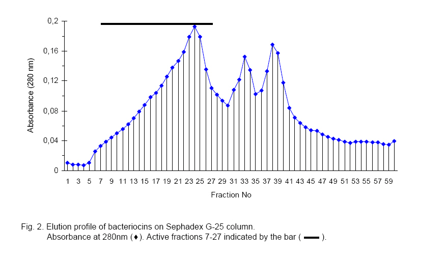
Legend: growth (+), no growth (-). On the basis of morphological and biochemical characteristics the isolates were assigned to the genus Enterococcus. Fermentation of melizitose and inability to ferment mellibiose helped in distinguishing between E. faecalis and E. faecium [10]. Based on the above tests, isolates R1, R6, R11, R19, R20 and R21 were identified as E. faecalis while isolates R7 and R12 were grouped into E. faecium, however, it was clear that they were different strains as they could be distinguished based on sugar fermentation pattern. Cell-wall protein extraction and analysis Cell wall protein profile for eight bacteriocin producing LAB isolates has been depicted in Fig. 1. The electrophoregram revealed great similarity among the eight isolates indicating that all of them belong to the same genus. In all isolates high molecular weight proteins in the range of 36–66 kDa were more prominent than others. Extraction and partial purification of bacteriocins Bacteriocins obtained from cell adsorption-desorption technique were subjected to Sephadex G-25 column chromatography and recovered in partially purified form in shoulder and peak regions of the elution profile as shown in Fig. 2. Active fractions obtained with all the strains were concentrated and equal quantity of protein was used to check antimicrobial activity against selected indicator organisms. The bacteriocins from R19 and R20 isolates were found to be most potent against S. aureus while R7 bacteriocin had the least inhibitory activity. All the strains were found to inhibit Pseudomonas sp., E. coli and L. diacetylactis to varying extents. Further studies are in progress for the purification and characterization of these bacteriocins from the isolates in order to ascertain their possible use against food borne pathogens, which are not inhibited by the bacteriocins reported till date. Acknowledgements. The authors are thankful to Dr. A. S. Bawa, Director, Defence Food Research Laboratory, References
The following images related to this document are available:Photo images[cc06008f2.jpg] [cc06008f1.jpg] | ||||||||||||||||||||||||||||||||||||||||||||||||||||||||||||||||||||||||||||||||||||||||||||||||||||||||||||||||||||||||||||||||||||||||||||||||||||||||||||||||||||||||||||||||||||||||||||||||||||||||||||||||||||||||||||||||||||||||||||||||||||||||||||||||||||||||||||||||||||||||||||||||||||||||||||||||||||||||||||||||||||||||||||||||||||||||||||||||||||||||||||||||||||||||||||||||||||||||||||||||||||||
| |||||||||
