 |
| About Bioline | All Journals | Testimonials | Membership | News |
|
||||||
|
||||||
Journal of Culture Collections, Vol. 6, No. 1, 2009, pp. 10-20 Distribution and ecobiology of antagonistic streptomycetes from agriculture andcoastal soil in Tamil Nadu, India Dharumadurai Dhanasekaran1*, Nooruddin Thajuddin1and Annamalai Panneerselvam2 1Department of Microbiology,
Bharathidasan University,
Tirchirappalli.620 024,
India; Code Number: cc09002 Summary Totally 189 Streptomyces isolates were obtained from eight different soils of Cuddalore, Tamil Nadu, India. Among them, only 78 isolates were morphologically distinct. The highest diversity in the Streptomyces populations was observed in agricultural soil with minimum occurrence of antagonistic organisms. The least number of Streptomyces strains was found in coastal soil with maximum antagonistic organisms. A connection between physicochemical properties of the soil and the Streptomycesabundance was established. There was a positive correlation between the total Streptomyces population and nitrogen, available phosphorus, ferrous and manganese, while the correlation with pH and sodium was negative. This study clearly revealed the occurrence of Streptomyces spp. and its correlation with the available nutrient in the environment. Key words: Streptomyces, diversity, antagonism, soil, Cuddalore Introduction The discovery of novel antibiotic and other potential compound lead molecules of pharmaceutical interest through microbial secondary metabolite screening is becoming increasingly fruitful. There is wide acceptance that microorganisms are virtually unlimited sources of novel substances with many therapeutic applications. Among them actinomycetes hold a prominent position due to their diversity and had proven their ability to produce new and novel substances. Among the various actinomycetes genera, Streptomyces is best recognized genus of actinomycetes. They are aerobic, spore forming and Gram positive bacteria that have DNA with a high GC content (69-73 %). They form extensive branching substrate, aerial mycelia and widely distributed in soil. Actinomycetes have provided many important bioactive compounds of high comercial value and continue to be routinely screened for new bioactive substances. These searches have been remarkably successful and approximately two-thirds of naturally occurring antibiotics, including many of medical importance, have been isolated from actinomycetes [34]. Actinomycetes are abundant in terrestrial soils, a source of the majority of isolates shown to produce bioactive compounds. Intensive screening programme carried out over the past several decades resulted in the production of known bioactive compounds [33]. However, actinomycetes are more abundant in terrestrial soils than in marine sediments, show varying degrees of salt tolerance and produce spores that are undoubtedly washed in large numbers from shore into the sea [14]. Inspite of the fact that they remain active in the marine environment, their role in the production of bioactive compounds is yet to be studied.The marine environment is an untapped source for many useful drugs and an assessment of this potential is imperative. It is well known that the antibiotics are medicinally valuable and that the actinomycetes are the potential sources of antibiotics, which could be profitably used in the pharmaceutical Industries; and the best known example, is the product of Streptomyces. There is a growing demand and need for new antibiotics to control many bacterial, fungal and neoplastic diseases of plants, animal and human beings. A perusal of the literature clearly indicates that reports on the antagonistic actinomycetes from the marine environment are very scanty. The marine soil of Tamil Nadu has rich potential of microbial diversity. Nevertheless, they have not been extensively explored for the registration of novel actinomycetes. Keeping these points in view, the present study has been undertaken to isolate and screen the antimicrobial compounds produced by Streptomyces sp from Cuddalore district, Tamil Nadu, India. In order to achieve this goal the present investigation has been planned to understand the Streptomyces population in different soil types (agricultural field, sea shore, estuarine and salt pan) of Cuddalore district, Tamil Nadu, to understand the relationship between the physicochemical properties of soil and population of Streptomyces. Materials and Methods Description of sampling sites. Totally eight locations belonging to four soil environs were selected in Cuddalore district, Tamil Nadu, India (Lat. 11°43’ N; Long. 79°49’ E), which include terrestrial soils (Keelpattampakkam and Vasanagkuppam), sea shore soils (Samiyarpettai and xPudhukkuppam), estuarine soil (Annangkoil and MGR Thittu) and salt pan soil (Porto Novo and Thaikkalthurai). Soil sample collection. The soil samples were collected from eight locations in Cuddalore district, Tamil Nadu, India during the period of June 2002. The soil samples were collected at random, brought to the laboratory in sterile polythene bags and used for further analysis. Physicochemical characterization. The soil moisture, temperature, pH, electric conductivity of various soils was determined by the methods described by Jackson [18]. Organic carbon content was determined by adopting chromic acid wet digestion method [50], nitrogen was estimated by alkaline permanganate method [45], available phosphorus was estimated by Bray I method [2]. The sodium and potassium content in the extract was determined by using flame photometer [46], calcium and manganese content was estimated by the method of Jackson [18], sulfur content in the soil was extracted with 0.15 % calcium chloride and the available sulphur content was estimated by a colorimetric method [37]. Cation exchange control (CEC) of the soil was determined by using 1 N ammonium acetate solution as described by Jackson [18], and the available micronutrients (Br, Zn, Cu, Mn and Fe) were determined in the diethylene triamine penta acetic extract of soil using Perkin-Elmer model 2280 atomic absorption spectrophotometer [27, 38]. Isolation
of Streptomyces. Starch casein agar[23] medium was
prepared and sterilized at 121 °C in
Screening of Streptomyces producing antimicrobial compounds. Antimicrobial property of the Streptomyces was screened by agar overlay method [17]. In this method spores of Streptomyces were streaked on petri plate containing 15 ml of starch casein agar and incubated for five days at 28 ± 2 °C. 10 ml of sterile soft nutrient agar (0.75 %) medium for the test bacteria and sterile soft potato dextrose agar medium for test fungi were mixed with 0.1 ml of cells/spore suspension separately and overlaid with five days old growth of Streptomyces isolates. The plates were further incubated at 28 ± 2 °C for 24 hours and 72 hours for bacteria and fungi, respectively. Based on the presence and absence of inhibition zone, Streptomyces producing antimicrobial compounds were screened. Extraction of extracellular antimicrobial compounds of Streptomyces. The most intense antagonistic activity of the Streptomyces was selected and its antimicrobial spectrum was tested against the pathogenic bacteria and fungi. The selected isolates were inoculated into a 500 ml conical flask containing 200 ml of starch casein liquid medium and shaken at 28 ± 2 °C and 200 rpm for three days. An equal volume of ethyl acetate was added into the cell free culture filtrates°and shaken for two hours, and the antimicrobial compounds were extracted. Extraction of intracellular antimicrobial compounds of Streptomyces. The Streptomyces strains were inoculated in starch casein broth in a 500 ml Erlenmeyer flask, then flasks were incubated at 28 ± 2 °C for seven days. The biomass was separated from the fermentation broth by filtration°and centrifugation. 200 ml of Triton X100 was mixed with cell biomass. It was incubated at 28 ± 2 °C for 20 min. 200 ml of 0.5 M EDTA was added to the above solution and kept at 65 °C for 45 min. Then cell suspension was centrifuged at 10,000 rpm for 10 min. Intracellular metabolites were present in the supernatant and it was used for antimicrobial assay.
Two clinically
important pathogenic bacteria namely, Escherichia coli and Staphylococcus
aureus were inoculated into nutrient broth and incubated at 37 °C for 24 hours. The
test organisms were spread over on Muller Hinton agar and wells made by using
Two types of yeast (Candida
albicans and Saccharomyces cerevisiae) and two filamentous fungi (Aspergillus
niger and Penicillium funiculosum)
were independently shaken in potato dextrose liquid medium at 25 °C for 3 days. A
mycelium suspension of each fungus was added to molten PDA medium, mixed and
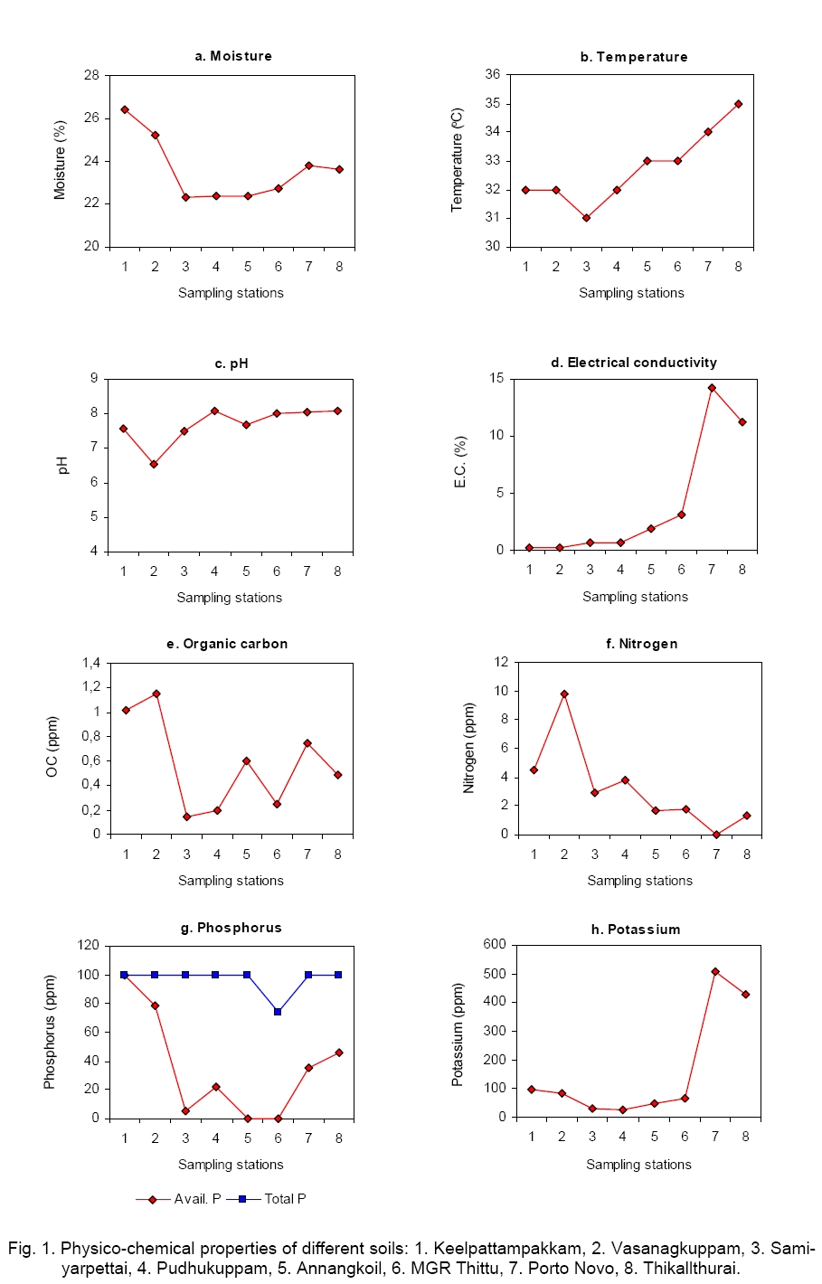
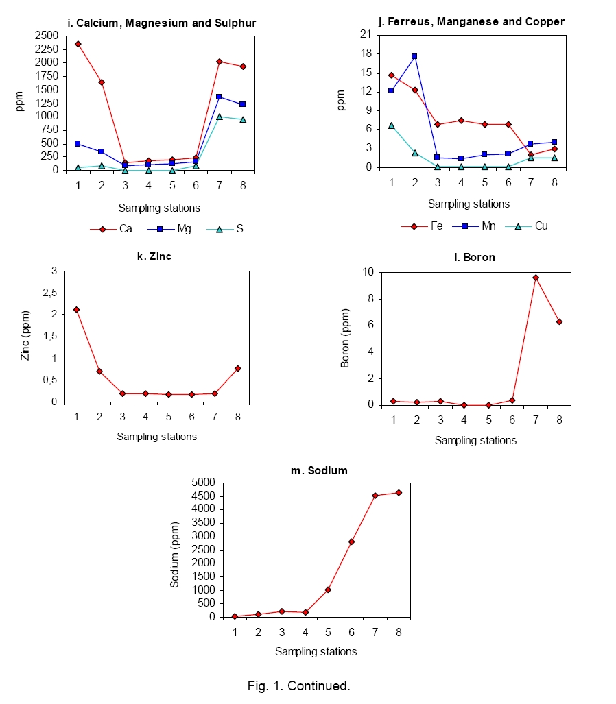
poured into sterile Petri plates. Wells were made on medium by using Results The moisture content of soil ranged from 22.3 to 26.4 %. The maximum moisture content was observed in the agricultural field soil and the minimum moisture content was recorded in the sea shore soil. The maximum soil temperature (35 °C) was established in the salt pan soil of Thaikkalthurai and minimum temperature (31 °C) was observed in the sea shore soil. The pH of the soil was maximum (8.1) in the salt pan soil and minimum (6.5) was recorded in the agricultural field soil. Maximum level of electrical conductivity of the soil (14.2) was established in the salt pan soil of Porto Novo and minimum (0.21) was recorded in the agricultural field soil of Vasanagkuppam (Figure 1, a-d). Maximum (1.15 ppm) organic carbon con-tent was observed in the agricultural field soil of Vasanagkuppam and minimum (0.25 ppm) was found in estuarine soil of Pudhukuppam. The organic nitrogen content was the least (below the detectable level) in salt pan soil of Porto Novo and maximum (9.8 ppm) was recorded from agricultural field soil of Vasanagkuppam. Available phosphorus content of soil was the least (below the detectable level) in estuarine soil and maximum (100 ppm) was recorded from agricultural field soil of Keelpattampakkam. Among the eight stations, seven stations showed maximum (100 ppm) total phosphorus content, and minimum phosphorus content was recorded from M.G.R. Thittu. Maximum content of potassium (508 ppm) was observed in salt pan soil of Porto Novo and minimum (26 ppm) was observed in sea shore soil of Pudhukuppam (Figure 1, e-h). The micronutrients of soil such as calcium (2360 ppm) and magnesium (1367 ppm) were recorded from agricultural field soil and salt pan soil, respectively. The sulphur content was maximum (997.8 ppm) in salt pan soil of Porto Novo. Nevertheless the maximum contents of zinc (2.11 ppm), Iron (14.6 ppm), manganese (17.62 ppm), copper (6.73 ppm) and boron (9.6 ppm) were established from the agricultural field soil, whereas maximum content of sodium (4620 ppm) was recorded from salt pan soil. The agricultural and sea shore soils did not show much variation in the micronutrient content (Figure 1, i-m). Streptomyces diversity of soil Totally 189 Streptomyces isolates including white, gray, brown and pink coloured colonies with different morphological types were isolated from eight different soil samples. Among them 111 isolates from agricultural field soil (51 from Keelpattampakkam and 60 from Vasanagkuppam), 57 isolates from sea shore soil (30 from Samiyarpettai and 27 from Pudhukuppam), 12 isolates from estuarine soil (8 from Annangkoil and 4 from MGR Thittu) and 9 isolates from salt pan soil (6 from Porto Novo and 3 from Thaikkalthurai) were isolated (Table 1). Among the 189 Streptomyces isolates, only 78 isolates were morphologically distinct, which included 30 isolates from agricultural field soil (13 from Keelpattampakkam and 17 from Vasanagkuppam), 27 isolates from sea shore soil (23 from Samiyarpettai and 4 from Pudhukuppam), 13 isolates from estuarine soil (8 from Annangkoil and 4 from M.G.R. Thittu) and 9 isolates from salt pan soil (6 from Porto Novo and 3 from Thaikkalthurai). These isolates were purified for further studies (Table 1 and Table 2). Table 1. Streptomyces populations in different soils of Cuddalore district.
*Data in parentheses are morphologically distinct isolates. Table 2. Cultural characteristics of Streptomyces isolates.
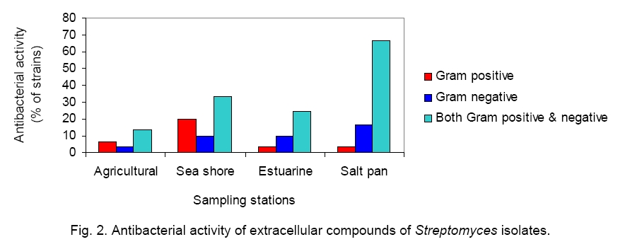
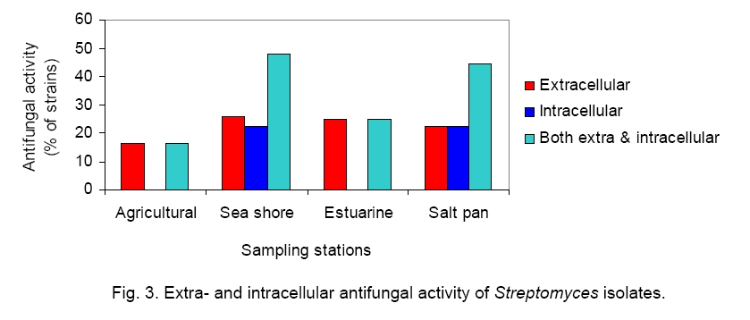
Streptomyces antagonism Totally 78 isolates of Streptomyces were screened for their antimicrobial activity against pathogenic bacteria and fungi by agar overlay assay method. Among the 78 isolates, 22 isolates showed antibacterial activity and 18 isolates showed antifungal activity. Among 22 isolates tested, 10 inhibited the Gram positive and 12 isolates inhibited the Gram negative bacteria. The maximum percentage (66.7 %) of antibacterial Streptomyces was found in salt pan soil (6/9 isolates) followed by sea shore soil (9/27 isolates, 33.33 %), estuarine soil (3/12 isolates, 25 %), and agricultural field soil (4/30 isolates, 13.3 %). The selected 22 isolates of antagonistic Streptomyces were further screened for extra and intracellular antibacterial compounds. All the 22 isolates showed extracellular activity, whereas 8 isolates showed both extra and intracellular antibacterial activity (Figure 2). The maximum percentage of the isolates of Streptomyces, which showed antifungal antagonistic activity, was found in sea shore soil (13/27 isolates, 48.14 %) followed by salt pan soil (4/9 isolates, 44.44 %), estuarine soil (3/12 isolates, 25 %) and agricultural field soil (5/30 isolates, 16.6 %). Among the 18 isolates tested, all the isolates showed extracellular antifungal activity including 8 isolates having both extra and intracellular antifungal activity (Figure 3). Discussion Actinomycetes are prokaryotes with extremely high metabolic potentiality. They produce numerous substances essential for health such as antibiotics, enzymes, immunomodulators, etc. During the last few decades actinomycetes have become the most fruitful source for antibiotics. In the 60s’ and 70s’ of the 20th Century 75 to 80 % of all the discovered antibiotics were derived from the order Actinomycetales, mainly from Streptomyces species. Considering the practically useful compounds, today about 130 to 140 microbial products and a similar number of derivatives (including semi synthetic antibiotics), are derived from the diverse group of actinomycetes, and hence the members of this group are proved to be of commercial importance. Furthermore, some 15 to 20 compounds are used in agriculture mainly as pesticides, plant protecting agents and food additives [29].
India,
with a long coastline of over Diversity of Streptomyces spp. In the present study, totally 78 strains of Streptomyces were isolated from four different soils (agricultural field, sea shore, estuarine and salt pan) in eight locations namely Keelpattampakkam, Vasanagkuppam (agricultural field soil), Samiyarpettai, Pudhukuppam (sea shore soil), M.G.R. Thittu, Annangkoil (estuarine soil), Porto Nova and Thaikkalthurai (salt pan soil). The total number of Streptomyces isolates was the highest in agricultural field soil (30 isolates) followed by that in sea shore soil (27 isolates), estuarine soil (12 isolates) and salt pan soil (9 isolates). The diversity of Streptomyces isolates was increased due to the nutritive status of the respective soil. Actinomycetes from agricultural soil habitat have been reported [4, 6, 7, 12, 20, 24, 26, 28, 32, 40, 48, 51, 52]. Thus it is obvious that Streptomyces is ubiquitous and adopted to diverse habitats which vary widely in space and time, and also to diverse environmental conditions. The first report on marine actinomycetes was made by Nadson [30] from the salt muds. Actinomycetes, especially Streptomyces, have been reported from the marine sub habitats such as, dead marine algae [34]; marine sediments [3, 5, 9, 10, 11, 14, 15, 16, 19, ,21, 22, 25, 51, 35, 39, 41, 47] and also from almost all parts of the world. Thus they have a worldwide distribution, which indicates their plasticity and adoptability to extremely varied environment. In spite of the fact that the actinomycetes have wide distribution they show variation in their population dynamics. In the present investigation it was found that there was correlation between physicochemical properties of soil and total Streptomyces population (TSP). It revealed a significant positive correlation between TSP and nitrogen (r = 0.8952; p<0.01); TSP and available phosphorus (r = 0.7057; p<0.05); TSP and Fe (r = 0.8742; p<0.01) and TSP and Mn (r = 0.8135; p<0.05). There was a significant negative correlation between TSP and pH (r = -0.7992; p<0.05) and TSP and Na (r = -0.7673; p<0.05) (Table 3). Similar study was reported by Saadoun and Al-Momani [44]. Jiang and Xu have studied the pH, organic matter, nitrogen and phosphorus content of the soils as correlated with actinomycetes population [20]. The correlation between salinity, pH and organic content of marine sediments and actinomycetes population have been reported by several workers [19, 31]. Jensen et al. reported that there was no correlation between the percentage of organic content of marine sediment and actinomycetes population [19]. Ghanem et al. found that the variation in temperature, pH and dissolved phosphate showed in-significant values, but that variation in total nitrogen and organic matter were significant in the population in Alexandria [13]. Lee and Hwang reported that the soil pH (5.1-6.5), moisture (9.1-13.0 MHC) and organic matter (9.1-11.0 %) influence the dominance of Streptomyces in the agricultural field soils of Western part of Korea [26]. Hence it could be concluded that though actinomycetes are ubiquitous, their population dynamics are often influenced by the available nutrients and the physicochemical conditions of the ecosystem. Table 3. Correlation coefficient between physicochemical properties of soil and total Streptomyces population (TSP).
Antagonistic activity of Streptomyces The antibiotic potentiality of the actinomycetes, especially of Streptomyces is well known. However, there are both quantitative and qualitative variations in the antibiotics produced by different genera and species. Substrate and micro habitats greatly influence the qualitative and quantitative differences in the production of antibiotics by different isolates of Streptomyces. It has been reported that among 47 strains of actinomycetes, only 19 showed antagonistic activity [29]. Thus, there are variations in the quality and quantity of the antibiotics produced even by the different isolates of the same species. In the present study among the 78 isolates of Streptomyces only 22 isolates were found to possess antimicrobial activity. The percenttage of occurrence of antagonistic organisms also varied and it was in salt pan (66.7 isolates), followed by sea shore (33.33 isolates), estuarine (25.00 isolates) and agricultural field soil (13.3 isolates). Among the 22 isolates, only 8 isolates had the potentiality to inhibit the growth of both Gram positive and Gram negative bacteria and all of them produced extracellular antimicrobial com-pounds, but only eight isolates produced both extra and intracellular antimicrobial compounds (Figure 3). It was also found that Streptomyces spp. varied in their antibacterial activities against differrent groups of bacteria, namely Gram positive bacteria, S. aureus and Gram negative bacteria, E. coli. Totally, 10 isolates have antibacterial activity against Gram positive bacteria - 6.6 % (2 isolates), 20 % (6 isolates), 3.3 % (1 isolate) and 3.3% (1 isolate) from agricultural field soil, sea shore, estuarine and salt pan soil, respectively. 12 isolates are active against Gram negative bacteria - 3.33 % (1 isolate), 10 % (3 isolates), 10 % (3 isolates) and 16.66 % (5 isolates) from agricultural field, sea shore, estuarine and salt pan soil respectively. Production of extra and intracellular anti-microbial compounds by Streptomyces from Bay of Bengal has been reported by Sambamurthy and Ellaiah [43]. Augustine et al. isolated 312 actinomycetes from Maharashtra soil (India) and screened them for the extracellular and intracellular antifungal compounds [1]. 22 % of the isolates were extracellular antifungal compound producers. The present investigation is focused on the extracellular antimicrobial compound production by Streptomyces spp. Thus the above findings supports the present study also concludes that the isolates of the Streptomyces species are varying in the quantitative and qualitative production of antimicrobial compounds, especially the extra and intracellular products. The compounds produced by the isolates of Streptomyces showed not only antibacterial but also antifungal properties. In the present investigation the maximum percentage of antifungal isolates of Streptomyces were isolated from sea shore soil (48.14 %) followed by salt pan (44.44 %), estuarine soil (25.0 %) and agricultural field soil (16.6 %). Among the 78 Streptomyces isolates, only 18 isolates produced extracellular antifungal compounds and 10 isolates produced both extra and intercellular anti-fungal compounds. Among the effective 28 antifungal isolates, 9 isolates produced both extra and intracellular antifungal compounds (Figure 3). It has been reported that out of 127 isolates only 26 (20.4 %) showed antifungal activity [19]. They were isolated from 47 soils of seven district of West Bengal. Ouhdouch et al.evaluated the antifungal actinomycetes from Atlas mountain soil, Sahara sand, dung, sludge, well, river, lake, sea water and sediments [36]. Twenty six out of 96 strains showed antifungal activity against C. albicans and C. tropicalis. Dhanasekaran et al. reported 107 antifungal actinomycetes from different coastal area of Tamil Nadu and found that out of 107 isolates 22 possessed antifungal activities [8]. Thus the present study gives an idea about the Streptomyces biodiversity in different soils (both marine and agricultural field soils) of Cuddalore district, Tamil Nadu. Although actinomycetes, especially Streptomyces are ubiquitous, they vary in their morphological, physiological, biochemical and genetical characteristics de-pending on the physicochemical environment of the habitats. References
Copyright 2009 - National Bank for Industrial Microorganisms and Cell Cultures - Bulgaria The following images related to this document are available:Photo images[cc09002f3.jpg] [cc09002f1a.jpg] [cc09002f2.jpg] [cc09002f1b.jpg] |
| |||||||||
