 |
| About Bioline | All Journals | Testimonials | Membership | News |
|
||||||
|
||||||
Journal of Culture Collections, Vol. 6, No. 1, 2009, pp. 21-27 Occurrence of different morphotypes of Streptomyces exfoliatus in mangrove ecosystems of Bhitarkanika, Orissa, India Nibha Gupta*, Srilekha Mishra and Uday Chand Basak Regional
Plant Resource Centre, Bhubaneswar – 751 015
(Orissa),
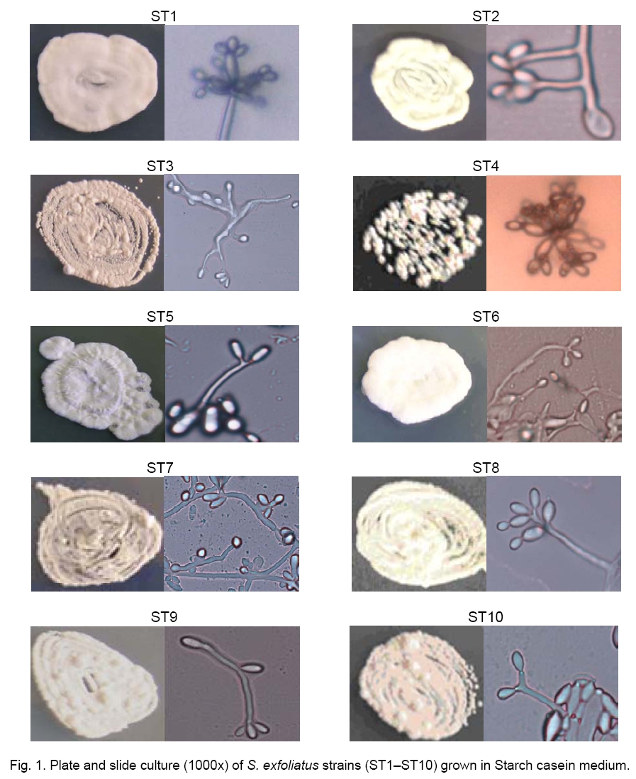
India Code Number: cc09003 Summary Ten strains of genus Streptomyces were isolated from phyllosphere of three mangrove tree species viz., Sonneratia caseolaris, Kandelai candel and Exoecocaria aggallocha found in Bhitarkanika mangrove ecosytem of Orissa. According to physiological and biochemical data, all ten isolates were taxonomically assigned to the species Streptomyces exfoliatus. However, all the strains varied morphologically and exhibited different extracellular activity. This is a unique report on this mangrove ecosystem as far as Streptomyces occurrence is concerned. Key words: mangroves, Streptomyces, salinity, Orissa, Bhitarkanika Introduction Streptomycetes are an important group of bacteria, which are very well studied for specific activity like antibiotic production etc [2] Now a day less explored area like caves, old mines, salty ecosystem, hot springs have attracted much scientific efforts in view of searching for novel organisms and products [1, 3]. In order to obtain new scientifically important and with potential applications streptomycetes, strains associated with mangrove plants of Bhitarkanika (Orissa, India) were isolated. They were identified and studied for specific growth, physiological and biochemical characteristics. Materials and Methods Source of materials. The leaf samples from three mangrove plants i.e. Kendalia candle, Sonneratia casealaris, Exocoecatia agallocha found in Khola region of Bhitarkanika, Orissa were collected for the isolation of Streptomyces. Isolation media. The dilution plate technique was followed for the isolation of Streptomyces strains on ISP 3, ISP4 and ISP5 me-dia. The isolates obtained were designated as ST1 - ST10. Growth characteristics: Streptomyces isolates were grown in Starch casein liquid medium of 4.5 and 7.2 pH and at 30 ºC and 37 ºC for 10 days in static culture conditions. Finally, their dry biomass was determined by keeping filtered and fresh biomass at 40 °C for 72 h and measuring the weight through electronic balance. At the same time, change in pH, color of filtrate, diffusible pigments were observed and recorded. The plate culture of all isolates prepared in Starch casein medium was observed for morphological studies like colony characteristics, coloration, margin etc. Morphological studies. Slide cultures of all the isolates of Streptomyces were prepared on Starch casein medium and ISP3 medium, and incubated at 30 ºC and 37 ºC by using cavity slides. Periodical observations regarding spore morphology, arrangements and mycelloid structure were recorded by using Nikon Japan Trinocular Research Microscope Model 50i. Taxonomic identification. The isolates were characterized by different biochemical tests for carbohydrate utilization, nitrogen utilization, growth in different stress conditions, antibiotic resistance, and amino acid degradation, enzyme activity like amylase, protease, asparaginase, antifungal activity and phosphate solubilization. Finally, data were used for the identification of Streptomyces isolates using Probabilistic identification of bacteria (PIB win). Results and Discussion Bhitarkanika mangrove ecosystem is un-explored till date as far as the occurrence of streptomycetes is concerned. Therefore, the present study is of prime importance. Overall 10 isolates of genus Streptomyces were isolated from S. caseolaris (ST1), E. agallocha (ST2) and K. candel (ST3 - ST10). Colony characteristics. All isolates produced white, cottony and raised colony. Reverse coloration is mostly reddish in all except ST2, ST3, ST4, and ST9 and ST10 that showed brown and white reverse coloration, respecttively. All the strains have entire or round margin except ST3, ST4, ST5, ST8 and ST9 that exhibited irregular margin (Table 1, Figure 1). Table 1. Colony characteristics of Streptomyces strains.
Spore chain morphology. The ST1 and ST8 have unicellular free spores and straight structure. It has fascicled spore arrangements. The mycelium appears as blue color in starch casein medium. It has single sporophore with five subsporophores in tri- to pentacular organization with single attached spores. The sporophore of ST2 and ST4 contains five subsporophores with 3 to 5 attached spores. It is alsofascicled type structure. It has hypha unsegmented when observed on starch casein media through slide culture. No spirals have been found. ST3, ST6, ST7 and ST9 have no coiling structure or spore bunch rather they form a flexous spore structure with hyphae segmented when observed on starch casein medium through slide culture. No spiral chain was found in these strains. They have three spores attached on straight sporophore. ST5 has no coiling structure or spore bunch rather it forms a flexous spore structure with hyphae segmented when observed on starch casein medium through slide culture. No spiral chain was found in this strain. It has two spores attached (Figure 1, Table 3). Growth characteristics. Effect of temperature and pH on growth of all isolates of Streptomyces is shown in Table 2. Five strains namely ST1, ST4, ST5, ST7 and ST8 preferred 4.5 pH and 30 ºC, ST2, ST6, ST8 and ST10 performed best at the same pH but at 37 ºC whereas ST3 grew well at both the pH and temperature used in this study. Table 2. Growth characteristics of Streptomyces strains in Starch caesin medium.
Biochemical characteristics. Melanoid pigments are not formed on Peptone yeast iron agar and Tyrosine agar. The organisms could be able to tolerate Potassium tellurite (0.01%) and phenol (0.1%) but could not grow in the presence of sodium azide (0.01%), NaCl (7%). They exhibited better growth in sodium pyruvate whereas inhibitory action of sodium acetate, sodium propionate and sodium citrate on their growth was observed (Table 3).
The
strains were not able to utilize many carbon sources like sucrose, meso-inositol,
raffinose, manitol, D-lactose, salicin, D-melizitose, D-xylose, adonitol, L-arabinose Table. 3. Morphological and biochemical characteristics of Streptomyces strains.
Table 3 (continued).
Growth characteristics. Effect of temperature and pH on growth of all isolates of Streptomyces is shown in Table 2. Five strains namely ST1, ST4, ST5, ST7 and ST8 preferred 4.5 pH and 30 ºC, ST2, ST6, ST8 and ST10 performed best at the same pH but at 37 ºC whereas ST3 grew well at both the pH and temperature used in this study. All the strains showed lipolytic activity and susceptibility to Oleandomycin, Neomycin, Rifampicin and Penicillin G. No allantoin degradation was found in any strain. No protease activity was found in ST1, ST5 and ST7 whereas ST1, ST4 and ST6 were positive for amylolytic activity. They were positive for nitrate reduction and xanthin degradation. Strains ST8, ST9 and ST10 gave positive reaction for H2S production. Special activities: All the strains showed antifungal activity against Fusarium sp. except ST1, ST2, ST8 and ST9. Three isolates i.e. ST1, ST3 and ST7 were good producers of both intracellular and extracellular L-asparaginase. TheStreptomyces strain ST7 was found to be an extracellular enzyme producer endowed with antifungal activity. This strain could also solubilize TCP and rock phosphate. A computerized database was used to compare the biological properties of all isolates of streptomycetes with those of Streptomyces sp. The results suggest that strains ST1 - ST10 are streptomycetes strongly related to S. exfoliatus. Though they have similarity with the type strain S. exfoliatus in the incapability to utilize inositol and manitol, at the same time they are marginally distinct with respect to non utilization of sucrose and raffinose. All biochemical and physiological characteristics specified these strains as S. exfoliatus. However, studied strains are morphologically distinct from the type strain S. exfoliatus [4]. Our isolates may be a new variant of S. exfoliatus. Acknowledgement Authors are grateful to Department of Ocean Development, Ministry of Earth Sciences, and Government of India for the financial support through DOD project no.11-MRDF/4/4/UNI/97(P-22). References
Copyright 2009 - National Bank for Industrial Microorganisms and Cell Cultures - Bulgaria The following images related to this document are available:Photo images[cc09003f1.jpg] | |||||||||||||||||||||||||||||||||||||||||||||||||||||||||||||||||||||||||||||||||||||||||||||||||||||||||||||||||||||||||||||||||||||||||||||||||||||||||||||||||||||||||||||||||||||||||||||||||||||||||||||||||||||||||||||||||||||||||||||||||||||||||||||||||||||||||||||||||||||||||||||||||||||||||||||||||||||||||||||||||||||||||||||||||||||||||||||||||||||||||||||||||||||||||||||||||||||||||||||||||||||||||||||||||||||||||||||||||||||||||||||||||||||||||||||||||||||||||||||||||||||||||||||||||||||||||||||||||||||||||||||||||||||||||||||||||||||||||||||||||||||||||||||||||||||||||||||||||||||||||||||||||||||||||||||||||||||||||||||||||||||||||||||||||||||||||||||||||||||||||||||||||||||||||||||||||||||||||||||||||||||||||||||||||||||||||||||||||||||||||||||||||||||||||||||||||||||||||||||||||||||||||||||||||||||||||||||||||||||||||||||||||||||||||||||||||||||||||||||||||||||||||||||||||||||||||||||||||||||||||||||||||||||||||||||||||||||||||||||||||||||||||||||||||||||||||||||||||||||||
| |||||||||
