 |
| About Bioline | All Journals | Testimonials | Membership | News |
|
||||||
|
||||||
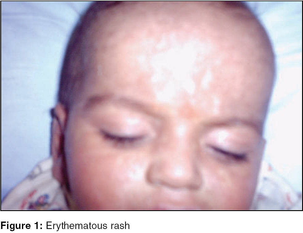
Indian Journal of Critical Care Medicine, Vol. 10, No. 1, January-March, 2006, pp. 47-49 Case Report Transfusion-associated graft-versus-host disease in an immunocompetent individual Surekha Devi Allanki, Shailesh R. Singi*, Ramana Dandamudi* From: Departments of Tranfusion Medicine and *Hemato-Oncology, Global Hospitals, Hyderabad - 500 004, India Correspondence Address:Dr. Surekha Devi Allanki, Consultant, Tranfusion Medicine, Global Hospitals, Hyderabad - 500 004, India. E-mail: dr.surekhadevi@gmail.com Code Number: cm06009 Abstract A 9 months old immunocompetent male baby was admitted with high grade fever, erythematous maculo-papular rash and frequent loose stools. He was unwell three weeks prior, with febrile illness and anemia, for which he was transfused 2 units of non irradiated whole blood from first-degree family donors (father and mother), in a private hospital. Seven days after transfusion, he became symptomatic. Investigations revealed elevated liver enzymes, elevated bilirubin levels and progressive pancytopenia. Clinical findings and results of skin biopsy and bone marrow examination were consistent with transfusion-associated graft-versus-host disease. He was treated with steroids, Cyclosporin, broad spectrum antibiotics and supported by irradiated blood components. He died on day 10, post admission and day 31, post transfusion, despite intensive medical care. Keywords: Irradiation, related donor, TA-GVHD. Transfusion associated graft-versus-host disease (TA-GVHD) is an under-diagnosed condition, in clinical practice. It can occur in immunocompromised as well as immunocompetent hosts and may follow allogenic bonemarrow transplant or transfusion from a related donor.[1] More than 90% of patients succumb to refractory infections.[2] The only effective preventive measure, is administration of irradiation of blood products.[2] A case of graft-versus-host disease following related donor transfusion, is presented here. Case Report A 9-months-old, immunocompetent male infant, was admitted with high grade fever, erythematous maculo-papular rash from 7 days and frequent loose stools from 2 days. He was unwell three weeks prior, with febrile illness and anaemia, for which he was transfused 2 units of non-irradiated whole blood donated, by his parents. Seven days after receiving transfusion, he developed erythematous rash all over the body, which increased in severity progressively. On admission, he appeared sick and toxic with high grade fever, pallor and icteric. There was erythematous rash all over the body with superficial peeling [Figure - 1], hepatomegaly, no lymph node enlargement, nor bleeding diathesis. His hemogram revealed hemoglobin of 7.9 gm% and total WBC count of 3500/cumm. The differential leucocyte count was-neutrophils-20%, lymphocytes-56%, eosinophils-1%, monocytes-3% and atypical lymphocytes-20%. The platelet count was 80,000/cumm. Biochemical investigations revealed total bilirubin of 5.3 mg/dl, direct bilirubin of 4.4 mg/dl, SGOT of 520 IU/lt, SGPT of 662 IU/lt, total proteins of 6.3 gm/dl, albumin of 3.7 gm/dl, alkaline phosphatase of 846 IU/lt and creatinine of 0.3 mg/dl. Immunohematology showed positive direct antiglobulin test (DAT) and indirect antiglobulin test (IAT). Auto-antibody workup revealed significant titers of warm agglutinis (1:32). Bone marrow examination revealed predominantly transformed and atypical lymphoid cells, with few myeloid precursors and normal megakaryocytes. Skin biopsy [Figure - 2] showed lymphocytic aggregates at the epidermal and dermal junction, with keratinocytes. Features were consistent with acute GVHD-horn′s grade II. The baby was started on methyl Prednisolone, broad spectrum antibiotics and supported by irradiated blood components (2 units of packed RBC and 1 unit of single donor platelet concentrate). He had progressive pancytopenia and did not respond to Cyclosporin either. Nine days later his hemogram showed hemoglobin of 5.8 gm%, total WBC count of 100/cumm and platelet count of 9000/cumm. He had respiratory failure, for which he was ventilated. He continued to deteriorate and died on day 10, post admission and day 31, post transfusion. Discussion Transfusion-associated graft-versus-host disease (TA-GVHD) is a fatal immunologic transfusion complication, caused by engraftment and clonal expansion of donor lymphocytes, in a susceptible host.[3] The engrafted lymphocytes mount an immunologic attack against recepient tissues, including hematopoietic cells, leading to refractory pancytopenia with bleeding and infectious complications, which are primarily responsible for 90-100% mortality rate in afflicted patients.[3] The total number of TA-GVHD cases reported in the world′s literature was fewer than 200.[3] TA-GVHD occurs in an immunocompetant individual, who receives cellular blood components from family members or random donors, from a genetically restricted community (genetically homogenous in Japan).[4] This is because of major HLA haplotype similarities between donor and recepient, specifically the donor is homo-zygous for class I haplotype, for which recepient is heterozygous.[4] TA-GVHD occurs with infusion of 8x10¹¹ lymphocytes per kg body weight.[4] GVHD has not been reported following infusions with FFP and cryoprecipitate or coagulation factor concentrates, as they are devoid of viable lymphocytes.[4] The possible risk factors for developing TA-GVHD in our patient, was the use of fresh blood transfusion from both father and mother. We made the diagnosis of TA-GVHD in our patient through association of the clinical manifestations (skin rash, fever and diarrhoea), combined with relevant laboratory findings like progressive pancytopenia, elevated transaminases and bilirubin levels, pathologic findings on skin biopsy and bone marrow aspirate. As treatment is ineffective, prevention of TA-GVHD is of paramount importance.[2] Gamma irradiation of cellular blood components is the standard method of preventing TA-GVHD.[3] The dose mandated by FDA is 2500 cGY, which renders T lymphocytes incapable of replication, without susbstantially affecting the function of red cells, platelets and granulocytes.[3] Diagnosis and treatment of TA-GVHD is frequently delayed, because presenting symptoms of the disease may be mistaken for bacterial sepsis, viral infection, or drug reactions.[5] In conclusion, clinicians should be aware of this rare but devastating complication of blood transfusion in the differential diagnosis of fever accompanied by pancytopenia, hepatitis, skin eruptions and diarrhea, in a patient with history of blood transfusion from related donors. References
Copyright 2006 - Indian Journal of Critical Care Medicine The following images related to this document are available:Photo images[cm06009f2.jpg] [cm06009f1.jpg] |
| |||||||||
