 |
| About Bioline | All Journals | Testimonials | Membership | News |
|
||||||
|
||||||
Indian Journal of Cancer, Vol. 39, No. 4, (October - December 2002) , pp. 139-142 Usefulness of Cytogenetics in Leukemias Frenny J. VinSheth, Jayesh J. Sheth, Ashwin I. Patel, Ami D. Shah, Alian Verhest* Foundation for Research in Genetics and Endocrinology,
Genetic Centre, 20/1, Bima Nagar,
Satellite, Ahmedabad 380 015.
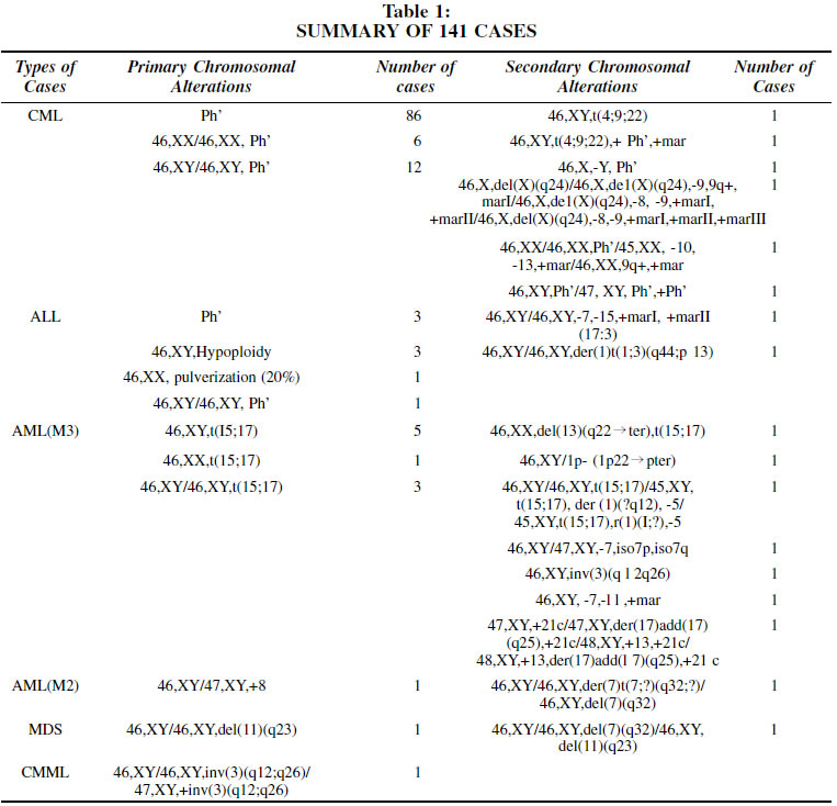
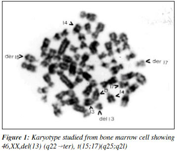
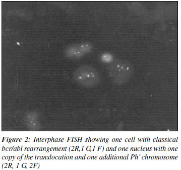
Code Number: cn02011 ABSTRACT Present study consists of cytogenetic evaluation in 141 cases referred to our centre for various leukemias. This includes 110 cases of CML, 10 of ALL, 16 of AML (M3), 2 of AML(M2), 2 of MDS & 1 of CMML. The conventional cytogenetic study was carried out in all the cases using G Banding technique. Of the 141 patients studied, 17 patients showed secondary chromosomal alterations along with primary chromosomal alterations. In two patients of CML with secondary chromosomal alteration t(4:9:22), molecular cytogenetic technique (FISH) has been carried out which has confirmed the primary observations revealed by the conventional cytogenetic technique. Other secondary alterations were numerous and would have been missed if only FISH or PCR technique would have been used for diagnosis. We observed from our study that advanced molecular techniques like FISH & PCR cannot replace the conventional cytogenetic study but are useful as supportive and confirmative diagnostic tools. Key Words: Leukemia, Cytogenetic, FISH. INTRODUCTION The development of cancerous cells is a multistep process that involves the transformation of normal cells to neoplastic cells and subsequent progression to tumor. This is a sequential, acquired or constitutional genetic defect that confer uncontrolled growth and proliferation advantages from individual tumor.1 With the advent of various chromosome-banding techniques, increased emphasis has been placed on specific chromosome alterations, especially translocations, deletions and inversion that may be responsible for proto-onco-gene/s activation and functional loss of certain tumor suppressor genes or anti-oncogenes. The established relationship between malignancy and chromosomal changes has made cytogenetic studies a significant part of a number of hematological disorders.2 It helps in the diagnosis, prognosis, monitoring and therapy.3 A major advance in human cytogenetics is the development and implementation of a variety of non-isotopic techniques like Fluorescence In Situ Hybridization (FISH) and molecular techniques such as PCR.4,5 These have provided the cytogeneticist with considerably increased ability to identify chromosome segment to correlate chromosome structure with gene location, to reveal cryptic abnormalities that are undetectable using standard banding techniques and to analyze and describe complex rearrangements. 6 Though FISH and molecular techniques (PCR) are becoming an important tool for location of minor alterations in the chromosomes mainly at gene level, conventional cytogenetic technique remains the method of choice for initial diagnosis of leukemia.2 The present study is aimed to confirm or refute the above observations. PATIENTS & Methods Cytogenetic Study The study consists of 141 patients with different type of leukemias. Of these, 110 were CML, 10 were ALL, 16 were AML (M3), 2 of AML (M2), 2 were MDS and I was CMML. Chromosome analysis was performed on unstimulated whole blood or from bone marrow cells collected in collection medium. The cells were grown in RPMI 1640 (Himedia, India) supplemented with 20% fetal calf serum (Centron, India) for 24 hours as per standard protocol. The chromosome banding was done by Trypsin - Giemsa staining. At least 20 well spread metaphase plates were analyzed from each sample and 3 to 4 well spread plates were photographed and karyotyped. FISH Study The study was carried out in two cases for the Philadelphia [Ph'] rearrangement using a LSI bcr/abl ES probe (Vysis, U.S.A). Slides were pretreated in RNase solution (Boehringer, Germany) followed by incubation in pepsin/HCI solution and fixed in a PBS/MgC12/formaldehyde solution (Fluka, Switzerland). The slides were hybridized overnight following the Vysis protocol. RESULTS AND DISCUSSION In this study, we describe secondary chromosomal alterations in various hematological malignancies along with primary aberrations by conventional cytogenetics study. Out of 141 hematologically confirmed leukemia cases referred to our centre, 17 cases were found to have secondary chromosomal alterations along with primary chromosomal anomalies. The cytogenetic results obtained in different leukemias are summarized in Table 1. In a case of AML (M3), we observed deletion of # 13 along with primary translocation15,17 which might have been undetected if only FISH or PCR had been applied as a diagnostic tool (Figure 1). In the case of CMML, inversion of #3 was observed by conventional cytogenetics. Even whole chromosome 3 probe painting is not likely to give any additional information to the cytogenetics finding. Conventional cytogenetic study is also helpful in cases where the clinical diagnosis is at the borderline. In present study, case AS was clinically diagnosed as ?AML/MDS-RAEBT with 29.0% blast cells.7,8 The cytogenetic study revealed inversion of 3, which confirms the clinical diagnosis of MDS. It has been reported that in CML, a low incidence of other chromosomal changes also occurs9 that could be detected only by cytogenetic study and may be missed if FISH or PCR study is employed to study target specific break points. In two cases of present series, a unique three way complex translocation involving #4, #9, #22 (q25;q34;q11) was observed by conventional cytogenetic technique. This was further confirmed by FISH using bcr/abl and whole chromosome 9 paint probe (data not shown) (Figure 2). If FISH had been employed as the only diagnostic tool, the translocations other than Ph' might have gone un-noticed. In these cases, though the patients were having CML in accelerated state, it is likely that because of balanced 4;9;22 translocation, they are healthy till date.10,11 Thus, failing to detect the secondary balanced translocation might have failed to reveal the prognostic value of such translocations. Molecular techniques like FISH and PCR would play an important role in confirming diagnosis only in some cases where: (i) Cytogenetic preparations do not reveal a
clear picture
These molecular biology techniques cannot be used as a routine diagnostic tool as they are much expensive and may not be available in all cytogenetic laboratories. Thus, cytogenetic study has its own importance and cannot be replaced either by FISH or PCR study. These techniques may be used as a supportive tool to conventional cytogenetics to give a detailed insight of the chromosomal rearrangements. ACKNOWLEDGEMENT We sincerely thank all referring doctors Dr. Urmish Chudgar, Dr. Mahendra Desai, Dr. Sandip Shah, Dr. Chirag Desai, Dr. Pankaj Shah for their reference. We are indebted to Ms. Francine Jacque for her skillful FISH contribution and Mrs Manisha Desai for her techical assistance. REFERENCES
Copyright 2002 - Indian Journal of Cancer. The following images related to this document are available:Photo images[cn02011f1.jpg] [cn02011t1.jpg] [cn02011f2.jpg] |
| |||||||||
