 |
| About Bioline | All Journals | Testimonials | Membership | News |
|
||||||
|
||||||
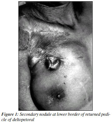
Secondaries at Deltopectoral Flap Donor Site Ashok B. C., Mahil T. Cherian, Honey Ashok Department of Plastic & Reconstructive Surgery, St. John's Medical Collee Hospital, Sarjapur Road, Bangalore 560 034. Code Number: cn02013 ABSTRACT Deltopectoral flap and pectoralis major faps form the basis of most reconstructions following ablative surgeries of head and neck region for cancers. There are many papers reporting acute complications but the literature on one late complication i.e. secondaries at flap donor site is rare. This is one such report and review of literature. Key Words: Secondaries, Deltopectoral Flap, Donor site. Even after the advent of free flaps, deltopectoral (DP) and pectoralis major myocutaneous flaps (PMMC) are the most common distant flaps used in the reconstruction of defects derived from surgical ablation head and neck neoplasms. There are a number of articles on complications that follow these flaps.1-3 But most of these address acute complications. Little is written about a delayed but dreaded complication i.e. secondaries in flap donor site. This is a clinical report and search in literature of the same. CASE REPORT A 55-year-old female tobacco user presented with 3x2 centimeter ulcer near the right upper gingivobuccal sulcus. There was neither skin involvement nor any clinically palpable cervical lymph nodes. Biopsy from ulcer proved to be poorly differentiated sqamous cell carcinoma. Tumor was resected widely, skin was spared. Modified radical neck dissection was done on the same side. Frozen section showed the margins to be tumor free, neck nodes were also metastasis free. The lining defect was primarily reconstructed with a deltopectoral flap. There was partial loss of flap due to necrosis, which was allowed to heal secondarily. At four weeks the pedicle was divided and inseted. The rest of the flap pedicle was returned to the donor site. The donor site healed well. In the final histopathology report there was a small sub capsular metastasis in level 2 lymph nodes. Patient refused radiotherapy and went home. Three months later she came with a 1x2 cm ulcerated nodule on the donor site (Figure 1). Biopsy proved it to be sqamous cell carcinoma. Nodule was excised and resurfaced by advancing the remaining flap pedicle. Patient was advised radiotherapy, which she refused. DISCUSSION DP and PMMC are most common flaps used in reconstruction of head and neck defects. They may be considered as gold standard even in the age of free faps. They alone or in combination can be used for both skin and mucosal resurfacing. They offer the following advantages, can be harvested in supine position in rapid time, cost effective (especially in third world countries) and does not require a great deal of expertise (compared to free flaps). Most complications are acute, but one ominous late complication is dermal metastasis in the flap donor site. It has poor prognosis with rapid dissemination and death. There are a number of papers on complications following reconstructions of head and neck defects with PMMC and DP flaps.1-3 There are a few papers on secondaries in flap donor site. In a review of 617 primary reconstructions using PMMC Kroll et al found one incidence of tumorspread.4 Greene D et al have reported metastasis from laryngeal sqamous cell carcinoma to pectoralis major donor site.5 There is a case report on chest wall mass after pectoralis major myocutaneous flap reconstruction,6 although it turned out to be a pseudocyst. Most of these reports of dermal metastasis are in PMMC flaps. This may due to larger number of PMMCs used in these kinds of reconstructions. This case demonstrates the donor site of DP flap may be as good a site for dermal metastasis as PMMC or any other flap. Theoretically it may be due to metastasis through lymphatics or blood or it may be direct tumor seeding during surgery. All spread to flap donor site in the absence of disseminated metastasis should be considered as seeding at the time of surgery. Utmost care should be taken while harvesting flaps and treating these dermal metastases. REFERENCES
Copyright 2002 - Indian Journal of Cancer. The following images related to this document are available:Photo images[cn02013f1.jpg] |
| |||||||||
