 |
| About Bioline | All Journals | Testimonials | Membership | News |
|
||||||
|
||||||
Indian Journal of Cancer, Vol. 40, No. 4, (October -December 2003) , pp. 135-139 Cytogenetics and Fluorescence In-Situ Hybridization in detection of Hematological Malignancies Frenny VJ, Antonella Z,* Luisa A,* Shah AD, Sheth JJ, Rocchi M* FRIGE, Genetic Centre, 20/1, Bima Nagar, Satellite, Ahmedabad - 380015, India;
and *DAPEG Sezione Di Genetica, Instituto Di GeneticaVia Amendola, 165/A, 70126
Bari, Italy.
Code Number: cn03023 ABSTRACT: BACKGROUND: The technique of Fluorescence In-Situ Hybridization (FISH), a hybrid of cytogenetics and molecular biology has increased the resolution and application of cytogenetics in various neoplastic processes. In various types of leukemias, primary investigation by conventional cytogenetic [CC] technique followed by FISH has increased our understanding of the abnormal clonal formation involving different gene region. AIMS: Present study is aimed to use different kinds of in-house FISH probes in various hematological malignancies and its correlation with conventional cytogenetic finding. MATERIAL AND METHODS: Cytogenetic study was carried out in 360 patients either from peripheral blood or from bone marrow cells suspected for various types of leukemias. Four of 360 cases were further selected for FISH study by using different types of in-house probes, such as BAC [Bacterial Artificial Chromosome], PAC [Phague Artificial Chromosome], alphoid, PCP [Partial Chromosome Paint] and WCP [Whole Chromosome paint]. RESULTS: The results confirmed breakpoints of inversion 16 and del 16 in case 2 and 3 respectively. Whereas, case 1 did not confirm the cytogenetic findings of t(15;17) by PML/RARa fusion signals as multiple cell lines were involved in the patients. PCP and WCP were helpful in the identification of the marker chromosome in case 1. Telomeric and centromeric probes confirmed the cytogenetic findings of t(5;7) in case 4. CONCLUSION: We observe from this study that, in addition to the conventional cytogenetic study, FISH study provide further confirmation of chromosomal rearrangements. This facilitates our understanding of the neoplastic process more precisely for the better prognostification of the patient. Key Words: Cytogenetic, Fluorescence in-situ hybridization, Leukemia, Bacterial artificial chromosome, Phague artificial chromosome, Alphoid, Partial chromosome paint, Whole chromosome paint. Introduction Conventional Cytogenetics [CC] studies are widely used to diagnose and manage patients with hematological malignancies.1,2 This has provided critical insight into the genetic changes that underlie malignant transformation of the cell. These advances have led to the establishment of chromosome pattern as diagnostic and prognostic indexes in acute and chronic leukemias, lymphomas and as a guide for the localization of oncogenes and tumor suppressor genes that are apparently responsible for the development of neoplastic process. The clues provided by the chromosome break points and multiple rearrangements have led to identify genes involved in these rearrangements by new molecular methods like FISH.3 The applications of FISH with chromosome specific DNA probes help to further define molecular subclass and cytogenetic risk categories for patients with leukemias. Recent WHO classification recognizes that genetic anomalies are one of the most reliable criteria for classification of hematological neoplasms.3 With CC, a confirmative method of FISH technique represents a major advance in chromosomal analysis.4 It has aided in identification of large number of genes involved in normal hematopoiesis by studying the structural chromosomal rearrangement in various hematopoietic process. The aim of the present study is to identify the usefulness of cytogenetic and further confirmation by molecular cytogenetic study using in-house FISH probes in various hematological malignancies. Material and Methods A total of 360 patients were investigated for cytogenetic analysis during the period of 1997 to 2002. These include Chronic Myeloid leukemia [CML], Acute Myeloid leukemia [AML], Acute lymphoblastic leukemia [ALL], Myeloid dysplastic syndrome [MDS] etc. Based on cytogenetic findings four selected cases of acute leukemia and/or MDS were further analyzed by FISH5 Table 1. Cytogenetic analysis CC analysis was performed on either unstimulated whole blood or from bone marrow cells for 24 to 48 hrs as per standard techniques and evaluated by Giemsa-Trypsin-Giemsa (GTG) banding at about 400-band level according to ISCN.6 At least 20 well spread metaphase plates were analyzed from each sample and 3 to 4 well spread plates were photographed and karyotyped. FISH study The study group comprises of four clinically diagnosed cases of acute leukemia and/or MDS having various chromosomal anomalies. The selection criteria were made in such a way that different kind of FISH probes [BACs (Bacterial Artificial Chromosome), PACs (Plague Artificial Chromosome), Alphoid, PCPs (Partial Chromosome Paint) and WCPs (Whole Chromosome Paint)] can be used and provide maximum information in metaphases as well as in interphase cells. The selection of the probes were carried out considering the various break points involved in the structural rearrangements that provide maximum information in the metaphase and interphase nuclei as well. PCP and WCP were used to identify the origin of the marker or derivative chromosomes. Telomeric and Centromeric probes together were used to confirm the telomeric rearrangements involving two different chromosomes that are not always visible by CC study. The probes used for FISH experiments were isolated from the BAC, PAC and Plasmids using Plasmid Mini Prep Kit from Biorad. BAC and PAC, belonging to RPC1 P. de Jong libraries were obtained from the Sanger Centre [http://www.sanger.ac.uk] or directly from P. de Jong [http://www.chori.org/bacpac/]. Prefix dJ or bA for library identification as used at Sanger Centre, has been maintained [http://www.biologia.uniba.it/rmc/0-1a_pagina/4-libraries.html]. WCP were obtained from sorted chromosome in collaboration with Sanger Centre [http://www.biologia.uniba.it/rmc/8-WCP-PCP%20new/WCP -SORTED.html] or [PCP and WCP] from somatic cell hybrids [http://www.biologia.uniba.it/rmc/0-1a_pagina/1_3_WCP.html]. Alphoid probes for chromosomes were obtained from [http://www.biologia.uniba.it/rmc/5-alfoidi/alf-15.html]. FISH analysis was performed on cytogenetic preparations obtained
from bone marrow cells from leukemia cases. Chromosome preparations were hybridized In-Situ with
probes labeled directly with Cy3 or Fluor-X by nick translation. In short,
500ng of labeled probes were used for FISH experiments, hybridization was performed
at 370C in 2xSSC, 50% (v/v) demonized formamide, 10% (w/v) dextran
sulphate, 5mg Cot-1 [DNA] BRL and 3mg sonicated Salmon Sperm DNA in a volume
of 15ml. Post hybridization washing was carried out by three washes in 0.1xSSC
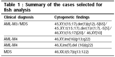
at 600C. Primers: CCGACTCGAGNNNNNNATGTGG Pre-treatment of metaphase spreads on the aged slide was done by incubating it with 0.005% pepsin in 10mM HCl for 30 mins at 370C, wash for 5 mins with PBS followed by PBS plus 50mM MgCl2. Post-fix the slide for 5mins at room temperature with 1% Para formaldehyde in PBS plus 50mM MgCl2, wash with PBS and dehydration with 70%, 90%, 100% ethanol. Chromosomes were identified by 4'-6-diaminido-2-phenylindole [DAPI] stain. Digital images were obtained using Leica DMRXA epifluorescence microscope equipped with a cooled CCD camera. [Princeton Instruments]. Cy3 [red, NEN], Fluor-X [green, Amersham] and DAPI [blue] fluorescence signals were detected using specific filters, which were recorded separately as gray scale images. Pseudocolouring and merging of images was performed using Adobe Photoshop software. A total number of 20 metaphases were studied and at least 200 interphase cells were analyzed using different probes. Results and Discussion The cytogenetic findings of the selective four cases revealed multiple cell line in case 1 [45,XY,t(15;17),der(1)(q12),-5[65]/ 45,XY,t(15;17),der(1)r(1;?),-5 [5]/ 46,XY,t(15;17)[20]/ 46,XY[10]], inv16 (p13;q22) in case 2, del 16(q22) in case 3 and t(5;7) (q13;q22) in case 4. All the cases were further studied using various FISH probes.
They are divided in to three groups.
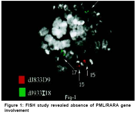
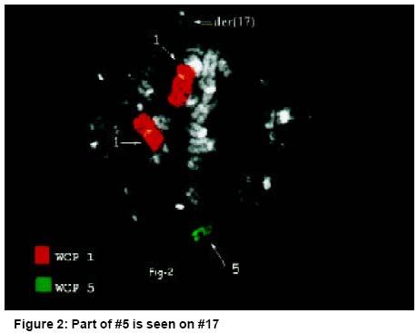
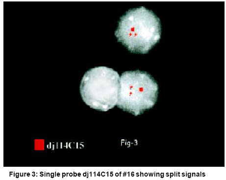
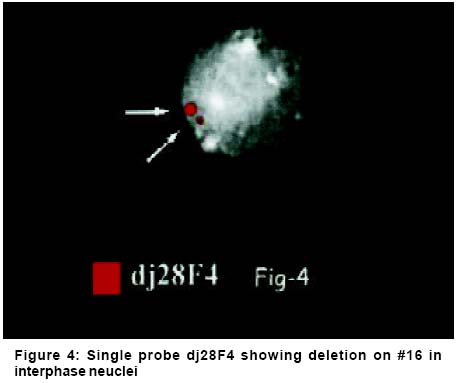
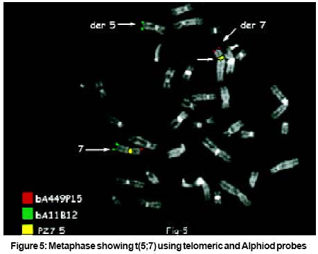
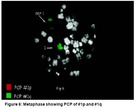
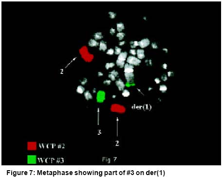
FISH was done in all four cases using unique DNA sequences. In case 1 of AML-M3/MDS, a locus specific probe for the PML gene at 15q labeled with red fluorochrome and a locus specific probe for the RAR-a gene at 17q labeled with green fluorochrome were used. After fusion this gives yellow signal on chromosome 17 [the combination of red and green].7 However in present case, though cytogenetically t(15;17) was apparent, FISH study revealed the absence of PML/RARa gene involvement as shown in Figure 1. Instead FISH has shown an additional material of #5 on the distal part of one #17 which was apparently normal by CC and deleted material of another #17 could not be characterized due to multiple cell lines Figure 2. Study carried out by Brockman et al in 2003 using dual colour probes showed that FISH method can detect all alternate translocation involving RAR-a and not PML.8 In the second case of AML-M4 with inversion of chromosome 16, dj114C15 FISH probe was used which spans the break point cluster region in MYH11 in band 16p13. Presence of inversion gave split signals9 and was observed in metaphases. Same split signals were seen in interphase cells with same intensity in another study Figure 3.10,11 In the third case of deletion 16q22, dj28F4 a site specific probe covering the break point cluster region of CBFB in band 16q22 and deleted chromosome showed small signal in comparison with normal chromosome 16 and can also be observed in non-dividing cells Figure 4.9 The probes used in case 2nd and 3rd are found to be very useful when cytogenetic picture is hazy and/or paucity of cells for cytogenetic analysis. Fourth case is of MDS having reciprocal translocation involving chromosome 5 and 7. This case was further studied using subtelomeric probes of chromosome 7 Figure 5. Telomeric region of the chromosome appeared pale or has unstained bands by G-banding methods and hence such probes are very informative where telomeric region are involved in translocation. Similarly, FISH technique using BACs or PACs probes in above cases comprise a unique DNA sequence, which are usually genomic clones. They are particularly useful for confirmation of cytogenetic findings suggestive of structural rearrangements such as recurring translocations/ inversions/ deletions using genomic probes that are derived from the breakpoint labeled with different fluorochromes.12,13 However FISH study has a limitation in the selection of specific probes. In group 2nd, case four of MDS was further studied having reciprocal translocation involving chromosome 5 and 7 using centromeric specific probe of chromosome 7 Figure 5. Here satellite DNA probe hybridize to multiple copies of the repeat unit present at the centromere. This results in a two bright fluorescent signals in metaphase and interphase cells as well. Such centromeric probes are of great help in newly diagnosed cases of leukemias to rule out trisomies, in monitoring the response to therapy and to detect minimal residual disease [MRD] with increased precision. Since hundreds of cells [including nondividing cells] can be examined in a short time, FISH is highly sensitive and can be performed rapidly in such cases.14,15 Last and 3rd group of PCP and WCP FISH probe was used in case 1 having clinical diagnosis of AML-M3/MDS with multiple chromosomal rearrangements. Here derived chromosomes were analyzed using PCP of 1p and 1q along with WCP of chromosome 1 and 3. Figure 6 & Figure 7 revealed that derived chromosome 1 is multiple rearranged along with a part of chromosome 3. Such detailed information was not possible only with CC analysis. This shows that PCP/WCP remains useful for confirming the rearrangement detected by routine banding techniques/or for uncovering abnormalities not seen by conventional banding technique. They are suitable for use in metaphase cells only, as painting probes are not helpful in the analysis of interphase cells because the signal domains are so large and diffuse. The overall observation made in our study clearly shows that primary cytogenetic investigation provides the knowledge and information about the known/unknown chromosomal rearrangements. While FISH study provides further confirmation of these arrangements and also information on minor cytogenetically undetectable rearrangements. This gives information on gene-gene fusion and abnormal clonal formation. Commercial FISH probes are useful in routine practice when only recurrent structural arrangements are to be studied. Where as if in-house probes are once established, they can be used in all the cases requiring further insight into the chromosome rearrangements. The uniqueness of these probes is, with two fluorochrome more than two probes can be labeled and can be studied in the same metaphase (Figure 5). Nonetheless FISH technique has its own limitations. It is highly sensitive for trisomy but less for chromosome loss. With FISH technique, data can be obtained only for the target chromosomes, while conventional cytogenetic analysis can detect the presence of virtually all-chromosomal abnormalities including multiple lesions by a single test.16 Together, the combination of cytogenetic and FISH analysis incorporates the screening potential of cytogenetics with accuracy of molecular genetic technique, which will lead to better understanding of neoplastic disease, more accurate diagnosis, stratification of patients in to genetic subgroups and eventually improvements in treatment and outcome. The importance of the present study is the critical selection of the patients so that all different kind of FISH probes can be studied in metaphase and can also be seen in interphase cells. These provide the utility of in-house FISH probes alone and in combination of different probes using minimum fluorochromes. Acknowledgements We sincerely thank the staff of DAPEG Sezione Di Genetica, Department of Genetics, Bari (Italy) for technical guidance in the present study, to Dr. Ashwin Patel and Dr. Trupti Patel for their critical suggestions in this article, to Dr Urmish Chudgar and other doctors of Ahmedabad for their reference and to Mrs. Manisha Desai for her technical assistance. The financial support of UICC (Geneva) is gratefully acknowledged. References
Copyright 2003 - Indian Journal of Cancer The following images related to this document are available:Photo images[cn03023f1.jpg] [cn03023f4.jpg] [cn03023f3.jpg] [cn03023t1.jpg] [cn03023f5.jpg] [cn03023f2.jpg] [cn03023f6.jpg] [cn03023f7.jpg] |
| |||||||||
