 |
| About Bioline | All Journals | Testimonials | Membership | News |
|
||||||
|
||||||
Indian Journal of Cancer, Vol. 41, No. 1, (January-March 2004) , pp. 37-40 Case Report Correlation of Radiological and Clinical Features of Metanephric Neoplasms in Adults Chaudhary H, Raghvendran M, Dubey D, Srivastava A, Mandhani A, Kapoor R, Kumar A Department of Urology, Sanjay Gandhi Post Graduate Institute of Medical Sciences,
Lucknow. India.
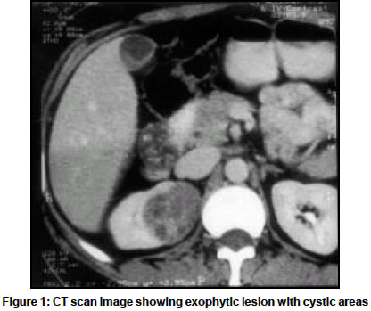
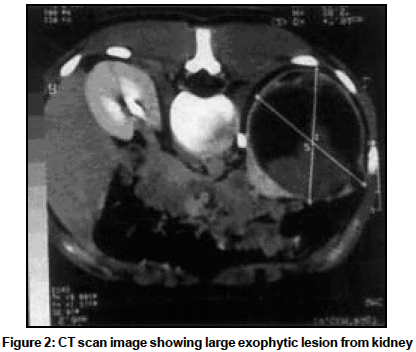
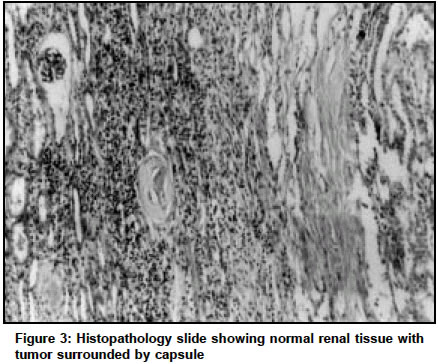
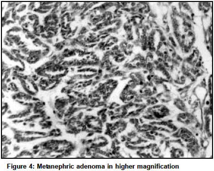
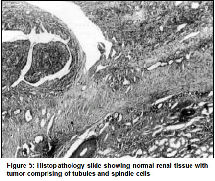
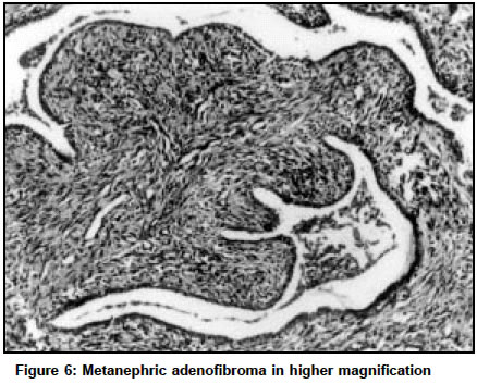
Code Number: cn04007 ABSTRACT The main objective was to determine the clinical and radiological features of metanephric neoplasms. The tumors were diagnosed on histopathological examination. The clinical data and imaging features were retrospectively analyzed. Between 1998 and 2003, 3 patients underwent radical nephrectomy for renal masses turning out as metanephric neoplasms on histopathology. Two of these tumors were metanephric adenoma (MA) and one was metanephric adenofibroma (MAF). Clinical and radiological features were reviewed. All patients were adult females who presented with flank pain. Tumor was detected on screening ultrasound as a hyperechoic lesion. In all cases CT showed a hyper-attenuating exophytic lesion with cystic areas that enhanced with IV contrast. Based on combination of clinical and imaging features it may be possible to prospectively identify metanephric neoplasms and thus avoid unnecessary radical nephrectomy in favor of conservative surgery. Key Words: Radical nephrectomy, Partial nephrectomy, Renal tumor, Benign epithelial tumor, Ultrasound, CT scan. INTRODUCTION Metanephric neoplasms are rare, benign tumors of the kidney, which have recently been included in the classification systems. Initially they were thought to be epithelial in origin.1 Subsequently they are considered as a separate entity.2 Relatively recent identification, rarity and lack of established clinical and radiographic features, make metanephric neoplasms a primarily pathologic diagnosis. The primary purpose of the study was to look at the clinical and radiological features of this tumor. Pre-operative diagnosis of this benign tumor would be ideal, since it might modify the treatment and assign a good prognosis. This was the secondary aim of this study. CASE REPORT The department of Urology, SGPGIMS, Lucknow undertook this study. A retrospective review of hospital records between 1998 and 2003 revealed three patients with a final pathological diagnosis of metanephric neoplasm. All patients underwent ultrasonography and CT scan of abdomen. Enhanced and unenhanced CT scans were performed. 10 mm sections were taken from dome of diaphragm to pubic-symphysis, with 5 mm sections in region of interest. 100-150 ml of 51.8% Iohexol was given IV bolus and images acquired 60 seconds later. Ultrasound and CT scans were reviewed to note the size, location, homogeneity and enhancement of tumor after IV contrast administration. Clinical presentation were reviewed and correlated with radiologic findings. All patients were females. Mean age was 38.33 Yrs. Patient profile and symptomatology are given in Table 1. Fever and flank pain were presenting features in 2, whereas 1 patient had only flank pain. Cytology in all patients was inconclusive. Ultrasound was non-diagnostic, though all tumors were hyperechoic compared to adjacent parenchyma. On unenhanced CT scan the tumors had higher attenuation compared to adjacent parenchyma. In all cases, peripheral areas of the mass had significantly higher attenuation compared to central portion, suggestive of central necrosis. CT also showed exophytic lesions enhancing on IV contrast in all cases. (Figures 1 and 2). Mean attenuation on unenhanced and enhanced scans were 42 (4046) HU and 96.3 (80-105) HU respectively. Mean diameter of the tumors on CT scan was 8.73 cm. Open and laparoscopic radical nephrectomy were done in 2 patients, whereas in 3rd partial nephrectomy was done. All patients are normal at mean follow up of 13.6 months. On gross examination the outer surface was lobulated and cystic. Thin capsule was identified in 1 and in other 2 tumor was non-encapsulated but well demarcated from adjoining renal tissue. Tumor protruded above renal surface in all cases. Cut surface varied from tan to gray to yellow and cystic areas were identified in all. Microscopic examination in 2 cases was consistent with metanephric adenoma (MA). (Figures 3 and 4). In third case a diagnosis of metanephric adenofibroma (MAF) was made (Figures 5 and 6). DISCUSSION MA is a rare benign tumor. Most of these are assymptomatic and incidentally detected on ultrasonography or CT.3 They have a female preponderance and commonly occur in fifth decade. In present series, all were females and presented with flank pain. There are basically 2 types of cystic RCCs: multilocular and unilocular. Multilocular cystic RCC is a distinctive subtype and accounts for only 2-3% of RCC.4 Unilocular cystic RCC can result from extensive necrosis in previously solid renal neoplasm. However these tumors have thick, irregular and calcified wall.5 In the present series all tumors were exophytic, unilocular cystic with clear walls. We postulate that when a young female presents with flank pain and imaging reveals exophytic, unilocular cystic renal mass with clear walls and normal adjoining parenchyma, consideration should be given to a possibility of metanephric neoplasm. Metanephric neoplasms are part of spectrum of lesions belonging to the Wilms' tumor group and are morphologically and immunophenotypically identical to maturing WT and nephrogenic rests.6 It is unclear whether these lesions are benign de-novo or result from spontaneous cyto-differentiation of nephroblastoma cells. Mean age of nephroblastoma in adult patients is 23 and all patients have tumors comprising of both elements.3 However in present series and literature, metanephric adenoma has occurred in fourth decade and for this tumor to arise from nephroblastoma cells this age of presentation seems unlikely. We feel that age of presentation negates the possibility of tumor arising from Wilms' tumor elements. MAF is a recently characterized renal neoplasm with only few reports in literature.2,7 Herein we report the first case from India. Status of immuno- histochemical staining is not clear.8 It has been shown that cytokeratin-7 labeling is often diffuse in papillary RCC and never in metanephric tumors. EMA has been found to be negative in metanephric neoplasms.2 `CD57' has been shown to be absent in papillary RCC, and present in metanephric adenomas.9 These immuno-histochemical markers have been suggested to be of use in differentiation of metanephric neoplasm from papillary RCC, when diagnosis can not be made on histopathology alone.2 However this problem was not encountered by us. Percutaneous renal aspiration in evaluation of renal masses has limited role due to improved diagnostic accuracy of imaging modalities and recognition of inaccuracy of aspiration because of sampling errors and difficulty in interpretation. 5-15 % of renal cell carcinomas are missed by fine needle aspiration biopsy.10 We found cytology to be inconclusive in all cases. These tumors have been described as hyperechoic on ultrasound due to presence of psammomatous calcifications and interfaces caused by numerous tubules.11 On unenhanced CT, metanephric adenomas have high attenuation compared with the adjacent parenchyma, as seen by us also. All 3 tumors were exophytic on imaging. Fielding reported that it is difficult to preoperatively identify these tumors based on imaging features alone.11 Coincidentally all three patients in their series had exophytic lesions.
The primary outcome measure was to look for any characteristic clinical or radiological features suggestive of metanephric adenoma. Our retrospective analysis suggests that there are characteristic clinico-radiological features suggestive of this tumor. Recognition of these tumors preoperatively is very important to offer nephron-sparing surgery (NSS) and assign a good prognosis. In the era of laparoscopic surgery, a bias is found among urological surgeons towards laparoscopic radical nephrectomy, as laparoscopic partial nephrectomy is technically difficult . Pre-operative recognition of metanephric neoplasms would thus favor NSS, thus being of immense benefit to the patient. This is the secondary outcome and main strength of the study. However this is a retrospective analysis and has all the limitations which goes with such an endeavor. REFERENCES
Copyright 2004 - Indian Journal of Cancer The following images related to this document are available:Photo images[cn04007f1.jpg] [cn04007f6.jpg] [cn04007f4.jpg] [cn04007f3.jpg] [cn04007f2.gif] [cn04007f5.jpg] [cn04007t1.jpg] |
| |||||||||
