 |
| About Bioline | All Journals | Testimonials | Membership | News |
|
||||||
|
||||||
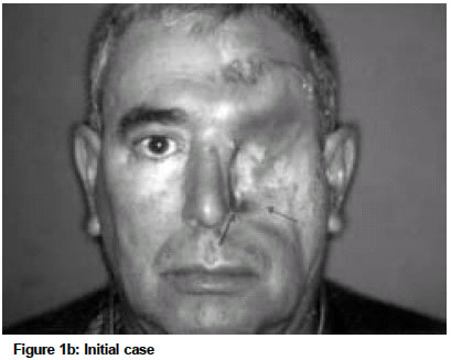
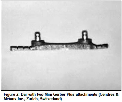
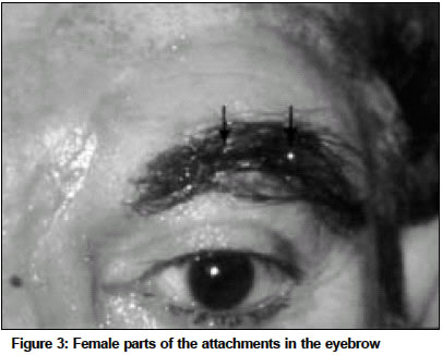
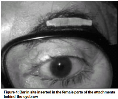
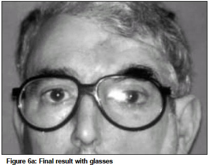
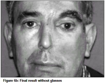
Indian Journal of Cancer, Vol. 41, No. 2, Apr-Jun, 2004, pp. 85-88 Case Report Residual facial disfigurement after the ablative surgery of a lachrymal gland carcinoma: A clinical report of the prosthetic rehabilitation Ciocca Leonardo, Scotti R Section of Oral and Maxillofacial Prosthetic Rehabilitation, Department of Oral Science, Alma Mater Sudiorum University of Bologna, Bologna Code Number: cn04015 ABSTRACT Facial disfigurement after ablative surgery of a massive adeno-carcinoma of the lachrymal gland is described. A rehabilitation with a maxillofacial prosthesis was proposed to restore the aesthetic appearance of the patient without inserting craniofacial implants. Retention of a maxillofacial prosthesis, that is not anchored to implants, depends on the use of adhesives or on mechanical devices like glasses. This clinic report describes a residual anatomic defect which allowed for the double choice of wearing the facial prosthesis both with or without glasses. A retentive backside of the prosthesis was developed to engage the facial defect undercuts, which enhanced retention when the patient used the skin glue without glasses. A unique foam silicone was utilized to reduce the weight of the prosthesis and to permit its retention only by skin adhesive. Keywords: Adeno-carcinoma, Lachrymal gland, Facial Prosthesis INTRODUCTION When maxillofacial ablative surgery involving aesthetic areas of the face is indicated for the removal of significant quantities of tissue due to cancer, often restorative plastic surgery is not able to recuperate the extensive area removed. Moreover, a long oncological observation period of the patient is sometimes necessary before any restorative plastic surgery can be performed. During this observation period, the patient often psychologically familiarizes himself with his changed appearance. This was the case for the patient presented in this paper, whose main wish was for a prosthetic rehabilitation without having additional surgery [Figure - 1a]. This report presents an experimental design conceived to allow the patient to manage his facial prosthesis both with and without glasses. A special technology, which makes use of precision intra-oral attachments available on the market, was used to alternate with the use of the skin-adhesive. A special foam silicone was utilised to reduce the weight of the prosthesis. This constructing feature allowed the patient to wear it without glasses, only by means of backside engaging of the defect undercuts, and by means of skin adhesive. MATERIAL AND METHODS Technique of facial prosthesis fabrication The sculpting, processing, materials and the surface make-up of the prosthesis were entrusted to the artistic technicians of Special Effects Creaturestudios Inc. (Viterbo, Italy), who provided an extremely light product (170 g) of custom-made foam silicone, aesthetically well integrated with the rest of the face. The eyeball was produced by the firm Cappagli S.r.l. of Bologna, Italy. During the try-on phases, great attention was paid to the undercut hook-up areas [Figure - 1a] and [Figure - 1b] and to the boundary of soft/hard tissues, especially next to the cheek and to the pre-maxillary areas. A backside of the prosthesis was realised to engage the available undercuts in the defect at maximal depth. This enhances retention when the prosthesis is utilised without glasses and only by means of skin adhesive. A metal plate, with two attachments to be joined to the glasses, was designed and manufactured. [Figure - 2] The resin (Vertex, Vertex Dental B.V., Zeist, The Netherlands) mesiostructure, with the female parts of the attachments, were incorporated in the silicon structure at the level of the arch of the eyebrows. [Figure - 3] The attachments chosen were the Mini Gerber Plus (Cendres and Metaux Inc., Zurich, Switzerland) for their characteristic of variable friction intensity management and for the long-term maintenance. The joint between the metal plate of the two attachments and the glasses was obtained by constructing the connection with a conventional resin (Splintline, Lang Dental MFG Co, Wheeling IL, USA) and then pigmenting it with the same colour as the glasses. [Figure - 4] and [Figure - 5] A female portion of transparent resin mesiostructure was prepared with female parts of the attachments inserted, so that its point of attachment would correspond to the eyebrow level and the female holes could be hidden by the eyebrows during use without glasses [Figure - 3] - arrows; [Figure - 4] and [Figure - 5]. During the appointment for delivery, the patient was given all the necessary instructions for insertion and removal as well as a leaflet indicating the directions for hygiene and home care maintenance. Along with the prosthesis, the patient was given a package of adhesive (In-Health ®, Health Care Technology Inc, Carpenteria, CA-USA) and hygienic towels for its removal (Remove ®, Smith and Nephew Inc., Largo, FL-USA). [Figure - 6a] and [Figure - 6b] CASE REPORT A 56-year-old male had undergone ablative surgery of a left lachrymal gland adeno-carcinoma 17 years earlier. Since then, the patient had worn an eye patch and a hat, together with dark glasses, to hide the defect. It involved the whole orbital area, the zygomatic zone and part of the nose area, both superficially and in depth, involving also the scalp due to a rotated flap used for the reconstructive plastic surgery [Figure - 1a]. In addition, the peculiarity of his facial defect forced him to cover the respiratory nose mucosa exposed, with a very thick band, to avoid excessive trigeminal stimulation due to the cold temperature during breathing. The patient continued to smoke, thus also exposing himself to high risks of degeneration, although he had received repeated warnings. Due to the patient′s request and to the poor quality of the available bone tissue, the maxillofacial surgeon could not insert implants. The defect was very deep in the area of the nasal choanas, where the left turbinate bones had been removed, while in the surface skin areas, the skin thickness was normal. In particular, the supporting points of the epithesis [Figure - 1a] and [Figure - 1b] - black arrows) were detected to obtain mechanical retention points. One of these was localized in the left area, internal to the glabella [Figure - 1a] and [Figure - 1b] - white arrow). DISCUSSION The retention of a maxillofacial prosthesis usually depends on the retention system anchored to implants. Earlier reports have shown that the failures and complications appear to be site specific, radiation and time dependent.[1],[2],[3],[4] Sometimes the retention depends on mechanical systems, like glasses for nasal and ocular prostheses, or on adhesive systems.[5],[6],[7],[8],[9],[10],[11] The approach and technology used in this research, took into consideration the patient′s chief request of "not always having to hide behind glasses". The clinical staff tried to resolve the important psychological aspects of the patient by conceiving a structure that would fulfil his wish. Advanced intra-oral technology for removable prosthetic attachments, to connect the prosthesis to the glasses was used. All defect undercuts were engaged by the backside of the prosthesis. A foam silicone was chosen to reduce the weight of the prosthesis and to allow for use without glasses, by means of only skin adhesive. The satisfaction of the patient, who immediately wanted to wear the newly fitted prosthesis without glasses, the positive reaction in his familiar and working environments as well as the subsequent clinical follow-up have demonstrated how this type of experimental design may be employed as a valid response to the psychological-functional need of the patients, by giving an acceptable aesthetic recovery [Figure - 6a] and [Figure - 6b]. REFERENCES
Copyright 2004 - Indian Journal of Cancer The following images related to this document are available:Photo images[cn04015f2.jpg] [cn04015f6a.jpg] [cn04015f3.jpg] [cn04015f6b.jpg] [cn04015f1a.jpg] [cn04015f1b.jpg] [cn04015f4.jpg] [cn04015f5.jpg] |
| |||||||||
