 |
| About Bioline | All Journals | Testimonials | Membership | News |
|
||||||
|
||||||
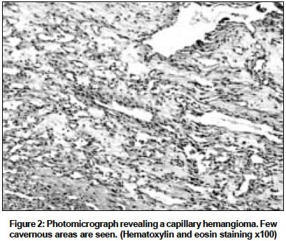
Indian Journal of Cancer, Vol. 41, No. 4, October-December, 2004, pp. 181-183 Case Report Hemangioma of base of tongue Qureshi SajidS, Chaukar DevendraA, Pathak KumarA, Sanghvi VikramD, Sheth Tanuja, Merchant NH, Dcruz AnilK Departments of Head and Neck Oncology, Tata Memorial Hospital. Ernest Borges Road, Parel. Bombay Code Number: cn04036 ABSTRACT Although vascular malformations of the tongue comprise a significant portion of head and neck angiodysplastic lesions, hemangioma of base of tongue is rare. We report a case of hemangioma of base of tongue extending to the supraglottis, which necessitated an extended supraglottic laryngectomy. Patient had an uneventful recovery and at three year, follow-up has a normal speech and no difficulty in swallowing or aspiration. More importantly, there was no recurrence of hemangioma or bleeding. Although hemangiomas may be treated by various conservative methods, occasionally patient may require surgical excision as in the present case due to the repeated bleeding episode and difficult access. A high index of suspicion and radiological investigations should be performed if the clinical presentation is atypical for malignancy, as in our case.Key Words: Hemangioma, base of tongue. INTRODUCTION Lingual hemangiomas are common but in base of tongue (BOT) hemangioma are extremely rare.[1],[2] They pose a difficult problem in view of the tongue being a mobile inquisitive organ is more prone for trauma and subsequent complications.[3] In this report, we document a case of hemangioma affecting the BOT and extending to involve the supraglottis. To the best of our knowledge, hemangioma simultaneously involving the BOT and the supraglottis has not been reported previously. The diagnostic difficulties and the factors coercing treatment decisions are discussed. CASE REPORT A 65-year-old male patient presented with complains of progressive dysphagia to solids of two months duration. In addition, he had two episodes of small amount of bleeding per orally. The patient was a chronic tobacco chewer. Clinical examination of the neck did not reveal any mass or evidence of neck nodes. Hopkins telescopic examination showed the presence of a mass at the BOT extending to the supraglottis and overhanging the glottis. A direct laryngoscopic examination showed a fleshy lesion over BOT extending across the vallecula to the epiglottis and left aryepiglottic fold (AEF). The cords were free and mobile. The tumor did not have the characteristic features of hemangioma. A biopsy resulted in profuse bleeding, (approximately 300 cc) which needed pressure and packing, but subsequently stopped spontaneously. Histopathology revealed a capillary hemangioma with fibrin deposits. The patient had a subsequent bleeding episode a day later. An emergency tracheostomy, to secure the airway, was performed. Computerized Tomography (CT) scan revealed a lobulated soft tissue mass involving the BOT and epiglottis and causing obliteration of the laryngeal airway. The mass was heterogeneous with hyperdense and hypodense areas. Magnetic Resonance (MR) angiography revealed a mass involving the BOT, vallecula, epiglottis and the left aryepiglottic fold. The mass was isointense to muscle on time (T) 1 weighted image. On T2 weighted and short tao inversion recovery (STIR) images the mass showed heterogeneous density with some areas of very bright signal. [Figure - 1]a and b The neck vessels were normal in course and caliber. In view of large lesion that was symptomatic (bleeding), a decision to resect the lesion surgically was made. At surgery, the lesion was localized to the left side of BOT extending across the vallecula, epiglottis and the left AEF. An extended supraglottic laryngectomy with excision of the lesion on BOT was performed, preserving as much normal tongue as possible. On gross examination, the lesion had a fleshy appearance with foci of ulceration. Microscopical analysis showed a capillary hemangioma. [Figure - 2] Inflammation was seen only in superficial area of ulceration. Lobules of delicate capillary structures were interspersed throughout the tumor. Occasional cavernous vascular structures were also seen. The patient had an uneventful intra-operative and post-operative recovery. Oral feeds were initiated on the fifth postoperative day and tracheostomy removed thereafter. There was no significant aspiration. At three year, follow-up patient has a normal speech and no difficulty in swallowing or aspiration. More importantly, there was no recurrence of hemangioma or bleeding. DISCUSSION Lingual hemangiomas pose distressing problems to the patients, producing cosmetic deformity, recurrent hemorrhage, and functional problems with speaking, deglutition, and mastication.[3] Lingual hemangiomas may remain indolent or may produce obstructive symptoms or alarming hemorrhage. Brown et al[3] reported a case of mixed capillary and cavernous hemangioma of tongue in a 76-year-old woman, which was present since birth and was asymptomatic for more than 50 years. The patient had dislocated jaw presumably due to mass effect of hemangioma, which started growing after 56 years. This hemangioma rapidly enlarged after an unrelated operation under spinal anesthesia causing severe functional and cosmetic deformity, for which surgical treatment was required. Most lingual tumors present as mucosal changes and tongue being superficially located and easily accessed, these can be diagnosed without imaging analysis. However, the characteristic and extent of lesions situated at deep portion of tongue, such as its base or submucosal lesions can be recognized only on cross-sectional CT scan or MRI.[4] Hemangiomas usually appear as a well-demarcated enhancing mass often containing phlebolith on CT scan. MRI shows the lesion as a solid mass with isointense or slightly high signal intensity to muscle on T1-weighted images and heterogeneous signal intensity on T2-weighted images. Post contrast T1-weighted imaging commonly demonstrates prominent enhancement.[4] A number of options exist for lesions that require therapy, including medical and surgical interventions. Medical management includes systemic and intralesional administration of corticosteroids.[5] However, only 30% respond to corticosteroids and they are not free from complications. Systemic corticosteroids carry well-documented risk, such as disseminated varicella, herpes infection, growth retardation and cushingoid habitus.[6] For lesions, which do not respond to steroids, surgical therapy is often necessary. Surgery may be complicated by extreme blood loss. Surgical resection may be facilitated by pre-operative embolisation in selected cases although embolisation has also been used as the sole form of treatment for unresectable lesion.[7],[8] Laser photocoagulation is the other modality of surgical treatment. Both surface and intralesional delivery of laser phototherapy are used for treatment of hemangiomas and vascular malformation, but recent interest has centered on the latter.[6],[9] Although laser therapy has fewer complications, the frequent numbers of treatment, variable response and regrowth of lesion are the disadvantages of this technique.[6] In the present case MR angiography did not show a tumor blush or feeding arteries hence a pre-operative embolisation was not done. The lesion was extending from the BOT to involve the supraglottis. Patient was having dysphagia due to mass effect of the lesion and was also having bleeding episodes. Surgery was elected over laser treatment as the lesion was large, involving the BOT and the supraglottis, laser excess was difficult and patient was having repeated bleeding post biopsy. As a lesson from the present case we would like to add that when patients′ present with bleeding and a tumor with features not definitive of malignancy, a high index of suspicion is required and imaging with either CT scan or MRI should be done before a biopsy is attempted. REFERENCES
Copyright 2004 - Indian Journal of Cancer The following images related to this document are available:Photo images[cn04036f1.jpg] [cn04036f2.jpg] |
| |||||||||
