 |
| About Bioline | All Journals | Testimonials | Membership | News |
|
||||||
|
||||||
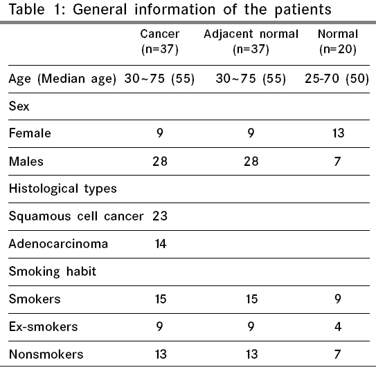
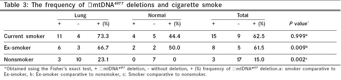
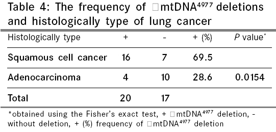
Indian Journal of Cancer, Vol. 43, No. 1, January-March, 2006, pp. 20-25 Original Article Mitochondrial DNA 4977 BP deletion mutations in lung carcinoma Dai JiGang, Xiao YingBin, Min JiaXin, Zhang GuoQiang, Yao Ke, Zhou RenJie Department of Thoracic Cardiovascular Surgery of Xinqiao
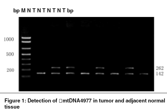
Hospital, The Third Military Medical University, Chongqing - 400037 Code Number: cn06004 Abstract BACKGROUND: The most common and also the most often assayed mtDNA deletion mutation, ΔmtDNA 4977sub has been demonstrated in various types of human cancer. However, knowledge about ΔmtDNA 4977 in lung carcinoma is poor.AIM: To study the 4977 bp deletions of mitochondrial DNA (ΔmtDNA 4977) in lung cancer, adjacent histologically normal and normal lung tissue and its potential roles in the development of cancer. MATERIALS AND METHODS: Thirty-seven matched lung cancer/adjacent histologically normal and 20 histologically normal lung tissue samples in subjects without lung cancer were analyzed by PCR technique. RESULTS: ΔmtDNA 4977 deletions were detected in 54.1% (20/37) of lung cancers, 59.5% (22/37) of adjacent normal and 30.0% (6/20) of normal lung tissue samples. No significant difference was found in the frequency of ΔmtDNA 4977 deletions between the tumor and adjacent normal lung tissues ( P value = 0.815). Moreover, no significant difference was found in the frequency of ΔmtDNA 4977 deletions between the tumor and histologically normal lung tissues in subjects without lung cancer ( P value = 0.101). However, the correlation between ΔmtDNA 4977 deletion and age and smoking factors was present in our data. STATISTICAL ANALYSIS: Fisher's exact test was used to assess the difference in different groups by the Scientific Package for Social Sciences (SPSS), version 10.0, Statistical analysis software. CONCLUSINS: Mitochondrial DNA 4977 bp deletion, which is not specific to lung cancer, may reflect the environmental and aging process influences operative during tumor progression. Keywords: Deletion, lung carcinoma, mitochondrial DNA, mutation Mitochondria are the intracellular organelles responsible for adenosine triphosphate (ATP) synthesis through the coupling of oxidative phosphorylation (OXPHOS) to mitochondrial respiration in human and animal cells. They contain their own genome in the form of mitochondrial DNA (mtDNA), which is the only extrachromosomal DNA in human cells. Mitochondria are involved in apoptosis and probably also tumorigenesis,[1],[2] which has led researchers to examine the potential roles of mtDNA alterations in the development and maintenance of cancers. Mammalian mitochondrial DNA (mtDNA) is a circular double-stranded DNA of 16.5 Kb in size. In contrast to the nuclear DNA, mtDNA is a naked compact DNA molecule without introns and is replicated at a much higher rate without an efficient DNA repair mechanism. Therefore, mtDNA is more vulnerable to attacks by reactive oxygen species and free radicals. Each nucleated human cell contains a few thousand copies of mtDNA, the somatic mutation rate of which is presumed to be 10 to 20 times higher than that of nuclear DNA.[3],[4],[5] Human mitochondrial DNA (mtDNA) is becoming the study hotspot for its alteration in correlation with its tumorigenesis.[6] mtDNA mutations were reported in different types of cancer and cancer cell lines. Reported sequence changes include point mutations (mostly transitions), multiple deletions and microsatellite instability in coding and noncoding regions.[7],[8],[9],[10],[11] Mitochondrial DNA 4977 bp deletion (ΔmtDNA 4977) is the most common change in mtDNA and has been detected in several types of human tumors including gastric cancer, esophageal carcinoma, hepatocellular carcinoma, thyroid tumors, etc.[12],[13],[14] However, little is known about this deletion of mtDNA in lung cancer. In this study, ΔmtDNA 4977 deletion mutation was detected in lung cancer, adjacent histologically normal and histologically normal lung tissue samples in subjects without lung cancer and the correlation between ΔmtDNA 4977 and age and smoking factors was analyzed. Materials and Methods Subjects Total cellular DNA containing the mtDNA and nucleus DNA (nDNA) was prepared using the routine method: (1) Small amounts of tissue (100 mg) were snipped off, suspended in cell lysis buffer and incubated for 1 h at 37°C. (2) The cell suspensions were mixed by vortexing and incubated for 3 h at 50°C after the addition of enzyme K (20 mg/ml). (3) They were mixed by vortexing for 10 min and centrifuged at 5000 r/min for 15 min at room temperature after the addition of equal volume of Tris-saturated phenol. (4) The aqueous fraction was extracted twice with an equal volume of phenol: chloroform: isopentanol (25:24:1). (5) The aqueous fraction was preserved at - 20°C for 1 h after the addition of 2 volume of ethanol and 0.1 volume of 3 mol/L sodium acetate. (6) Total DNA was collected as a pellet by centrifugation at 12 000 r/min for 10 min at 4°C. (7) DNA was washed once in 75% ethanol, collected by centrifugation at 12 000 r/min for 5 min and dried at room temperature. (8) DNA was resuspended in TE (10 mmol/L Tris-HCl, PH 7.5, 1 mmol/L EDTA) and stored at - 20°C. Detection of mitochondrial common deletion PCR reactions were carried out using TaKaRa PCR kit (TaKaRa, Japan) in a 50 μl reaction volume with 200 ng DNA template, 2 U TaKaRa Taq DNA polymerase (TaKaRa, Japan), 2.5 mmol/L MgCl2, 250 μmol/L each dNTP and 0.5 μmol/L of each primer. After denaturation at 94°C for 30 s, the reaction mixture was cycled 30 times at 94°C for 30 s, 59°C for 30 s and 72°C for 1 min, finally extended at 72°C for 10 min. PCR products were analyzed by 1% agarose gel electrophoresis at 60V (the buffer fluid was 1 x TAE buffer). The electrophoresis gels were observed under ultraviolet and photographed. Sequencing analysis Statistical analysis Results ΔmtDNA 4977 deletions in lung cancer, adjacent normal and histologically normal lung tissue samples in subjects without lung cancer. The detection of ΔmtDNA 4977 between the origins of replication of light and heavy mtDNA strands was performed by PCR amplification with two sets of primers. To control the ability to PCR-amplify mtDNA, one primer pair (P1/P2) localized inside of the region referred to as ΔmtDNA 4977 that amplified a 142-bp amplicon corresponding to wild-type mtDNA was used as an amplification control. In case the 4977-bp deletion is present, a 262-bp PCR product is generated as determined by another pair of primers (P3/P4) annealing to the fragments flanking the deleted region [Figure - 1]. These bands were further sequenced and confirmed to be mitochondrial in origin. ΔmtDNA 4977 deletions were detected in 54.1% (20/37) of lung cancers, 59.5% (22/37) of adjacent normal and 30.0% (6/30) of histologically normal lung tissues in subjects without lung cancer [Table - 2]. No significant difference was found in the frequency of ΔmtDNA 4977 deletions between the tumor and adjacent normal lung tissues ( P value = 0.815). The frequency of ΔmtDNA 4977 deletions in lung cancers and adjacent normal tissues appeared to be higher than that in histologically normal lung tissue samples in subjects without lung cancer. However, because of limited number of samples, no significant difference was found between the tumor and normal lung tissues ( P value = 0.101) and between adjacent normal and normal lung tissues ( P value = 0.052). The detection of ΔmtDNA 4977 was performed by PCR amplification with two sets of primers. One primer pair (P1/P2) yields a 142 bp amplicon. In case the 4977 bp deletion is present, a 262-bp PCR product is generated as determined by another pair of primers (P3/P4). 142 and 262 bp bands, suggesting the presence of heteroplasmy, were amplified in 4977 bp deleted mtDNA and only 142 bp band was observed in wild mtDNA with primer sets P1/P2 and P3/P4. T: lung cancer tissues, N: matched normal tissues, M: PCR Markers. Frequency of ΔmtDNA 4977 and the age of patients Frequency of ΔmtDNA 4977 and cigarette smoke Frequency of ΔmtDNA 4977 deletions and histologically type of lung cancer Discussion The most common and also the most often assayed mtDNA deletion mutation, ΔmtDNA 4977 is a deletion that occurs between nucleotides 8 470 and 13 477 of the human mtDNA. It has been established as responsible for or associated with several human diseases, including ocular myopathy, Pearson′s syndrome, diseases that progress with age.[16] Slipped-strand mispairing (SSM), DNA damage and defective DNA repair are the causes producing ΔmtDNA 4977. The mispairing between two 13 bp direct repeats (positions 8 470-8 482 and 13 447-13 459) after a single-strand break caused by ROS or electron species produces fragment-deleted mtDNA. The accumulation of somatic mtDNA mutations could contribute to the progression of mitochondrial diseases, the occurrence of various types of degenerative diseases and aging[17],[18],[19] and could be associated with external factors such as radiation and cigarette smoking.[20],[21] Cortopassi et al[22] reported that mtDNA 4977 deletions accumulate in normal individuals during aging, particularly in postmitotic tissues such as muscle and brain. The ΔmtDNA 4977 deletions in human lung tissues were found to be related to smoking habit or lifetime cigarette consumption.[23] Cigarette smoke is a complex mixture of more than 3800 compounds, including both free radicals in high concentrations and chemical compounds that readily react to form other reactive substances.[24] The free radicals cause peroxidation of membrane lipids, accumulation of oxidized dysfunctional proteins and increased DNA damage. In a study of the level of ΔmtDNA 4977 in bronchoalveolar tissues from smokers and non smokers a seven-fold higher frequency of this deletion was found in smokers.[25] In this study, 37 matched lung cancer/adjacent histologically normal and 20 normal lung tissue samples from patients without lung cancer were analyzed by PCR technique and the results showed that the frequency of ΔmtDNA 4977 deletions is related to cigarette smoke and age. These data suggested that environmental and aging factors play an important role in the accumulation of mtDNA deletion mutations. The incidence of ΔmtDNA 4977 deletions in squamous cell cancers was higher than in those of adenocarcinoma samples. We suspected it may be just a reflection of the cigarette smoke influences operative during the accumulation of ΔmtDNA 4977, because the proportion (19/23) of smokers (including current smokers and ex-smokers) in patients with squamous cell cancer was higher than that (5/14) of adenocarcinoma. Carcinogenesis is a multi-step process involving the accumulation of genetic changes that end in malignant cell transformation. Contribution of mtDNA mutations to carcinogenesis was postulated when wide spectra of the mtDNA alterations were reported in different types of cancers: colon, lung, pancreatic, liver, thyroid, bladder, prostate, esophageal and gastric cancer.[7],[8],[9],[10],[11] Cavalli et al[26] reported that the tumor cells were depleted of mitochondrial DNA by treatment with ethidium bromide. These rho(-) respiratory-deficient cells showed a distinct change in the tumorigenic phenotype, including loss of ability to grow in an anchorage-independent fashion and a substantial increase in sensitivity to cytotoxic drugs. Their results indicate that mitochondria/mitochondrial DNA play a direct role in modulating aspects of the tumorigenic phenotype. However, the biological impact of ΔmtDNA 4977 deletion on tumors is not entirely clear. Zhu et al[27] found that the ΔmtDNA 4977 was present in 33% of adjacent histologically normal specimens from a cancerous breast and 46% of breast cancers. The 4977 bp mtDNA deletion was detected in 12 of 13 (92.3%) gastric tumor cell lines, 38 of 52 (73.1%) of gastric tumors and 27 of 52 (52%) adjacent normal tissues and was thought to play an important role in the carcinogenesis of human gastric tumor by Shen et al[28] In this investigation, we found that ΔmtDNA 4977 was present in lung cancer, adjacent histologically normal and normal lung tissue samples and in eight cases of patients, the deletion was found in adjacent nontumoral tissues but not in cancerous tissues. These results suggest that ΔmtDNA 4977 deletion is not specific mutation to lung cancer. The ΔmtDNA 4977- which affects important genes involved in OXPHOS, such as ATPase 6, ATPase 8, cytochrome oxidase III, NADH subunits ND3, ND4, ND4L and ND5 and 5 of 22 tRNAs that are essential for protein synthesis of the mitochondria, may have a strong metabolic disadvantage so that cells carrying this mutation are selected against. Since each cell contains many mitochondria with multiple copies of mtDNA, it is possible that wild-type and mutant mtDNA can co-exist in a state called heteroplasmy. The mtDNA deletions accumulated in cells may result in impaired mitochondrial respiration and decreased ATP synthesis and the cells harboring high proportion of mtDNA deletions may not survive and lead to dropout from the population. Dani et al considered[29] that though the metabolic effect of mtDNA 4977 deletion may be minimal in tissue, in tissue with active cell division, such as in tumors, even low levels of pmtDNA 4977 deletions may be intolerable. One could argue that tumor cells have a higher capacity for glycolysis and do not rely entirely on OXPHOS to survive and hence that mutations such as °mtDNA 4977 deletion would not be metabolically detrimental. Nevertheless, the evidence[30],[31],[32] indicates that, as glycolysis, OXPHOS is important to the survival and growth of tumors so that the cells harboring high proportion of mtDNA deletions cannot survive and lead to dropout from the population. "mtDNA4977 deletions were detected by Dani et al[33] in 24% of the breast tumors, 52% of the colorectal tumors, 79% of the gastric tumors and 40% of the head and neck tumors as compared with 77, 83, 100 and 90% of the adjacent respective nontumoral tissues. Real-time Quantitative PCR experiments were further performed to quantify the number of "mtDNA4977 deletions per cell in selective nine cases of cancers, by determining the mitochondrial-to-nuclear DNA ratio. The average number (303.32) of deletions/cell of "mtDNA4977 in tumors was also found to be significantly lower than that (6.73) of the respective nontumoral tissue. Our data support a prior report[34] indicating that the 4977 bp deletion is present at similar frequency in both normal and tumor tissue. Therefore, we conclude that ³%mtDNA 4977 deletion, which is not specific to lung cancer, may reflect the environmental and aging-process influences operative during lung cancer progression. As regards whether this deletion mutation is directly associated with the development and progression of lung cancer is still unclear and was not the purpose of our current study but is an area which we plan to investigate in the future. References
Copyright 2006 - Indian Journal of Cancer The following images related to this document are available:Photo images[cn06004t4.jpg] [cn06004t2.jpg] [cn06004t1.jpg] [cn06004f1.jpg] [cn06004t3.jpg] |
| |||||||||
